Biology 12 - The Reproductive System!
Biology 12 - The Reproductive System!
Biology 12 - The Reproductive System!

Create successful ePaper yourself
Turn your PDF publications into a flip-book with our unique Google optimized e-Paper software.
Name: Block: Date:<br />
<strong>Biology</strong> <strong>12</strong> - <strong>The</strong> <strong>Reproductive</strong> <strong>System</strong>!<br />
• Part A: Definitions: Please define or explain the following terms, in your OWN WORDS, in as few words as<br />
clarity allows.<br />
a) testes male gonads which produce sperm and testosterone<br />
b) scrotum sac in which testes are suspended, hang underneath penis outside male’s body<br />
c) seminiferous tubules 3 coiled tubes within each testis in which sperm is produced.<br />
d) epididymis tubular storage sac on top of each testis in which sperm mature.<br />
e) sperm the male gamete, consists of head, middle piece, and tail. Released during<br />
ejaculation.<br />
f) vas deferens tube the carries sperm from epididymis to urethra during ejaculation.<br />
g) acrosome cap on sperm head that contains enzymes which can dissolve the outer barriers of<br />
egg.<br />
h) spermatogenesis development of sperm: involves meiosis<br />
i) penis cylindrical shaped organ that hangs in front of scrotum that functions in sexual<br />
intercourse. Increased blood flow causes it to become erect.<br />
j) Interstitial cells cells that lie between the seminiferous tubules that produce testosterone<br />
k) Sertoli cells cells in the seminiferous tubules that support, nourish, and regulate the<br />
spermatogenic cells<br />
l) Semen thick whitish fluid ejaculated from the penis (about 3.5 ml per ejaculation). Can<br />
contain as many as 400 million sperm plus secretions from the prostat, Cowper’s<br />
and seminal vesicles.<br />
m) seminal fluid non-sperm part of semen that nourishes (contains fructose), protects, lubricates<br />
sperm and helps it to move. Also contains prostaglandins that cause the uterus to<br />
contract.<br />
n) seminal vesicles pair of organs that each have duct to join to each vas deferens. Produce secretion<br />
contain fructose to nourish sperm.<br />
o) prostate gland donut shaped organ that surrounds upper part of urethra just below bladder.<br />
Secretes a milky alkaline fluid that helps sperm survive in the acidic vaginal canal.<br />
p) Cowper’s glands pea-sized organs that lie below the prostate on either side of urethra<br />
q) urethra tube that carries urine from bladder during urination and semen from vas deferens<br />
(though never at the same time)<br />
r) testosterone the principal male sex hormone that is needed for the development of the primary<br />
male sexual characteristics, maturation of sperm in spermatogenesis and also<br />
causes secondary sexual characteristics. It is also responsible largely for the sex<br />
drive and aggressive behavior.<br />
s) FSH (in males) Follicle stimulating hormone. Released by the anterior pituitary in response to<br />
GnRH. Promotes spermatogenesis in the seminiferous tubules by causing the<br />
seminiferous tubules to take up testosterone. <strong>The</strong> release of FSH is stopped by<br />
the hormone inhibin, also released by seminiferous tubules.<br />
t) LH (in males) Luteinizing hormone (sometimes called ICSH (interstitial cell stimulating hormone)<br />
in males. LH stimulates the interstitial cells to release testosterone.<br />
u) ovaries female gonads that produce eggs and the female sex hormones. About 3 cm by 1<br />
cm in size.<br />
v) oviducts “Fallopian tubes” Cilia-lined tubes that transport eggs from ovaries to uterus. Are<br />
the usual site of fertilization.<br />
w) uterus thick-walled muscular organ, shaped like an upside-down pear, when fetus<br />
develops. Normally about 5 cm wide.<br />
x) cervix opening of uterus from vagina (joins vagina at nearly right angles).<br />
y) vagina muscular tube with mucus-secreting lining; receives penis during intercourse, and<br />
serves as a birth canal.<br />
RAYCROFT Worksheet - Reproduction - Key.doc - Page 1 of 4
z) follicles sac-like structures within ovaries (about a million per ovary at time of birth -- only<br />
about 400,000 remain by puberty); each follicle contains an immature egg (oocyte).<br />
Only about 400 follicles ever mature. Oogenesis takes place in follicle.<br />
aa) oocyte an immature egg cell<br />
bb) zona pellucida mucoprotein that surrounds the secondary oocyte.<br />
cc) ovulation the release of a secondary oocyte (egg) from the ovary. It occurs once per month.<br />
dd) corpus luteum a follicle that has released its egg. Corpus luteum produces estrogen and<br />
progesterone. If no pregnancy occurs, it breaks down in about 10 days. If<br />
pregnancy does occur, it breaks down in 3 - 6 months.<br />
ee) clitoris small erectile organ in females above urethral opening that has many sense<br />
receptors and functions in arousal and organsm.<br />
ff) hypothalamus part of brain that ultimately controls release of sex hormones. Releases GnRH<br />
gg) FSH Hormone released by anterior pituitary. In females, causes development of follicle,<br />
in males, promotes spermatogenesis.<br />
Part B: Fill In <strong>The</strong> Blanks<br />
1. <strong>The</strong> part of the male reproductive system that creates an antacid secretion is called the PROSTATE<br />
GLAND.<br />
2. Semen is composed of SPERM, which is made in the SEMINIFEROUS tubules, and secretions from the<br />
PROSTATE gland, COWPER’S glands, and SEMINAL vesicles.<br />
3. <strong>The</strong> seminal vesicles secrete a fluid that is rich in the monosaccharide FRUCTOSE, which serves as FOOD<br />
for the sperm.<br />
4. Cowper’s glands secrete a fluid that acts as a LUBRICANT.<br />
5. <strong>The</strong> INTERSTITIAL cells in the testes produce testosterone in response to the hormone LH.<br />
6. <strong>The</strong> hormone FSH promotes spermatogenesis.<br />
7. <strong>The</strong> male hormone testosterone is a STEROID hormome, meaning that it is LIPID-soluble. Like all steroid<br />
hormones, it is derived from the steroid hormone CHOLESTEROL.<br />
8. SPERM is made inside the seminiferous tubules and sent from there to the EPIDIDYMIS for storage.<br />
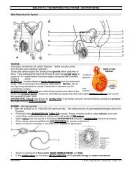
9. Label the diagram of the male reproductive system.<br />
1. BLADDER<br />
2. SEMINAL VESICLE<br />
3. VAS DEFERENS<br />
4. PROSTATE GLAND<br />
5. COWPER’S GLAND<br />
6. URETHRA<br />
7. EPIDIDYMIS<br />
8. SEMINIFEROUS TUBULES<br />
9. INTERSTITIAL CELLS<br />
10. PENIS<br />
11. TESTIS<br />
<strong>12</strong>. SCROTUM<br />
13. EPIDIDYMIS<br />
10. List the structures through which sperm passes in order, from the following list: epididymis, seminiferous<br />
tubules, urethra, penis, vas deferens. SEMINIFEROUS TUBULES, EPIDIDYMIS, VAS DEFERENS,<br />
URETHRA, PENIS<br />
11. Label the parts of the diagram of the sperm cell and list a function for each part:<br />
RAYCROFT Worksheet - Reproduction - Key.doc - Page 2 of 4
Name: Block: Date:<br />
Name Function<br />
W HEAD CONTAINS THE MALE’S 23 CHROMOSOMES<br />
X ACROSOME CAP CONTAINS ENZYMES FOR ENTERING EGG<br />
Y MID-PIECE CONTAINS MITOCHONDRIA FOR ENERGY FOR MOVEMENT<br />
Z TAIL WIGGLES BACK AND FORTH TO PROPEL SPERM<br />
<strong>12</strong>. List 3 function of testosterone<br />
A PROMOTES NORMAL DEVELOPMENT AND FUNCTION OF PRIMARY SEXUAL ORGANS<br />
B CAUSES DEVELOPMENT OF SECONDARY SEXUAL CHARACTERISTICS DURING PUBERTY<br />
C TESTOSTERONE IS NECESSARY FOR THE DEVELOPMENT OF SPERM<br />
13. <strong>The</strong> HYPOTHALAMUS produces the hormone GnRH when testosterone and INHIBIN levels are LOW.<br />
14. This causes the ANTERIOR pituitary gland to release FSH and LH.<br />
15. LH causes INTERSTITIAL cells in the testes to release more TESTOSTERONE.<br />
16. FSH causes the seminiferous tubules to absorb more TESTOSTERONE, which in turn causes them to<br />
produce more SPERM. As it makes more sperm, it also releases more of the hormone INHIBIN. High<br />
levels of this hormone feedback to the HYPOTHALAMUS and PITUITARY, causing them to release less of<br />
their hormones.<br />
17. Label the following diagram and give a function for each labeled part.<br />
Name Function<br />
UTERUS SITE WHERE EMBRYO DEVELOPS<br />
OVIDUCT CONDUCTS EGG TO UTERUS<br />
OVARY PRODUCE HORMONES AND RELEASE EGGS<br />
VAGINA RECEIVES PENIS AND SERVES AS BIRTH CANAL<br />
18. List 3 functions of estrogen:<br />
A STIMULATES THE GROWTH OF THE UTERUS AND VAGINA, NECESSARY FOR EGG MATURATION<br />
B CAUSES AND MAINTAINS SECONDARY SEX CHARACTERISTICS AT PUBERTY<br />
C RESPONSIBLE FOR PROLIFERATIVE PHASE OF UTERINE CYCLE<br />
19. <strong>The</strong> entrance to the uterus is called the CERVIX.<br />
20. <strong>The</strong> female erectile organ containing many sensory nerve receptors is called the CLITORIS.<br />
21. the menstrual cycle lasts on average 28 days. Day 1 is the first day that MENSTRUATION starts, and<br />
usually finishes by day 5.<br />
22. During menstruation, levels of female HORMONES are low.<br />
23. In the follicular phase (days 1 – 14), low levels of hormones are detected by the hypothalamus, which<br />
releases GnRH. This is sent to the pituitary gland, which releases FSH and LH.<br />
24. FSH causes several immature EGGS, along with their surrounding FOLLICLE cells, in the ovaries to begin<br />
to develop. <strong>The</strong> developing follicle cells release increasing amounts of ESTROGEN.<br />
RAYCROFT Worksheet - Reproduction - Key.doc - Page 3 of 4
25. This hormone is responsible for the PROLIFERATIVE phase of the uterine cycle. In the uterus, BLOOD<br />
vessels and GLANDS proliferate.<br />
26. Rising levels of estrogen cause the release of a large amount of LH on about day 13 which causes<br />
OVULATION.<br />
27. Ovulation normally occurs on day 14. In ovulation, the EGG is released from the ovary, leaving behind the<br />
FOLLICLE cells, which go on to form the CORPUS LUTEUM. This structure continues to release the<br />
hormones estrogen and progesterone. Of these two hormones, PROGESTERONE is most important for<br />
the luteal phase of the ovarian cycle. This hormone cause the SECRETORY phase of the uterine cycle.<br />
<strong>The</strong> uterine glands mature and release a thick mucus, and the endometrium DOUBLES in thickness.<br />
28. High levels of PROGESTERONE cause NEGATIVE feedback to the anterior pituitary, shutting down the<br />
release of LH. Lower levels of LH cause the CORPUS LUTEUM to disintegrate. Since it is breaking down,<br />
it can no longer release estrogen and progesterone.<br />
29. Low levels of female HORMONES by day 28 will cause the uterine LINING to be shed, and the cycle will<br />
start anew.<br />
30. However, if fertilization happens, the MENSTRUAL cycle will be interrupted. Fertilization usually occurs in<br />
the upper OVIDUCT. <strong>The</strong> fertilized egg is first called a ZYGOTE and then an EMBRYO as it divides<br />
through mitosis.<br />
31. <strong>The</strong> embryo, upon reaching the UTERUS, will embed itself into the endometrium. This is called<br />
IMPLANTATION.<br />
32. A shared set of membranes called the placenta forms around the embryo. This will begin to secrete the<br />
hormone HCG, which temporarily maintains the corpus luteum.<br />
33. As the placenta develops and matures, it makes its own ESTROGEN and PROGESTERONE. This will<br />
maintain the uterine lining so that MENSTRUATION does not occur during pregnancy.<br />
34. After 9 months, the fetus is ready to be born. <strong>The</strong> pressure of the baby’s head against the cervix causes a<br />
nerve impulse to be sent to the hypothalamus. This causes the hypothalamus to release the hormone<br />
OXYTOCIN to the pituitary, which releases it into the blood. This hormone causes LABOUR. It operates<br />
on a POSITIVE feedback loop. <strong>The</strong> hormone causes the uterine muscles to CONTRACT with ever greater<br />
intensity until the baby is pushed out of the uterus through the VAGINA, which serves as the birth canal.<br />
35.<br />
RAYCROFT Worksheet - Reproduction - Key.doc - Page 4 of 4