Light-harvesting chlorophyll c-like pigment in Prochloron
Light-harvesting chlorophyll c-like pigment in Prochloron
Light-harvesting chlorophyll c-like pigment in Prochloron

You also want an ePaper? Increase the reach of your titles
YUMPU automatically turns print PDFs into web optimized ePapers that Google loves.
680 Microbiology: Larkum et al.<br />
Table 1. The ratios of Chl a/b- and Chl a/c-<strong>like</strong> <strong>pigment</strong> found<br />
by reverse-phase HPLC analysis (method 1) for <strong>Prochloron</strong> sp.<br />
from five didemnid ascidians collected at One Tree Reef,<br />
Capricorn Section, Great Barrier Reef<br />
Species of ascidian host for <strong>Prochloron</strong> sp. Chl a/b Chl a/c-<strong>like</strong><br />
Didemnum molle (Herdmann, 1886) 3.1 15.8<br />
Diplosoma virens (Hartmeyer, 1908) 2.9 5.0<br />
Lissocl<strong>in</strong>um patella (Gottschaldt, 1898) 3.1 6.2<br />
Lissocl<strong>in</strong>um bistratum (Sluiter, 1905) 7.6 9.6<br />
Trididemnum paracyclops (Kott, 1980) 3.55 6.1<br />
Quantitation based on absorbance of <strong>in</strong>dividual peaks and ext<strong>in</strong>ction<br />
coefficients <strong>in</strong> methanol (12). Note that 90% acetone/10%o water<br />
extracts of the symbionts gave similar values for the Chl a/b ratio<br />
and, surpris<strong>in</strong>gly, also for the Chl a/c-<strong>like</strong> ratio (13), despite the<br />
difference <strong>in</strong> absorption maximum of Chl c and MgDVP <strong>in</strong> the red<br />
region of the spectrum (14).<br />
c-<strong>like</strong> <strong>pigment</strong>s. Thus, carefully prepared frozen or freezedried<br />
samples of <strong>Prochloron</strong> from L. patella were further<br />
analyzed <strong>in</strong> Sydney (the frozen samples came from a site at<br />
15-m depth near Lizard Island, Great Barrier Reef; the<br />
freeze-dried samples were a gift to G.C.C. from R. Lew<strong>in</strong> and<br />
were collected <strong>in</strong> Palau, Micronesia). TLC analysis (14, 19)<br />
revealed the presence of two Chl c-<strong>like</strong> compounds that had<br />
a significantly greater Rf value than those of either Chl c1 or<br />
Chl c2 (Fig. 2) and migrated <strong>in</strong> the relative position ofMgDVP<br />
(14, 19). There is also evidence from the fluorescence spectra<br />
of two related <strong>pigment</strong>s (see below). The presence of a Chl<br />
c-<strong>like</strong> <strong>pigment</strong> was also confirmed by reverse-phase HPLC<br />
with a polyethylene column, but only one broad peak was<br />
resolved (Fig. 3). The spectral peaks of the compound(s)<br />
eluted at 7.9 m<strong>in</strong> (Fig. 3 Lower, solid curve) (438 nm, 576 nm,<br />
and 626 nm-<strong>in</strong> 90% acetone/10% water) agree well with<br />
those of MgDVP (14, 19) <strong>in</strong> the same solvent. With extracts<br />
of brown algae or d<strong>in</strong>oflagellates, Chl c1 or Chl c2 or both<br />
were eluted later (not shown). The spectral peaks of the<br />
demetallated Chl c-<strong>like</strong> <strong>pigment</strong> (Table 2) (419 nm, 568 nm,<br />
and 588 nm) match well with those of demetallated MgDVP<br />
(11, 20). In addition, cells ofM. pusilla were extracted <strong>in</strong> 90%<br />
0<br />
0<br />
.0<br />
0<br />
.0<br />
0.2<br />
ol 1<br />
Time, m<strong>in</strong><br />
300 500<br />
Wavelength, nm<br />
700<br />
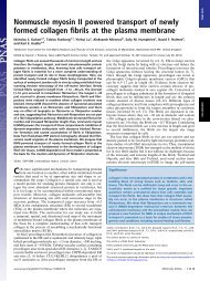
FIG. 1. (Upper) An elution profile (Adet = 440 mm) of the 90%o<br />
acetone extract of <strong>Prochloron</strong> sp. from Diplosoma virens separated<br />
by reverse-phase HPLC (method 1; see Table 1) on a Novapak C18<br />
column (Table 1). Bands: a, Chl a; b, Chl b; c, Chl c-<strong>like</strong> <strong>pigment</strong>;<br />
others, carotenoids. (Lower) On-l<strong>in</strong>e spectrum of Chl c-<strong>like</strong> <strong>pigment</strong><br />
eluted as band c at 14.4 m<strong>in</strong>. [This band was also detected by<br />
fluorescence emission <strong>in</strong> the red region of the spectrum (excitation,<br />
440 nm) with a maximum at 635 nm (not shown).]<br />
0<br />
co<br />
0 .0<br />
Q<br />
Proc. Natl. Acad. Sci. USA 91 (1994)<br />
O carotenoids<br />
* phaeophyt<strong>in</strong>s<br />
0 chi a(±b)<br />
@ chl c-<strong>like</strong><br />
e chl c-<strong>like</strong><br />
* chl c1<br />
0 chl c2<br />
* Orig<strong>in</strong><br />
A B C D E F<br />
o'<br />
. 0<br />
0 0 @ 0<br />
* 0 0 0 - -<br />
% a) 440 nm<br />
.07 . b)438 nm<br />
.05<br />
.05 .04 a<br />
.03b .03<br />
I 1 500 600<br />
Wavelength, nm<br />
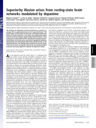
FIG. 2. (Upper) Diagram of a developed th<strong>in</strong>-layer chromatogram<br />
(14) of the follow<strong>in</strong>g 90% acetone extracts. Lanes: A, Endarachne<br />
b<strong>in</strong>ghamiae (Phaeophyta); B, Colpomenia s<strong>in</strong>uosa (Phaeophyta); C,<br />
<strong>Prochloron</strong> sp. (L. patella from Palau); D, <strong>Prochloron</strong> sp. (L. patella<br />
from Lizard Island); E, Pavlova lutheri (Prymnesiophyta); F, Amphid<strong>in</strong>ium<br />
carterae (Pyrrophyta). The develop<strong>in</strong>g solvent was acetone.<br />
In lane C only the lower Chl c-<strong>like</strong> <strong>pigment</strong> was clearly visible,<br />
s<strong>in</strong>ce the upper Chl c-<strong>like</strong> <strong>pigment</strong> was overla<strong>in</strong> by an orange<br />
<strong>pigment</strong>, probably a carotenoid. (Lower) Spectra of two Chl c-<strong>like</strong><br />
<strong>pigment</strong>s <strong>in</strong> 90% acetone from lane D <strong>in</strong> Upper. Spectra: a, spectrum<br />
of lower Chl c-<strong>like</strong> (stippled) <strong>pigment</strong>; b, spectrum ofupper Chl c-<strong>like</strong><br />
(hatched) <strong>pigment</strong>. (Amax for the major peak of a and b spectra are<br />
shown.)<br />
acetone/10% water and analyzed by HPLC with a C18<br />
column (method 1). A Chl c-<strong>like</strong> <strong>pigment</strong> was present, which<br />
was coeluted with the major Chl c-<strong>like</strong> <strong>pigment</strong> from <strong>Prochloron</strong>.<br />
It also had similar spectral properties to the Chl c-<strong>like</strong><br />
<strong>pigment</strong> of <strong>Prochloron</strong> (Table 2). The Chl c-<strong>like</strong> <strong>pigment</strong> <strong>in</strong><br />
Micromonas spp. has been tentatively identified as MgDVP<br />
(12, 14, 19) on the basis of its spectral properties and elution<br />
characteristics on TLC and HPLC.<br />
MgDVP has a fully unsaturated porphyr<strong>in</strong> macrocycle and<br />
does not carry a long-cha<strong>in</strong> esterify<strong>in</strong>g alcohol at C-17 and<br />
differs from Chl c2 only <strong>in</strong> the presence at C-17 of a propionic<br />
acid <strong>in</strong>stead of an acrylic acid (14). Evidence suggests that<br />
MgDVP is an <strong>in</strong>termediate <strong>in</strong> the pathway of Chl a [and<br />
bacterio<strong>chlorophyll</strong> (BChl)] biosynthesis (21). It was first<br />
documented <strong>in</strong> the photosynthetic bacterium Rhodobacter<br />
sphaeroides grown <strong>in</strong> the presence of 8-hydroxyqu<strong>in</strong>ol<strong>in</strong>e,<br />
but it probably occurs also <strong>in</strong> some mutants ofR. sphaeroides<br />
as a result of a lesion <strong>in</strong> the normal synthesis of BChl (21).<br />
MgDVP was also identified as a <strong>pigment</strong> ofunknown function<br />
<strong>in</strong> several mar<strong>in</strong>e flagellates (22) <strong>in</strong>clud<strong>in</strong>g the pras<strong>in</strong>ophyte<br />
M. pusilla, where it has s<strong>in</strong>ce been shown to be present <strong>in</strong> an<br />
LHC as a light-<strong>harvest<strong>in</strong>g</strong> <strong>pigment</strong> (23, 24). However, it<br />
should be po<strong>in</strong>ted out that the precise structure of the Chl<br />
c-<strong>like</strong> <strong>pigment</strong> <strong>in</strong> M. pusilla and the related alga Mantoniella<br />
squamata is debated (12, 20) [both algae are placed <strong>in</strong> the<br />
special group, the micromonadophytes (25)]. MgDVP is also<br />
probably present <strong>in</strong> the prochlorophyte Prochlorococcus<br />
mar<strong>in</strong>us (11), although full details have yet to be documented.<br />
On the basis of the present evidence, we conclude that a chl<br />
c-<strong>like</strong> <strong>pigment</strong>, similar to MgDVP, occurs <strong>in</strong> <strong>Prochloron</strong> sp.<br />
together with a closely related <strong>pigment</strong>; however, both <strong>pigment</strong>s<br />
need to be further exam<strong>in</strong>ed by nuclear magnetic