Familial benign chronic pemphigus (Hailey-Hailey Disease ... - SciELO
Familial benign chronic pemphigus (Hailey-Hailey Disease ... - SciELO
Familial benign chronic pemphigus (Hailey-Hailey Disease ... - SciELO

Create successful ePaper yourself
Turn your PDF publications into a flip-book with our unique Google optimized e-Paper software.
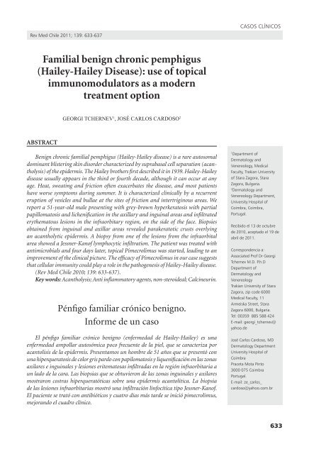
Rev Med Chile 2011; 139: 633-637<br />
<strong>Familial</strong> <strong>benign</strong> <strong>chronic</strong> <strong>pemphigus</strong><br />
(<strong>Hailey</strong>-<strong>Hailey</strong> <strong>Disease</strong>): use of topical<br />
immunomodulators as a modern<br />
treatment option<br />
ABSTRACT<br />
GeorGi tcherneV 1 , José carlos cardoso 2<br />
Benign <strong>chronic</strong> familial <strong>pemphigus</strong> (<strong>Hailey</strong>-<strong>Hailey</strong> disease) is a rare autosomal<br />
dominant blistering skin disorder characterized by suprabasal cell separation (acantholysis)<br />
of the epidermis. The <strong>Hailey</strong> brothers first described it in 1939. <strong>Hailey</strong>-<strong>Hailey</strong><br />
disease usually appears in the third or fourth decade, although it can occur at any<br />
age. Heat, sweating and friction often exacerbates the disease, and most patients<br />
have worse symptoms during summer. It is characterized clinically by a recurrent<br />
eruption of vesicles and bullae at the sites of friction and intertriginous areas. We<br />
report a 51-year-old male presenting with grey-brown hyperkeratosis with partial<br />
papillomatosis and lichenification in the axillary and inguinal areas and infiltrated<br />
erythematous lesions in the infraorbitary region, on the side of the face. Biopsies<br />
obtained from inguinal and axillar areas revealed parakeratotic crusts overlying<br />
an acantholytic epidermis. A biopsy from one of the lesions from the infraorbital<br />
area showed a Jessner-Kanof lymphocytic infiltration. The patient was treated with<br />
antimicrobials and four days later, topical Pimecrolimus was started, leading to an<br />
improvement of the clinical picture. The efficacy of Pimecrolimus in our case suggests<br />
that cellular immunity could play a role in the pathogenesis of <strong>Hailey</strong>-<strong>Hailey</strong> disease.<br />
(Rev Med Chile 2010; 139: 633-637).<br />
Key words: Acantholysis; Anti inflammatory agents, non-steroideal; Calcineurin.<br />
Pénfigo familiar crónico <strong>benign</strong>o.<br />
informe de un caso<br />
El pénfigo familiar crónico <strong>benign</strong>o (enfermedad de <strong>Hailey</strong>-<strong>Hailey</strong>) es una<br />
enfermedad ampollar autosómica poco frecuente de la piel, que se caracteriza por<br />
acantolisis de la epidermis. Presentamos un hombre de 51 años que se presentó con<br />
una hiperqueratosis de color gris pardo con papilomatosis y liquenificación en las zonas<br />
axilares e inguinales y lesiones eritematosas infiltradas en la región infraorbitaria a<br />
un lado de la cara. Las biopsias que se obtuvieron de las zonas inguinales y axilares<br />
mostraron costras hiperqueratóticas sobre una epidermis acantolítica. La biopsia<br />
de las lesiones infraorbitarias mostró una infiltración linfocítica tipo Jessner-Kanof.<br />
El paciente se trató con antibióticos y cuatro días más tarde se inició pimecrolimus,<br />
mejorando el cuadro clínico.<br />
CASOS CLíNiCOS<br />
1 Department of<br />
Dermatology and<br />
Venereology, Medical<br />
Faculty, Trakian University<br />
of Stara Zagora, Stara<br />
Zagora, Bulgaria.<br />
2 Dermatology and<br />
Venereology Department,<br />
University Hospital of<br />
Coimbra, Coimbra,<br />
Portugal.<br />
Recibido el 13 de octubre<br />
de 2010, aceptado el 19 de<br />
abril de 2011.<br />
Correspondencia a:<br />
Associated Prof Dr Georgi<br />
Tchernev M.D. P.h.D<br />
Department of<br />
Dermatology and<br />
Venereology<br />
Trakian University of Stara<br />
Zagora, zip code 6000<br />
Medical Faculty, 11<br />
Armeiska Street, Stara<br />
Zagora 6000, Bulgaria.<br />
Tel: 00359 885 588 424<br />
E-mail: georgi_tchernev@<br />
yahoo.de<br />
José Carlos Cardoso, MD<br />
Dermatology Department<br />
University Hospital of<br />
Coimbra<br />
Praceta Mota Pinto<br />
3000 075 Coimbra<br />
Portugal.<br />
E-mail: ze_carlos_<br />
cardoso@yahoo.com.br<br />
633
CASOS CLíNiCOS<br />
<strong>benign</strong> <strong>chronic</strong> familial <strong>pemphigus</strong> (haileyhailey<br />
disease [hhd]) is a hereditary<br />
blistering skin disease, transmitted in an<br />
autosomal dominant way with variable genetic penetrance<br />
1 . it was described for the first time in 1939<br />
by the two brothers hugh and howard hailey 2 . it<br />
is hypothesized that the main reason for the pathologic<br />
changes is an altered protein composition<br />
of desmosomes leading to acantholysis, deriving<br />
from ATP 2 C 1 gene mutation localized on chromosome<br />
3q 2 . The ATP 2 C 1 gene codes a Ca 2+ -pump that<br />
regulates the transportation of calcium from the<br />
cytosol into the Golgi apparatus 2,3 . in approximately<br />
70% of the cases a positive family history may<br />
be elicited. more frequently, the disease becomes<br />
apparent during puberty, and intertriginous<br />
areas are preferentially affected, namely axillary,<br />
inguinal and neck folds. mucous membranes are<br />
far less affected. longitudinal white lines on the<br />
nails are frequently noticed. bacterial and fungal<br />
super infection, maceration and frequent sweating<br />
(axillary and inguinal hyperhidrosis) are considered<br />
to be important aggravating factors 4 . however,<br />
some authors consider mechanical irritation to<br />
be the only provoking factor. Vesicular lesions<br />
covered by crusts, erosions and wart-like papules<br />
are other possible clinical findings. histologically,<br />
there is acantholysis of large areas of the epidermis,<br />
giving rise to the appearance that was compared to<br />
a “dilapidated brick wall”. dyskeratosis can also be<br />
noticed 5 . decomposition of the desmosome-keratin<br />
filament complex is ultrastructurally proved.<br />
Clinically, the disease has a fluctuating course, and<br />
both the activity and the affected areas may vary 2,5 .<br />
Case report<br />
A 51-year-old patient came to the clinic with<br />
skin lesions localized to the axillary and inguinal<br />
areas that were present since he was 18 years of age<br />
(figures 1-4). Topically applied therapy, including<br />
corticosteroids, antimycotics and antibiotics had<br />
been ineffective. he did not have any relatives<br />
suffering from similar lesions.<br />
on observation, grey-brown hyperkeratosis<br />
with partial papillomatosis and lichenification<br />
were noticed in the axillary and inguinal areas (figures<br />
1-4). Additionally, infiltrated erythematous<br />
lesions were found in the infraorbitary region, on<br />
the side of the face.<br />
634<br />
<strong>Familial</strong> <strong>benign</strong> <strong>chronic</strong> <strong>pemphigus</strong>. Report of one case - G. Tchernev et al<br />
laboratory findings: differential blood count:<br />
neutrophils: 40.2%; monocytes: 11%; eosinophils:<br />
9.8%; triglycerides: 2.19 mmol/l, Ca: 2.2 mmol/l;<br />
blood sugar: 6.0 mmol/l.<br />
erythrocyte sedimentation rate, hemoglobin,<br />
haematocrit, cholesterol with all subfractions,<br />
aminostransferases, GGT, electrolytes and iron<br />
were all normal. Serological test for syphilis and<br />
antistreptolysin o titer were negative.<br />
direct immunofluorescence from lesional skin<br />
and indirect immunofluorescence studies were<br />
both negative.<br />
histological analysis of biopsies obtained from<br />
inguinal and axillar areas revealed parakeratotic<br />
crusts overlying an acantholytic epidermis, with<br />
acantholytic cells and rare diskeratotic cells. A<br />
biopsy from one of the lesions from the infraorbital<br />
area was in keeping with Jessner-Kanof<br />
lymphocytic infiltration.<br />
Smears for microbiologic analyses from the<br />
axillary and inguinal areas disclosed Staphylococcus<br />
aureus, E. coli and Pseudomonas aeruginosa.<br />
Treatment and outcome<br />
initially, intravenous antibiotic therapy was<br />
started with flucloxacillin 2 g three times daily<br />
and ampicillin 1 g three times daily, due to the<br />
proved superinfection with Gram-positive and<br />
Gram-negative bacteria, as well as local therapy<br />
with chlorhexidin 1% aqueous solution. four days<br />
after beginning antibiotic therapy, specific therapy<br />
with topical pimecrolimus twice a day was started,<br />
leading to a significant and fast improvement of<br />
the clinical picture.<br />
Discussion<br />
The differential diagnosis of familial <strong>benign</strong><br />
<strong>chronic</strong> <strong>pemphigus</strong> includes all the most frequent<br />
skin diseases manifesting at the axillary and inguinal<br />
areas.<br />
Pemphigus vegetans, Neumann type, is not<br />
frequently considered in the differential diagnosis.<br />
in this disease direct immunofluorescence proves<br />
deposition of lgG and C -fraction of the comple-<br />
3<br />
ment, and indirect immunofluorescence shows<br />
the presence of anti-desmoglein 3 antibodies. in<br />
hailey-hailey disease, alterations of the humoral<br />
immunity are not found.<br />
The clinical differentiation from inverse psoriasis<br />
is often very difficult, particularly in macerated<br />
Rev Med Chile 2011; 139: 633-637
<strong>Familial</strong> <strong>benign</strong> <strong>chronic</strong> <strong>pemphigus</strong>. Report of one case - G. Tchernev et al<br />
and superinfected lesions 2,6 . histological features<br />
provide essential clues to the differentiation between<br />
the two diseases, as the typical acantholysis<br />
and occasional dyskeratosis found in hhd are not<br />
features of psoriatic lesions.<br />
The diagnosis of acanthosis nigricans may<br />
also be evoked, especially in the case of hyperpigmented<br />
lesions of hhd that may elude clinical<br />
diagnosis.<br />
CASOS CLíNiCOS<br />
Figures 1-4. Clinical manifestation of <strong>Hailey</strong>-<strong>Hailey</strong> disease in the intertriginous areas: brown macerated and hyperpigmented<br />
ranges at the axillary and inguinal areas.<br />
Rev Med Chile 2011; 139: 633-637<br />
The histopathological and clinical differentiation<br />
between hhd and darier’s disease is not<br />
always easy 2,5,7 . in these cases the clinical picture<br />
is decisive, as is the presence of dyskeratotic cells<br />
and more focal acantholysis in darier’s disease,<br />
contrasting with the more diffuse acantholysis and<br />
less or inexistent dyskeratosis in hhd.<br />
Candidal superinfection seems to be a frequent<br />
concomitant infection in lesions of hhd, the same<br />
635
CASOS CLíNiCOS<br />
as in inverse psoriasis and in different forms of<br />
<strong>pemphigus</strong> vegetans.<br />
during the last years, the genetic cause and molecular<br />
pathogenesis of hailey-hailey disease have<br />
been elucidated. The distribution of intracellular<br />
Ca 2+ plays an important role in the regulation of<br />
cell-cell interactions in the epidermis 3 . damage<br />
of desmosomes gives rise to acantholysis, the<br />
characteristic finding in hailey-hailey disease. it<br />
is considered that the increase in cytosolic calcium,<br />
as well as its reduction in the Golgi apparatus leads<br />
to reduced glycosylation and incorrect arrangement<br />
of the intercellular adhesion molecules, i.e. of<br />
the desmosomal proteins 2 . The exact cause of the<br />
mutation of the ATP 2 C 1 gene, which is responsible<br />
for the metabolic disorders at both cellular and sub<br />
cellular levels, remains unclear 8,9 .<br />
The disclosure of the pathogenic mechanisms<br />
in hailey-hailey disease, as well as the creation<br />
of new medications shall probably lead to better<br />
therapeutic effect. The therapies that are usually<br />
applied, like topical corticosteroids, antibiotics,<br />
both topical and oral antimycotics, and isotretinoin<br />
are not always a successful treatment option.<br />
Chronic treatment with corticosteroids may lead<br />
to skin atrophy, telangiectasia, striae distensae and,<br />
possibly, to increased vulnerability to infections in<br />
the affected areas 2 . Calcineurin inhibitors (cyclosporin<br />
A, tacrolimus and pimecrolimus), initially<br />
introduced for post-transplant immunosupression,<br />
proved also useful for some inflammatory<br />
skin diseases, namely atopic eczema 2 .<br />
during last years, tacrolimus and pimecrolimus<br />
were studied and showed a very favorable<br />
clinical effect both in short and long duration<br />
treatments in patients with atopic dermatitis and,<br />
consequently, these molecules gained recognition<br />
as good topical immunomodulators 2,11 . The topical<br />
application of both preparations is not associated<br />
with any of the undesirable side effects arising after<br />
oral treatment with cyclosporin A. Pimecrolimus<br />
penetrates less through the skin and this leads, at<br />
least theoretically, to less risk of systemic effects<br />
compared to tacrolimus 2 . Pimecrolimus shows<br />
a higher affinity to epithelial structures, but its<br />
affinity to lymphoid structures is lower compared<br />
to tacrolimus 2 .<br />
Tacrolimus, pimecrolimus and cyclosporin A<br />
block calcineurin in the cytoplasm and lead to<br />
suppression of T-cell function. Tacrolimus and<br />
pimecrolimus achieve these effects through the<br />
636<br />
<strong>Familial</strong> <strong>benign</strong> <strong>chronic</strong> <strong>pemphigus</strong>. Report of one case - G. Tchernev et al<br />
initial linkage to the cytosolic receptor fKbP-12,<br />
designated also as macrophillin-12 2,10 . in contrast<br />
to them, cyclosporin A binds to another cytosolic<br />
receptor from the macrophillin group, the<br />
cyclophillin 2 . The macrophillin-12 – Cl complex<br />
blocks the calcineurin phosphatase and this<br />
leads to activation (through dephosphorylation)<br />
of NfAT (nuclear factor of activated T-cells).<br />
This blocks the transportation of NfAT to the<br />
nucleus, inhibiting the synthesis of immunomodulatory<br />
cytokines: il-2, il-4, il-8, TNf-α and<br />
γ-interferon. Consequently, there is impairment<br />
in the proliferation of T-cells that participate and<br />
maintain the inflammatory process. Pimecrolimus<br />
also blocks the expression of the receptors that<br />
take part in the processes of differentiation of the<br />
T-cell population 2,12 .<br />
An important advantage of both medications<br />
is that they do not block the synthesis of collagen<br />
in skin fibroblasts and do not lead to atrophy, in<br />
contrast to topical corticosteroids. The immunosuppressive<br />
effect of pimecrolimus is weaker than<br />
that of tacrolimus 2 .<br />
This allowed also the application of a modern<br />
pathogenic approach leading to a quick control<br />
of clinical skin abnormalitie without inducing<br />
the side effects usually associated with corticosteroids<br />
11,12 . A successful treatment of patients<br />
suffering from hailey-hailey disease applying tacrolimus<br />
has been described in the literature 12 , and<br />
this makes us provide this information about the<br />
favorable local treatment held with pimecrolimus.<br />
in untreated skin lesions of hailey-hailey disease,<br />
a significant infiltration with T-lymphocytes<br />
has been demonstrated in previous works, considerably<br />
decreasing after the therapy not only<br />
with tacrolimus and pimecrolimus, but also with<br />
cyclosporin-A 2 .<br />
The cause of T-cell infiltration in hailey-hailey<br />
disease is not clear yet. The fast beneficial impact<br />
of immune regulators on skin symptoms suggests,<br />
however, that T-lymphocytes play some role in the<br />
pathogenesis of the disease.<br />
The specific effect of tacrolimus and pimecrolimus,<br />
blocking the pathologic T-cellular response<br />
shows that in the pathogenesis of hailey-hailey<br />
disease, additionally to the genetic defect, a dysregulation<br />
of the cellular immune response is also<br />
present. Suppression of the proliferation of T-cells<br />
by pimecrolimus quickly controls the acantholysis<br />
in this disease 2 .<br />
Rev Med Chile 2011; 139: 633-637
<strong>Familial</strong> <strong>benign</strong> <strong>chronic</strong> <strong>pemphigus</strong>. Report of one case - G. Tchernev et al<br />
Topical pimecrolimus treatment applied in<br />
the form of cream twice a day led, in the present<br />
case, to a quick response of skin lesions. during<br />
the therapy no side effects were noticed, neither<br />
local, such as itching or burning sensations, nor<br />
any systemic complaints.<br />
References<br />
1. ding YG, fang h, lao lm, Jiang XJ, Chen hC. Genetic<br />
diagnosis of hailey-hailey disease in two Chinese families:<br />
novel mutations in the ATP2C1 gene. Clin exp<br />
dermatol 2009; 34: e968-71.<br />
2. Tchernev G, Zouboulis CC, orfanos C. Treatment of<br />
morbus hailey-hailey with pimecrolimus. bulgarian J<br />
dermatol Venereol 2004; 153: 37-40.<br />
3. leinonen PT, hägg Pm, Peltonen S, Jouhilahti em, melkko<br />
J, Korkiamäki T, et al. reevaluation of the normal<br />
epidermal calcium gradient, and analysis of calcium<br />
levels and ATP receptors in hailey-hailey and darier<br />
epidermis. J invest dermatol 2009; 129: 1379-87. epub<br />
2008 dec 4.<br />
4. hamada T, fukuda S, Sakaguchi S, Yasumoto S, Kim SC,<br />
hashimoto T. molecular and clinical characterization in<br />
Japanese and Korean patients with hailey-hailey disease:<br />
six new mutations in the ATP2C1 gene. J dermatol<br />
Sci 2008; 51: 31-6.<br />
5. wilgram Gf, Caufield Jb, lever wf. electron microscopic<br />
studies in skin diseases with acantholysis (pemphi-<br />
Rev Med Chile 2011; 139: 633-637<br />
CASOS CLíNiCOS<br />
gus vulgaris, <strong>pemphigus</strong> familiaris <strong>benign</strong>us <strong>chronic</strong>us,<br />
darier’s disease). dermatol wochenschr 1964; 147:<br />
281-92.<br />
6. Stoianov S, ivanov i. [Case of hailey-hailey disease<br />
(<strong>pemphigus</strong> <strong>chronic</strong>us <strong>benign</strong>us familiaris).] izv meditsinskite<br />
inst bulg Akad Naukite Sofia otd biol meditsinski<br />
Nauki 1955; 11-12: 565-74.<br />
7. Kovaks Z. [on the relationship between bullous form of<br />
darier’s disease (dyskeratosis follicularis typ bullosus)<br />
and hailey-hailey disease (<strong>pemphigus</strong> <strong>benign</strong>us <strong>chronic</strong>us<br />
familiaris)]. borgyogy Venerol Sz 1960; 36: 7-13.<br />
8. Cheng TS, ho Km, lam Cw. heterogeneous mutations<br />
of the ATP2C1 gene causing hailey-hailey disease in<br />
hong Kong Chinese. J eur Acad dermatol Venereol.<br />
2010 mar 4. [epub ahead of print].<br />
9. ma Ym, Zhang XJ, liang Yh, ma l, Sun ld, Zhou fS,<br />
et al. Genetic diagnosis in a Chinese hailey-hailey disease<br />
pedigree with novel ATP2C1 gene mutation. Arch<br />
dermatol res 2008; 300: 203-7. epub 2008 feb 8.<br />
10. Persić-Vojinović S, milavec-Puretić V, dobrić i, rados<br />
J, Spoljar S. disseminated hailey-hailey disease treated<br />
with topical tacrolimus and oral erythromycin: Case<br />
report and review of the literature. Acta dermatovenerol<br />
Croat 2006; 14: 253-7.<br />
11. ferraro V, Adamski h, le Gall f, Chevrant-breton J.<br />
[efficacy of topical tacrolimus in hailey-hailey disease].<br />
Ann dermatol Venereol 2006; 133 (5 Pt 1): 475-6.<br />
12. rocha Paris f, fidalgo A, baptista J, Caldas ll, ferreira<br />
A. Topical tacrolimus in hailey-hailey disease. int J<br />
Tissue react 2005; 27: 151-4.<br />
637