Spina Bifida (myelodysplasia) - MDC Faculty Home Pages
Spina Bifida (myelodysplasia) - MDC Faculty Home Pages
Spina Bifida (myelodysplasia) - MDC Faculty Home Pages

Create successful ePaper yourself
Turn your PDF publications into a flip-book with our unique Google optimized e-Paper software.
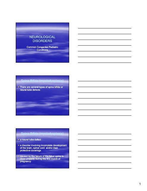
NEUROLOGICAL<br />
DISORDERS<br />
Common Congenital Pediatric<br />
Conditions<br />
<strong>Spina</strong> <strong>Bifida</strong> (<strong>myelodysplasia</strong>)<br />
There are several types of spina bifida or<br />
neural tube defects<br />
<strong>Spina</strong> <strong>Bifida</strong> (<strong>myelodysplasia</strong>)<br />
a neural tube defect<br />
a disorder involving incomplete development<br />
of the the brain brain, spinal cord and/or their<br />
protective coverings<br />
caused by the failure of the fetus’ spine to<br />
close properly during the first month of<br />
pregnancy<br />
1
Types of <strong>Spina</strong> <strong>Bifida</strong>: Occulta<br />
Posterior vertebral arches fail to fuse<br />
No herniation<br />
<strong>Spina</strong>l cord remains intact<br />
Usually not visible<br />
Meninges are not exposed on skin surface<br />
Types of <strong>Spina</strong> <strong>Bifida</strong>: Occulta<br />
Skin depression or dimple<br />
Tufts of hair at base of spine<br />
Port wine angiomatus nevi<br />
Neruologic defects not usually present<br />
May have gait / foot weakness, bowel and bladder<br />
disturbance<br />
2
Types of <strong>Spina</strong> <strong>Bifida</strong>: Cystica<br />
Protrusion of spinal cord and/or it’s<br />
meninges<br />
A visible i ibl d defect f t<br />
Types of <strong>Spina</strong> <strong>Bifida</strong>: Cystica<br />
Meningocele<br />
Protrusion involves meninges and a sac -<br />
like cyst that contains CSF but no neural<br />
elements<br />
l t<br />
Located in the mid-line mid line of the back<br />
Associated with neurological deficits.<br />
Types of <strong>Spina</strong> <strong>Bifida</strong>: Cystica<br />
Myelomeningocele<br />
Protrusion of meninges,CSF, and nerves<br />
Sac is covered with a thin membrane that is<br />
prone to leakage or rupture<br />
Neurological deficits evident, usually in<br />
lumbar sacral area<br />
3
Types of <strong>Spina</strong> <strong>Bifida</strong>: Cystica<br />
Encephalocele<br />
Brain and meninges herniated through<br />
d defect f t in i the th skull k ll i into t a sac<br />
Types of <strong>Spina</strong> <strong>Bifida</strong>: Cystica<br />
Hydrocephalus<br />
Occurs in 90% of cases<br />
Associated with Neural Tube Defect (NTD)<br />
due to downward displacement of the<br />
cerebellum through foramen magnum<br />
Etiology<br />
Specific cause unknown<br />
Factors include:<br />
Heredity<br />
Medication such as Valporic Acid<br />
4
Etiology<br />
Poor maternal nutrition<br />
Radiation<br />
Research indicates that Folic Acid<br />
deficiency of mother is also indicated<br />
Pathophysiology<br />
Fusion failure of vertebral laminae of spinal<br />
column during the 4th week of gestation<br />
V Varying i d degree of f neurological l i l d deficits fi it<br />
present related to the portion of the spinal<br />
cord involved<br />
Pathophysiology<br />
The higher the defect, the greater the<br />
neurologic dysfunction<br />
B Below l 2nd 2 d lumbar l b vertebrae: t b partial ti l<br />
paralysis of lower extremity, varying degrees<br />
of sensory deficits, bowel and bladder<br />
disturbances<br />
5
Complications of NTD<br />
Joint deformities produced in utero causes<br />
hip dislocation, scoliosis, and foot<br />
d deformities f iti<br />
Hydrocephalus may be present<br />
Cerebral paralysis<br />
Complications of NTD<br />
Mental retardation<br />
Fracture related to decreased muscle mass<br />
Painless ulcerations<br />
Injuries<br />
Burns decubitus<br />
Diagnostics<br />
Increased alfa alfa- fetal protein from leaking CSF in<br />
maternal serum and amniotic fluid at 14-16 14 16 weeks<br />
(obtained by amniocentesis)<br />
Ult Ultra sound d of f th the f fetus, t MRI, MRI CT CT scan and d flat fl t<br />
films of spinal column after delivery<br />
Usually evident at newborn exam<br />
Sac examination<br />
6
Therapeutic Management:<br />
medications<br />
Antibiotics: prophalactically to prevent<br />
infection (UTI/s)<br />
Anticholinergics<br />
Anticholinergics-Probanthine: Anticholinergics Probanthine: decreases<br />
bladder tone<br />
Direct Acting Cholinergics<br />
Cholinergics-Urecholine: Urecholine: to<br />
manage urinary incontinence related to<br />
contraction of the bladder<br />
Therapeutic Management:<br />
medications<br />
Antispasmodics<br />
Antispasmodics-Uripas: Uripas: to control bladder<br />
spasm<br />
L Laxatives ti and d stool t l softners softners-Dulcolax ft D DDulcolax l l and d<br />
Colace: to achieve bowel continence<br />
Therapeutic Management:<br />
treatment<br />
Correction of orthopedic deformities:<br />
Casting<br />
Bracing<br />
Traction<br />
Bowel and bladder: surgery and training<br />
program.<br />
7
Therapeutic Management:<br />
surgery<br />
Various neurological and plastic surgical<br />
procedures can be used for skin closure<br />
without disturbing the neural elements or<br />
removing any portion of the sac<br />
Vesicostomy: stoma created on abdominal<br />
wall for urinary drainage<br />
Pre Pre-Operative Operative Care: if<br />
menigocele or mylomeningocele<br />
Assess spinal column for defect<br />
Monitor vital signs and neuro status<br />
Protect the sac: cleanse using sterile<br />
technique and sterile normal saline saline-cover cover<br />
with sterile saline moistened gauze<br />
Pre Pre-Operative Operative Care<br />
No diapers diapers-padding padding underneath<br />
Gentle handling with feeding<br />
Avoid pressure on sac<br />
8
Pre Pre-Operative Operative Care<br />
Prone or side –lying lying<br />
Keep sac free of infection: gentle cleansing<br />
around dth the sac<br />
Avoid contamination with feces or urine<br />
Pre Pre-Operative Operative Care<br />
Observe for signs and symptoms of infection<br />
(meningitis ) and increased ICP (hydrocephalus)<br />
Measure head circumference: high risk for<br />
hd hydrocephalus hl<br />
Avoid exposure to all products that contain latex:<br />
catheters, elastic bandages, baby bottle nipples,<br />
pacifier and balloons<br />
Pre Pre-Operative Operative Care<br />
Symptoms of latex allergy<br />
Watery eyes<br />
Wheezing<br />
Hives<br />
Rash<br />
Swelling<br />
Severe anaphylaxis<br />
9
Post Post-Operative Operative Care<br />
Provide routine post post-op op care<br />
Check vital signs, neuro status, hydration,<br />
intake and output<br />
Check incision<br />
Monitor for urinary retention and stress<br />
incontinence (may diaper)<br />
Post Post-Operative Operative Care<br />
Perform crede’s maneuver to empty<br />
bladder or clean intermittent catheter q 3<br />
hrs hrs- 4 hrs, according to MD’s order<br />
Teach signs and symptoms of UTI<br />
Provide orthopedic appliances if necessary<br />
Post Post-Operative Operative Care<br />
Prevent constipation, develop bowel<br />
program<br />
Teach procedure to child and family<br />
Promote independence<br />
Refer to community agencies (SB<br />
Association)<br />
10
Nursing Diagnoses<br />
High risk for infection<br />
High risk for impaired skin integrity<br />
Altered urinary elimination<br />
Bowel incontinence/constipation<br />
Impaired physical mobility<br />
HYDROCEPHALUS<br />
Increased amount of CSF within the<br />
ventricles of the brain<br />
May be caused by obstruction of CSF flow<br />
or by by overproduction overproduction or inadequate<br />
reabsorption of CSF<br />
May result from congenital malformation or<br />
be secondary to injury, infection, or tumor<br />
Hydrocephalus: Classifications<br />
Noncommunicating:<br />
– Flow of CSF from the ventricles to subarachnoid<br />
space is obstructed<br />
Communicating:<br />
– Flow is not obstructed, but CSF is inadequately<br />
reabsorbed in the subarachnoid space<br />
11
Hydrocephalus: Assessment<br />
Assessment findings depend on age of<br />
onset and amount of CSF in the brain<br />
Infant to 2 years:<br />
Enlarging head size size, bulging bulging, non non-pulsating pulsating<br />
fontanels, downward rotation of eyes<br />
(sunset), poor feeding, vomiting, lethargy,<br />
irritability, high high-pitched pitched cry and abnormal<br />
muscle tone<br />
Hydrocephalus: Assessment<br />
Older Children:<br />
– Changes in head size less common<br />
– Signs of increased ICP (vomiting, ataxia,<br />
headache) common<br />
– Alteration in consciousness and papilledema<br />
late signs<br />
12
SHUNTS<br />
Insertion of a flexible tube into the lateral ventricle<br />
of the brain<br />
Catheter is threaded under the skin and the distal<br />
end positioned positioned in the peritoneum (common) or the<br />
right atrium<br />
Shunt drains excess CSF from the lateral<br />
ventricles; fluid is absorbed by the peritoneum or<br />
absorbed in the general circulation via the right<br />
atrium<br />
Nursing Interventions<br />
Pre Pre-operative operative<br />
– Monitor head circumference<br />
– Monitor for signs of ICP<br />
– S Small ll f frequent t feedings f di<br />
Nursing Interventions<br />
Post Post-operative operative<br />
– Position on opposite side of surgery or back<br />
– Avoid sedation<br />
– Monitor M MMonitor it f for signs i of f ICP<br />
ICP<br />
– Educate parents concerning signs and<br />
symptoms of shunt infection or shunt<br />
malfunction<br />
13
Cerebral Palsy<br />
Cerebral Palsy<br />
A non non-progressive progressive motor disorder of the<br />
CNS resulting in alteration in movement and<br />
posture<br />
C Cause i is t trauma, h hemorrhage, h anoxia i or<br />
infection before, during or after birth<br />
1/3 of children have some degree of mental<br />
retardation<br />
Cerebral Palsy<br />
Classified as:<br />
Spastic<br />
– Spasticity (hypertonicity of muscle groups)<br />
Athetoid<br />
–Worm Worm-like like movements of extremities<br />
Ataxic<br />
– Disturbed coordination<br />
Mixed<br />
14
Cerebral Palsy - Assessment<br />
May have hypertonicity or hypotonia of varying<br />
degrees on different extremities<br />
May have scissoring of the legs<br />
Absence of expected reflexes or presence of<br />
reflexes that extend beyond expected age<br />
Failure to meet developmental milestones<br />
Difficulty swallowing<br />
Altered speech<br />
Nursing Diagnoses<br />
Impaired physical mobility<br />
Self Self-care care deficit<br />
Altered nutrition: less than body<br />
requirements<br />
High risk for injury related to neuromuscular,<br />
perceptual or cognitive impairments<br />
Treatment<br />
Self Self-care care is a goal for all children<br />
– Team approach<br />
Nutrition<br />
– Increased caloric intake<br />
– Special feeding devices<br />
Community referrals<br />
Emotional support<br />
15
ACQUIRED NEUROLOGICAL<br />
DISORDER<br />
Meningitis<br />
Meningitis<br />
Inflammation of the meninges<br />
Most common infection of the CNS<br />
Two primary classifications<br />
–Viral Viral<br />
– Bacterial<br />
Assessment of Meningitis<br />
Viral meningitis<br />
– Infants and toddlers<br />
Infants and toddlers<br />
Irritability, lethargy, vomiting<br />
Change in appetite<br />
– Older children<br />
Usually preceded by a nonspecific febrile illness<br />
Headache, malaise, muscle aches, nausea/vomiting,<br />
photophobia, nuchal/spinal rigidity<br />
16
Assessment of Meningitis<br />
Bacterial meningitis<br />
– Infants and toddlers<br />
Poor feeding/suck, vomiting, high high-pitched pitched cry,<br />
bulging fontanel, fever or hypothermia, poor muscle<br />
tone<br />
– Children and adolescents<br />
Abrupt onset<br />
Fever, chills, headache, nuchal rigidity, vomiting,<br />
changes in LOC, photophobia, extreme irritability<br />
Figure 49 49-6 Opisthotonic position<br />
To test for Kernig sign , raise the child’ s leg with the knee flexed. Then extend the the child’s leg at the knee. If<br />
any resistance is noted or pain is felt , the result is positive Kernig sign. This is a common finding in meningitis.<br />
London / Ladewig / Ball / Bindler<br />
Maternal Maternal-Newborn Newborn and Child Nursing: Family Family-Centered Centered Care<br />
Nursing Diagnoses<br />
Risk for ineffective breathing<br />
Pain<br />
Risk for injury<br />
Risk for ineffective thermoregulation<br />
17
Pressure<br />
Appearance<br />
WBC WBCs (mm (mm³) ³)<br />
Protein (mg/dL)<br />
Sugar (mg/dL)<br />
LP Results<br />
Normal Viral Bacterial<br />
5-15 15<br />
Clear<br />
00-5 5<br />
10 10-30 30<br />
40 40-80 80<br />
Normal/sl ⇑<br />
Clear<br />
Sli Slightly htl ⇑<br />
Slightly ⇑<br />
Normal or ⇓<br />
Nursing Interventions<br />
Elevated<br />
Cloudy<br />
Elevated El Elevated t d<br />
Elevated<br />
Decreased<br />
Place child in isolation until 24 hours of<br />
antibiotic therapy has completed<br />
Administer antibiotics (7-14 (7 14 days)<br />
Fever Fever control<br />
control<br />
Monitor for signs of ICP<br />
Monitor for fluid overload<br />
Viral meningitis is treated symptomatically<br />
Mental Retardation<br />
18
Mental Retardation<br />
Significant below average intellectual functioning<br />
which is associated with impaired learning<br />
difficulties<br />
3% of US population<br />
Causes<br />
– Genetic<br />
–Pre Pre-natal natal<br />
– Perinatal<br />
–Post Post-natal natal<br />
MR - Classifications<br />
Mild<br />
– Slow learner, can work, marry, have children,<br />
may need assistance with crisis<br />
Moderate<br />
– Needs life supervision<br />
Severe<br />
– Needs a caretaker for basic needs<br />
Profound<br />
Interventions<br />
Goal is to promote optimal development<br />
Family support<br />
Community referrals<br />
19