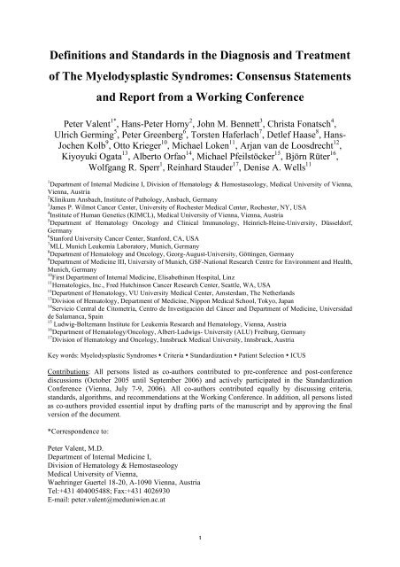
Definitions and Standards in the Diagnosis and ... - Hematologics Inc.
Definitions and Standards in the Diagnosis and ... - Hematologics Inc.
Definitions and Standards in the Diagnosis and ... - Hematologics Inc.

Create successful ePaper yourself
Turn your PDF publications into a flip-book with our unique Google optimized e-Paper software.
<strong>Def<strong>in</strong>itions</strong> <strong>and</strong> St<strong>and</strong>ards <strong>in</strong> <strong>the</strong> <strong>Diagnosis</strong> <strong>and</strong> Treatment<br />
of The Myelodysplastic Syndromes: Consensus Statements<br />
<strong>and</strong> Report from a Work<strong>in</strong>g Conference<br />
Peter Valent 1* , Hans-Peter Horny 2 , John M. Bennett 3 , Christa Fonatsch 4 ,<br />
Ulrich Germ<strong>in</strong>g 5 , Peter Greenberg 6 , Torsten Haferlach 7 , Detlef Haase 8 , Hans-<br />
Jochen Kolb 9 , Otto Krieger 10 , Michael Loken 11 , Arjan van de Loosdrecht 12 ,<br />
Kiyoyuki Ogata 13 , Alberto Orfao 14 , Michael Pfeilstöcker 15 , Björn Rüter 16 ,<br />
Wolfgang R. Sperr 1 , Re<strong>in</strong>hard Stauder 17 , Denise A. Wells 11<br />
1 Department of Internal Medic<strong>in</strong>e I, Division of Hematology & Hemostaseology, Medical University of Vienna,<br />
Vienna, Austria<br />
2 Kl<strong>in</strong>ikum Ansbach, Institute of Pathology, Ansbach, Germany<br />
3 James P. Wilmot Cancer Center, University of Rochester Medical Center, Rochester, NY, USA<br />
4 Institute of Human Genetics (KIMCL), Medical University of Vienna, Vienna, Austria<br />
5 Department of Hematology Oncology <strong>and</strong> Cl<strong>in</strong>ical Immunology, He<strong>in</strong>rich-He<strong>in</strong>e-University, Düsseldorf,<br />
Germany<br />
6 Stanford University Cancer Center, Stanford, CA, USA<br />
7 MLL Munich Leukemia Laboratory, Munich, Germany<br />
8 Department of Hematology <strong>and</strong> Oncology, Georg-August-University, Gött<strong>in</strong>gen, Germany<br />
9 Department of Medic<strong>in</strong>e III, University of Munich, GSF-National Research Centre for Environment <strong>and</strong> Health,<br />
Munich, Germany<br />
10 First Department of Internal Medic<strong>in</strong>e, Elisabeth<strong>in</strong>en Hospital, L<strong>in</strong>z<br />
11 <strong>Hematologics</strong>, <strong>Inc</strong>., Fred Hutch<strong>in</strong>son Cancer Research Center, Seattle, WA, USA<br />
12 Department of Hematology, VU University Medical Center, Amsterdam, The Ne<strong>the</strong>rl<strong>and</strong>s<br />
13 Division of Hematology, Department of Medic<strong>in</strong>e, Nippon Medical School, Tokyo, Japan<br />
14 Servicio Central de Citometría, Centro de Investigación del Cáncer <strong>and</strong> Department of Medic<strong>in</strong>e, Universidad<br />
de Salamanca, Spa<strong>in</strong><br />
15 Ludwig-Boltzmann Institute for Leukemia Research <strong>and</strong> Hematology, Vienna, Austria<br />
16 Department of Hematology/Oncology, Albert-Ludwigs- University (ALU) Freiburg, Germany<br />
17 Division of Hematology <strong>and</strong> Oncology, Innsbruck Medical University, Innsbruck, Austria<br />
Key words: Myelodysplastic Syndromes • Criteria • St<strong>and</strong>ardization • Patient Selection • ICUS<br />
Contributions: All persons listed as co-authors contributed to pre-conference <strong>and</strong> post-conference<br />
discussions (October 2005 until September 2006) <strong>and</strong> actively participated <strong>in</strong> <strong>the</strong> St<strong>and</strong>ardization<br />
Conference (Vienna, July 7-9, 2006). All co-authors contributed equally by discuss<strong>in</strong>g criteria,<br />
st<strong>and</strong>ards, algorithms, <strong>and</strong> recommendations at <strong>the</strong> Work<strong>in</strong>g Conference. In addition, all persons listed<br />
as co-authors provided essential <strong>in</strong>put by draft<strong>in</strong>g parts of <strong>the</strong> manuscript <strong>and</strong> by approv<strong>in</strong>g <strong>the</strong> f<strong>in</strong>al<br />
version of <strong>the</strong> document.<br />
*Correspondence to:<br />
Peter Valent, M.D.<br />
Department of Internal Medic<strong>in</strong>e I,<br />
Division of Hematology & Hemostaseology<br />
Medical University of Vienna,<br />
Waehr<strong>in</strong>ger Guertel 18-20, A-1090 Vienna, Austria<br />
Tel:+431 404005488; Fax:+431 4026930<br />
E-mail: peter.valent@meduniwien.ac.at<br />
1
Summary<br />
The classification, scor<strong>in</strong>g systems, <strong>and</strong> response criteria for myelodysplastic<br />
syndromes (MDS) have recently been updated <strong>and</strong> have become widely accepted. In<br />
addition, several new effective targeted drugs for patients with MDS have been<br />
developed. The current article provides a summary of updated <strong>and</strong> newly proposed<br />
markers, criteria, <strong>and</strong> st<strong>and</strong>ards <strong>in</strong> MDS, with special reference to <strong>the</strong> diagnostic<br />
<strong>in</strong>terface <strong>and</strong> ref<strong>in</strong>ements <strong>in</strong> evaluations <strong>and</strong> scor<strong>in</strong>g. Concern<strong>in</strong>g <strong>the</strong> diagnostic<br />
<strong>in</strong>terface, m<strong>in</strong>imal diagnostic criteria for MDS are proposed, <strong>and</strong> for patients with<br />
unexpla<strong>in</strong>ed cytopenia who do not fulfil <strong>the</strong>se criteria, <strong>the</strong> term ´idiopathic cytopenia<br />
of uncerta<strong>in</strong> significance´ (ICUS) is suggested. In addition, new diagnostic <strong>and</strong><br />
prognostic parameters, histopathologic <strong>and</strong> immunologic determ<strong>in</strong>ants, proposed<br />
ref<strong>in</strong>ements <strong>in</strong> scor<strong>in</strong>g systems, <strong>and</strong> new <strong>the</strong>rapeutic approaches are discussed.<br />
Respective algorithms <strong>and</strong> recommendations should facilitate diagnostic <strong>and</strong><br />
prognostic evaluations <strong>in</strong> MDS, selection of patients for <strong>the</strong>rapies, <strong>and</strong> <strong>the</strong> conduct of<br />
cl<strong>in</strong>ical trials.<br />
2
Introduction<br />
Myelodysplastic syndromes (MDS) represent a heterogeneous group of myeloid<br />
neoplasms characterized by abnormal differentiation <strong>and</strong> maturation of myeloid cells,<br />
bone marrow (bm) failure, <strong>and</strong> a genetic <strong>in</strong>stability with enhanced risk to transform to<br />
acute myeloid leukemia (AML). MDS are classified accord<strong>in</strong>g to <strong>the</strong>ir etiology<br />
(primary = de novo; or follow<strong>in</strong>g a known mutagenic event = secondary), cytologic<br />
features of bm <strong>and</strong> blood cells, <strong>and</strong> specific karyotypes.<br />
A most useful classification system, that has been applied successfully, was <strong>the</strong><br />
proposal of <strong>the</strong> French-American-British (FAB) cooperative study group [1]. This<br />
proposal is primarily based on morphologic criteria. More recently, <strong>the</strong> World Health<br />
Organization (WHO) has worked out an updated classification that represents an<br />
extension of <strong>the</strong> FAB proposal, with several modifications, which <strong>in</strong>clude <strong>the</strong> removal<br />
of RAEB-T (now considered to belong to <strong>the</strong> AML section) <strong>and</strong> of chronic<br />
myelomonocytic leukemia (now <strong>in</strong> MDS/MPD-<strong>in</strong>terface group), recognition of <strong>the</strong><br />
impact of multil<strong>in</strong>eage dysplasia <strong>in</strong> RA <strong>and</strong> RARS, <strong>and</strong> del<strong>in</strong>eation of a<br />
cytogenetically def<strong>in</strong>ed subvariant, <strong>the</strong> 5q- syndrome [2,3].<br />
However, <strong>in</strong> any particular WHO category, <strong>the</strong> prognosis <strong>and</strong> cl<strong>in</strong>ical course vary<br />
among patients. Whereas some transform to leukemia or die from complications of bm<br />
failure with<strong>in</strong> a short time, o<strong>the</strong>r MDS patients survive for many years without major<br />
cl<strong>in</strong>ical problems. Therefore, dur<strong>in</strong>g <strong>the</strong> past few decades, a number of attempts have<br />
been made to establish scor<strong>in</strong>g systems that can more accurately predict <strong>the</strong> prognosis<br />
concern<strong>in</strong>g survival <strong>and</strong> evolution to AML [3-5]. These systems were based on<br />
3
multiple prognostic parameters such as <strong>the</strong> percentage of blasts, karyotype, <strong>and</strong><br />
number of cytopenias. In 1997, <strong>the</strong> ´International Prognostic Scor<strong>in</strong>g System´ (IPSS)<br />
has been <strong>in</strong>troduced [4]. This score system has become <strong>the</strong> gold st<strong>and</strong>ard for risk<br />
assessment <strong>in</strong> patients with de novo MDS, <strong>and</strong> is widely used for stratification <strong>in</strong><br />
cl<strong>in</strong>ical trials as well as for patient-selection <strong>in</strong> cl<strong>in</strong>ical practice.<br />
However, despite <strong>the</strong> availability of <strong>the</strong> WHO classification <strong>and</strong> <strong>the</strong> IPSS, <strong>the</strong>re<br />
rema<strong>in</strong>s a need to fur<strong>the</strong>r improve diagnostic <strong>and</strong> prognostic scores <strong>and</strong> to def<strong>in</strong>e<br />
st<strong>and</strong>ards for evaluations, patient selection, <strong>and</strong> for <strong>the</strong> use of targeted drugs <strong>in</strong> <strong>the</strong><br />
various subgroups of MDS. This is important because of <strong>the</strong> complexity of <strong>the</strong> disease<br />
<strong>and</strong> <strong>the</strong> <strong>in</strong>creas<strong>in</strong>g number of emerg<strong>in</strong>g targets <strong>and</strong> <strong>the</strong>rapeutic approaches. In addition,<br />
many concepts <strong>and</strong> algorithms are based on FAB variants, <strong>and</strong> may not count <strong>in</strong> <strong>the</strong><br />
same way when patients are evaluated us<strong>in</strong>g <strong>the</strong> WHO or IPSS score.<br />
To address <strong>the</strong>se issues, a number of efforts <strong>and</strong> projects are currently <strong>in</strong> progress –<br />
many of <strong>the</strong>m conducted <strong>in</strong> well-recognized expert panels, such as <strong>the</strong> US National<br />
Comprehensive Cancer Network (NCCN), <strong>the</strong> International Work<strong>in</strong>g Group (IWG), or<br />
<strong>the</strong> European Leukemia Net (ELN). We report on a Work<strong>in</strong>g Conference on MDS,<br />
convened <strong>in</strong> <strong>the</strong> Year 2006, which <strong>in</strong>cluded representatives from <strong>the</strong>se groups. In this<br />
workshop, current criteria <strong>and</strong> st<strong>and</strong>ards <strong>in</strong> MDS were discussed <strong>and</strong> are presented as a<br />
consensus here<strong>in</strong>. In addition, potential forthcom<strong>in</strong>g st<strong>and</strong>ards were discussed.<br />
Def<strong>in</strong>ition of MDS <strong>and</strong> M<strong>in</strong>imal Diagnostic Criteria<br />
MDS are def<strong>in</strong>ed as a group of myeloid neoplasms characterized by bm failure with<br />
peripheral cytopenia <strong>and</strong> morphologic dysplasia <strong>in</strong> one or more of <strong>the</strong> follow<strong>in</strong>g<br />
4
hematopoietic cell l<strong>in</strong>eages: i. erythroid cells (also r<strong>in</strong>ged sideroblasts >15%<br />
considered diagnostic), ii. neutrophils <strong>and</strong> <strong>the</strong>ir precursors, <strong>and</strong> iii. megakaryocytes.<br />
Respective criteria were orig<strong>in</strong>ally established by <strong>the</strong> FAB study group [1] <strong>and</strong> later<br />
were adopted with modifications by <strong>the</strong> WHO [2].<br />
In most patients, it is thus straightforward to diagnose MDS on <strong>the</strong> basis of WHO<br />
criteria. However, <strong>in</strong> many cases with cytopenia(s), it may be quite difficult to<br />
establish (or exclude) <strong>the</strong> diagnosis MDS. These may be patients without a cytogenetic<br />
abnormality <strong>and</strong> only mild cytopenia, patients with a typical karyotype <strong>and</strong> cytopenia<br />
but only slight or absent dysplasia, or patients with transfusion-dependent macrocytic<br />
anemia without karyotype abnormalities <strong>and</strong> without diagnostic dysplasia.<br />
To assist <strong>in</strong> <strong>the</strong>se situations, m<strong>in</strong>imal diagnostic criteria sufficient to call a condition<br />
MDS, were discussed <strong>and</strong> presented as a consensus-proposal. This proposal is based<br />
on two ´prerequisite-type criteria´ (both must be fulfilled), at least one (out of 3)<br />
additional MDS-related (decisive) criterion, <strong>and</strong> several co-criteria (Table 1).<br />
Diagnostic prerequisites are a) marked <strong>and</strong> constant cytopenia (≥6 months unless<br />
cytogenetic studies reveal MDS) <strong>in</strong> at least one of <strong>the</strong> follow<strong>in</strong>g hematopoietic cell<br />
l<strong>in</strong>eages: erythroid cells (
eported to occur <strong>in</strong> MDS), iii. a constant blast cell count of 5-19%. In patients with<br />
´subdiagnostic´ or questionable results <strong>in</strong> i.-iii. (e.g. atypical chromosome aberration,<br />
dysplasia <strong>in</strong>
MDS or an MDS-prephase. These patients should <strong>the</strong>n be carefully monitored <strong>and</strong><br />
repeated tests be performed <strong>in</strong> <strong>the</strong> follow up to confirm or exclude MDS. If suspicion<br />
for MDS becomes evident from <strong>the</strong> rout<strong>in</strong>e follow up, a repeat bm exam<strong>in</strong>ation should<br />
be performed.<br />
At <strong>in</strong>itial presentation, it is st<strong>and</strong>ard to perform a sufficient hematologic <strong>in</strong>vestigation<br />
<strong>in</strong> all patients, <strong>in</strong>clud<strong>in</strong>g a bm treph<strong>in</strong>e biopsy with appropriate histology <strong>and</strong><br />
immunohistochemistry, bm smear-exam<strong>in</strong>ations with Romanowsky sta<strong>in</strong> <strong>and</strong> iron<br />
sta<strong>in</strong> as well as cytogenetics (Table 2).<br />
The bone marrow <strong>and</strong> peripheral blood smear<br />
The exam<strong>in</strong>ation of an appropriately prepared <strong>and</strong> sta<strong>in</strong>ed bm <strong>and</strong> peripheral blood<br />
smear rema<strong>in</strong>s <strong>the</strong> most important diagnostic approach <strong>in</strong> patients with (suspected)<br />
MDS [1-3]. For proper morphologic assessment, well prepared th<strong>in</strong> films (bm films<br />
conta<strong>in</strong><strong>in</strong>g particles at <strong>the</strong> fea<strong>the</strong>red edge) are required. An excellent Romanowsky<br />
sta<strong>in</strong> (MGG or WG sta<strong>in</strong>) should be utilized that has a good balance between <strong>the</strong> <strong>the</strong><br />
azur dyes to identify <strong>the</strong> cytoplasmic granules <strong>and</strong> basophilic cytoplasm. Oversta<strong>in</strong><strong>in</strong>g<br />
on thick smears results <strong>in</strong> all cells resembl<strong>in</strong>g each o<strong>the</strong>r, <strong>and</strong> understa<strong>in</strong><strong>in</strong>g prevents<br />
<strong>the</strong> separation of <strong>the</strong> major cell l<strong>in</strong>es as well (<strong>and</strong> may mimic hypogranulated<br />
neutrophils). An iron sta<strong>in</strong> of <strong>the</strong> aspirate (Perl’s reagent) with a nuclear countersta<strong>in</strong><br />
should be performed <strong>in</strong> all cases to identify sideroblasts. For an accurate differential<br />
count, at least 500 nucleated cells should be counted <strong>in</strong> <strong>the</strong> bm smear. In each case, <strong>the</strong><br />
cellularity of <strong>the</strong> smear, erythroid-to-myeloid (E:M) ratio, <strong>and</strong> <strong>the</strong> percentage of blasts<br />
should be reported. In cases where <strong>the</strong> E:M ratio is 1:1 or greater, one should count<br />
7
500 nonerythroid cells, elim<strong>in</strong>at<strong>in</strong>g lymphocytes, plasma cells, <strong>and</strong> mast cells. Also,<br />
when <strong>the</strong> E:M ratio is 1:1 or greater, <strong>the</strong> percentage of blasts is based on <strong>the</strong> non-<br />
erythroid component.<br />
If more than 10% of cells <strong>in</strong> <strong>the</strong> erythroid, neutrophil granulocytic, or megakaryocytic<br />
l<strong>in</strong>eage <strong>in</strong> <strong>the</strong> bm smear show dysplasia, <strong>the</strong> diagnosis MDS can be established<br />
provided that cytopenia is present <strong>and</strong> o<strong>the</strong>r diseases have been excluded as sole<br />
reason for dysplasia/cytopenia (note, however, that a co-exist<strong>in</strong>g myelogenous disease<br />
per se does not exclude MDS!). The demonstration of >15% r<strong>in</strong>ged sideroblasts is also<br />
considered a diagnostic sign of erythroid dysplasia [1]. The diagnosis MDS can be<br />
established <strong>in</strong> such cases even if morphologic dysplasia is found <strong>in</strong> less than 10% of<br />
cells.<br />
A number of morphologic criteria for <strong>the</strong> various cells recorded <strong>in</strong> MDS are available.<br />
Myeloblasts can be divided <strong>in</strong>to non-granulated blast cells <strong>and</strong> blast cells with<br />
granules. In rout<strong>in</strong>e diagnostics, it is not required to report on <strong>the</strong> various subtypes of<br />
blast cells.<br />
Apart from <strong>the</strong> bm, it is also important to review <strong>the</strong> peripheral blood smear <strong>in</strong> all<br />
cases with MDS <strong>and</strong> to report morphologic features of myeloid cells (Pseudo-Pelger-<br />
Huet forms, hypogranulated neutrophils, o<strong>the</strong>rs) <strong>and</strong> <strong>the</strong> differential count <strong>in</strong> all cases.<br />
Bone Marrow Histology <strong>and</strong> Immunohistochemistry <strong>in</strong> MDS: St<strong>and</strong>ards<br />
<strong>and</strong> Recommendations<br />
A histologic exam<strong>in</strong>ation of <strong>the</strong> bm is recommended <strong>in</strong> all patients with suspected<br />
MDS [6-9]. In those <strong>in</strong> whom ICUS is diagnosed, <strong>the</strong> bm histology is essential to<br />
8
exclude an underly<strong>in</strong>g ´occult´ myelogenous neoplasm (e.g. mastocytosis, lymphoma,<br />
o<strong>the</strong>rs) or o<strong>the</strong>r non-hematopoietic disorders (e.g. gelat<strong>in</strong>ous transformation of <strong>the</strong><br />
bone marrow; certa<strong>in</strong> <strong>in</strong>fectious diseases such as leishmaniosis; metastasis).<br />
In patients with established MDS, <strong>the</strong> bm histology may yield important diagnostic<br />
<strong>in</strong>formation <strong>and</strong> features, such as bm fibrosis, small clusters of immature (CD34+)<br />
progenitor cells, <strong>in</strong>creased angiogenesis, or a hypocellular marrow (Table 3A) [6-12].<br />
The IHC-based determ<strong>in</strong>ation of <strong>the</strong> percentage of CD34+ progenitor cells <strong>in</strong> <strong>the</strong> bm is<br />
important when <strong>the</strong> bm smear is contam<strong>in</strong>ated with peripheral blood cells.<br />
For diagnostic evaluation, <strong>the</strong> bm biopsy specimen is usually obta<strong>in</strong>ed from <strong>the</strong><br />
posterior iliac sp<strong>in</strong>e <strong>and</strong> should be of adequate length (≥1.5 cm). The specimen should<br />
be fixed <strong>in</strong> neutral formal<strong>in</strong>, decalcified <strong>in</strong> editic acid for at least 8 hours, <strong>and</strong><br />
embedded <strong>in</strong> paraff<strong>in</strong>-wax. Recommended st<strong>and</strong>ard rout<strong>in</strong>e sta<strong>in</strong>s <strong>in</strong>clude H&E,<br />
Giemsa, Prussian blue, naphtol AS-D chloroacetate esterase (CAE), <strong>and</strong> Gömöri´s<br />
silver impregnation. CAE is of superior value for detection of even m<strong>in</strong>or alterations<br />
of <strong>the</strong> microarchitecture of <strong>the</strong> bm <strong>in</strong>clud<strong>in</strong>g m<strong>in</strong>ute <strong>in</strong>filtrates of CAE-negative cells<br />
(e.g. blasts).<br />
The bm cellularity should be determ<strong>in</strong>ed as percentage of bm section-area accord<strong>in</strong>g to<br />
<strong>the</strong> st<strong>and</strong>ard proposed by Tuzuner <strong>and</strong> Bennett with recognition of age-dependent<br />
differences <strong>in</strong> cellularity [13,14]. It is <strong>the</strong>refore recommended that <strong>the</strong> pathologist<br />
determ<strong>in</strong>es <strong>the</strong> cellularity as ´normocellular´, ´hypocellular´, or ´hypercellular´, based<br />
on an age-adapted estimate.<br />
The application of immunohistochemical (IHC) markers is recommended <strong>in</strong> all<br />
patients with (suspected) MDS. The m<strong>in</strong>imal panel recommended <strong>in</strong>cludes CD34<br />
(progenitor cells), a megakaryocyte marker (CD31, CD42, or CD61), <strong>and</strong> tryptase<br />
9
(mast cell-related antigen). In difficult cases, additional l<strong>in</strong>eage-specific antibodies<br />
such as CD3, CD20, CD25, CD117, or o<strong>the</strong>rs, should be employed depend<strong>in</strong>g on <strong>the</strong><br />
differential diagnosis (Table 3B).<br />
When employ<strong>in</strong>g CD34 as a progenitor-related IHC marker <strong>in</strong> MDS [8-10], <strong>the</strong><br />
additional labell<strong>in</strong>g of larger <strong>and</strong> smaller blood vessels has to be taken <strong>in</strong>to account,<br />
which enables report<strong>in</strong>g on angiogenesis. If <strong>the</strong> microvessel density is high, it may<br />
<strong>the</strong>n be difficult to differentiate blast cells from endo<strong>the</strong>lium, i.e. to give an exact<br />
estimate on <strong>the</strong> percentage of CD34+ progenitor cells. Ano<strong>the</strong>r limitation of CD34 is<br />
that <strong>in</strong> a few patients with MDS, progenitor cells may be CD34-negative. In such<br />
cases, CD117 can be applied as an alternative (Table 3B). Never<strong>the</strong>less, <strong>in</strong> most cases,<br />
it is a straightforward approach to count progenitor cells (blasts) us<strong>in</strong>g antibodies<br />
aga<strong>in</strong>st CD34 [8-10]. Thus, such antibodies can be used for detection of even a slight<br />
diffuse <strong>in</strong>crease <strong>in</strong> blast cells, <strong>and</strong> for assessment of small compact blast cell <strong>in</strong>filtrates<br />
that may escape cytological <strong>in</strong>vestigation <strong>in</strong> bm smears. The estimated percentage of<br />
CD34+ progenitors, known to be of prognostic significance, should be reported <strong>in</strong> each<br />
case. This is also of importance when <strong>in</strong>vestigations of bm aspirates did not show<br />
conclusive results.<br />
A number of previous studies have reported on <strong>the</strong> diagnostic <strong>and</strong> prognostic value of<br />
an ´atypical localization of immature progenitor cells´ (ALIP) <strong>in</strong> MDS [7]. These<br />
studies have recently been confirmed us<strong>in</strong>g antibodies aga<strong>in</strong>st CD34. Thus, antibodies<br />
aga<strong>in</strong>st CD34 can assist <strong>in</strong> report<strong>in</strong>g on ALIP. However, <strong>the</strong> term ALIP may not be<br />
optimal because contrast<strong>in</strong>g <strong>the</strong> <strong>in</strong>itial def<strong>in</strong>ition of ALIP (no vic<strong>in</strong>ity to vessels or<br />
endosteal surfaces), <strong>the</strong> vic<strong>in</strong>ity of a vessel or endosteal surface can usually not be<br />
10
excluded. Therefore, we recommend to avoid <strong>the</strong> term ´ALIP´, <strong>and</strong> to replace it by<br />
describ<strong>in</strong>g ´multifocal accumulations of CD34+ progenitor cells´ <strong>in</strong> pathology reports.<br />
Megakaryocyte markers (e.g. CD31, CD42, or CD62) enable <strong>the</strong> detection of an<br />
atypical accumulation (group<strong>in</strong>g or cluster<strong>in</strong>g) <strong>and</strong> cytomorphological atypia of<br />
megakaryocytes. In fact, <strong>in</strong> almost all patients with MDS, megakaryocytes show cell<br />
atypia <strong>and</strong> abnormal distribution [6]. In contrast to o<strong>the</strong>r bm cells, it is possible to<br />
assess cytological atypia of megakaryocytes <strong>in</strong> adequately processed bm sections.<br />
Tryptase IHC is useful to detect loosely scattered mast cells which are <strong>in</strong>creased <strong>in</strong><br />
almost all cases of MDS <strong>and</strong> may show sp<strong>in</strong>dle-shape appearance [15,16]. Serum<br />
tryptase levels are also elevated <strong>in</strong> a group of patients with MDS [17]. If mast cells<br />
form compact clusters <strong>in</strong> <strong>the</strong> bm <strong>and</strong>/or express CD25, or tryptase levels are very high,<br />
it is appropriate to perform mutation analysis of KIT. In such cases, a coexist<strong>in</strong>g<br />
mastocytosis (occult mastocytosis) may be detected [15,16].<br />
Karyotyp<strong>in</strong>g <strong>in</strong> MDS: Current St<strong>and</strong>ards <strong>and</strong> Recommended Procedures<br />
Conventional karyotyp<strong>in</strong>g employ<strong>in</strong>g different chromosome-b<strong>and</strong><strong>in</strong>g techniques (G-,<br />
Q-, <strong>and</strong> R-b<strong>and</strong><strong>in</strong>g) rema<strong>in</strong>s an <strong>in</strong>tegral component <strong>and</strong> st<strong>and</strong>ard <strong>in</strong> <strong>the</strong> diagnostic<br />
work up of patients with (suspected) MDS [18-20]. By consensus, at least 20-25 bm<br />
metaphases should be exam<strong>in</strong>ed. In certa<strong>in</strong> <strong>in</strong>stances (clear-cut demonstration of<br />
clonal aberrations), 20 or even 10 metaphases may be sufficient.<br />
Karyotypes should be reported accord<strong>in</strong>g to ISCN guidel<strong>in</strong>es [21]. Based on <strong>the</strong>se<br />
guidel<strong>in</strong>es, a clone is def<strong>in</strong>ed by two bm cells show<strong>in</strong>g <strong>the</strong> same ga<strong>in</strong> of chromosomal<br />
11
material or <strong>the</strong> same structural aberration, or by at least three bm cells show<strong>in</strong>g loss of<br />
<strong>the</strong> same chromosome [21].<br />
In questionable cases (e.g. low number of metaphases <strong>in</strong> conventional karyotyp<strong>in</strong>g; or<br />
ICUS versus low risk MDS), additional fluorescence <strong>in</strong> situ hybridization (FISH) is<br />
recommended as a second step <strong>and</strong> should <strong>the</strong>n count <strong>in</strong> <strong>the</strong> demonstration of a clone<br />
[22-24] with <strong>the</strong> same diagnostic criteria that are used for conventional karyotyp<strong>in</strong>g.<br />
Such FISH <strong>in</strong>vestigations should <strong>in</strong>clude probes cover<strong>in</strong>g (at least) <strong>the</strong> follow<strong>in</strong>g<br />
regions: 5q31, CEP7, 7q31, CEP8, 20q, CEPY, <strong>and</strong> p53. Clonality by FISH is proven<br />
on <strong>the</strong> basis of <strong>the</strong> diagnostic cut off def<strong>in</strong>ed by <strong>the</strong> sensitivity of <strong>the</strong> probe. In case of<br />
small percentages (5-10%) of ´FISH-positive´ cells, a repeat analysis of <strong>the</strong> bm should<br />
be recommended. If available, multicolor FISH (mFISH; 24-color FISH, SKY) can be<br />
performed to better characterize marker chromosomes <strong>and</strong> complex aberrations [24].<br />
However, mFISH must be performed on metaphases after cultur<strong>in</strong>g bm cells, whereas<br />
<strong>in</strong>terphase FISH can also be performed on bm smears. In select cases, i.e. if no bm<br />
material is available, peripheral blood cells may be exam<strong>in</strong>ed. Note, however, that if a<br />
negative result is obta<strong>in</strong>ed from <strong>the</strong> blood, this does not exclude <strong>the</strong> presence of<br />
karyotype abnormalities <strong>in</strong> bone marrow cells.<br />
In a group of patients, clonal evolution with development of one or more subclones is<br />
reported. A subclone is def<strong>in</strong>ed by <strong>the</strong> presence (occurrence) of additional (so called<br />
secondary) chromosome aberrations <strong>in</strong> at least 2 or 3 cells (of <strong>the</strong> entire clone)<br />
accord<strong>in</strong>g to <strong>the</strong> def<strong>in</strong>ition of clonality (see above). The occurrence of a population of<br />
bm cells with <strong>in</strong>dependent chromosomal abnormalities is rarely seen. In <strong>the</strong>se cases it<br />
is usually impossible to def<strong>in</strong>e whe<strong>the</strong>r <strong>the</strong>se cells represent a subclone (of an orig<strong>in</strong>al<br />
12
neoplastic stem cell clone that did not exhibit any of <strong>the</strong> later acquired chromosomal<br />
abnormalities) or represent a new clone.<br />
A complex aberrant karyotype is def<strong>in</strong>ed by at least three <strong>in</strong>dependent chromosome<br />
aberrations <strong>in</strong> at least two cells [18-21]. The complex karyotype may also <strong>in</strong>clude<br />
subclones or even <strong>in</strong>dependent clones which by <strong>the</strong>mselves may show a complex<br />
chromosome pattern (three or more abnormalities). In this regard it is also noteworthy<br />
that <strong>the</strong> ´complex karyotype group´ of MDS patients may consist of several subgroups<br />
reflect<strong>in</strong>g different ´degrees of complexity´, <strong>and</strong> thus a variable prognosis.<br />
Dur<strong>in</strong>g <strong>the</strong> follow up of patients with MDS, karyotyp<strong>in</strong>g should be considered <strong>in</strong> case<br />
of suspected progression. In certa<strong>in</strong> <strong>in</strong>stances (e.g. when it is important to recognized<br />
progression as soon as possible) it may be preferable to perform karyotyp<strong>in</strong>g every 6-<br />
12 months. A comparison with <strong>the</strong> orig<strong>in</strong>al karyotype may reveal cytogenetic evidence<br />
of clonal evolution. Although <strong>the</strong> exact prognostic impact of each of <strong>the</strong> acquired<br />
chromosomal defect <strong>in</strong> MDS rema<strong>in</strong>s unknown, it is generally appreciated that<br />
karyotype evolution is associated with disease progression.<br />
Moreover, karyotyp<strong>in</strong>g at diagnosis is a most important prognostic approach <strong>and</strong><br />
<strong>the</strong>refore has been <strong>in</strong>cluded <strong>in</strong> several prognostic scor<strong>in</strong>g systems <strong>in</strong>clud<strong>in</strong>g <strong>the</strong> IPSS.<br />
The use of new effective drugs has recently led to an updated formulation of response<br />
criteria <strong>in</strong> MDS, <strong>in</strong>clud<strong>in</strong>g a ´complete cytogenetic response´ that may e.g. be seen <strong>in</strong><br />
5q- patients treated with lenalidomide or those who receive <strong>in</strong>tensive chemo<strong>the</strong>rapy.<br />
Molecular Typ<strong>in</strong>g <strong>and</strong> Po<strong>in</strong>t Mutation Analysis <strong>in</strong> MDS<br />
13
In recent years, gene expression profil<strong>in</strong>g (GEP) based on microarray analysis has<br />
been <strong>in</strong>troduced as a powerful new tool <strong>in</strong> leukemia research. However, little is known<br />
so far about <strong>the</strong> potential value of gene chip profil<strong>in</strong>g <strong>in</strong> MDS. First data suggest that<br />
microarray-based GEP (performed with CD34+ or CD133+ cells) can def<strong>in</strong>e specific<br />
<strong>and</strong> prognostically relevant gene signatures that may correlate with FAB-, WHO-, or<br />
IPSS subtypes [25,26]. However, <strong>the</strong>re may be a considerable overlap <strong>in</strong> gene<br />
expression profiles, when high risk MDS are compared with secondary AML, or low<br />
risk MDS with control samples (normal bm). Never<strong>the</strong>less, <strong>in</strong> a group of patients,<br />
microarray analysis may help <strong>in</strong> reach<strong>in</strong>g <strong>the</strong> conclusion <strong>the</strong> patient is suffer<strong>in</strong>g from a<br />
clonal myeloid disorder. In patients with o<strong>the</strong>rwise typical cl<strong>in</strong>ical features (e.g.<br />
transfusion-dependent macrocytic anemia), such ´monoclonal´ pattern toge<strong>the</strong>r with<br />
o<strong>the</strong>r co-criteria may lead to <strong>the</strong> conclusion <strong>the</strong> condition is ´highly suspective of<br />
MDS´. In addition, GEP may help to predict responses to <strong>the</strong>rapies <strong>in</strong> MDS <strong>in</strong> <strong>the</strong><br />
future. All <strong>in</strong> all, GEP is an excit<strong>in</strong>g new tool <strong>and</strong> may become a future diagnostic<br />
approach <strong>in</strong> MDS, but fur<strong>the</strong>r studies are required to def<strong>in</strong>e <strong>the</strong> exact diagnostic <strong>and</strong><br />
prognostic impact of GEP.<br />
In certa<strong>in</strong> cl<strong>in</strong>ical situations, mutation analysis seems useful as a diagnostic approach<br />
<strong>in</strong> MDS. Examples for such <strong>in</strong>vestigations are suspected associated systemic<br />
mastocytosis (screen for KIT mutation D816V) [15,16] or MDS with marked<br />
thrombocytosis, where molecular analysis may reveal <strong>the</strong> V617F Jak-2 mutation<br />
[27,28]. This mutation has been described to occur <strong>in</strong> a few patients with MDS<br />
<strong>in</strong>clud<strong>in</strong>g a small subgroup of 5q- patients [28]. In o<strong>the</strong>r cases, <strong>the</strong> detection of Jak-2<br />
V617F (toge<strong>the</strong>r with o<strong>the</strong>r features) may lead to <strong>the</strong> conclusion <strong>the</strong> patient is<br />
suffer<strong>in</strong>g from an overlap syndrome (MDS/MPD).<br />
14
Flow Cytometry <strong>in</strong> MDS<br />
A number of recent data suggest that flow cytometry can assist <strong>in</strong> <strong>the</strong> diagnosis <strong>and</strong><br />
prognostication <strong>in</strong> MDS [29-32]. In <strong>the</strong> diagnostic work up <strong>in</strong> suspected MDS, flow<br />
cytometry is of value <strong>in</strong> <strong>the</strong> quantitative <strong>and</strong> qualitative assessment of CD34+<br />
progenitor cells (blasts), matur<strong>in</strong>g myeloid cells, <strong>and</strong> monocytes. Results from<br />
quantitative assessments may be of particular value when bm smears are of suboptimal<br />
quality or miss<strong>in</strong>g, or monocytic cells are extremely immature (CMML versus AML).<br />
Qualitative features may help <strong>in</strong> reach<strong>in</strong>g <strong>the</strong> conclusion <strong>the</strong> patient is suffer<strong>in</strong>g from a<br />
clonal myeloid disorder [29-32]. In fact, although no marker-abnormality <strong>and</strong> no<br />
abnormal marker profile is specific for MDS, such changes may help <strong>in</strong> discrim<strong>in</strong>at<strong>in</strong>g<br />
a normal/reactive bm from a clonal myeloid malignancy (e.g. ICUS versus low risk<br />
MDS/RA). Whereas a s<strong>in</strong>gle phenotypic abnormality should not be regarded as<br />
<strong>in</strong>dicative, <strong>the</strong> likelihood of a myeloid neoplasm <strong>in</strong>creases with <strong>the</strong> number of<br />
phenotypic deviations. However, <strong>the</strong> f<strong>in</strong>al diagnosis of MDS has to be based on<br />
additional (cl<strong>in</strong>ical <strong>and</strong> laboratory) features/criteria. A summary of repeatedly<br />
described phenotypic abnormalities <strong>in</strong> <strong>the</strong> various myeloid l<strong>in</strong>eages <strong>in</strong> MDS is<br />
depicted <strong>in</strong> Table 4.<br />
Apart from <strong>the</strong> value of flow cytometry as a diagnostic tool, phenotyp<strong>in</strong>g may also<br />
assist <strong>in</strong> prognostication <strong>in</strong> MDS [30-32]. In particular, a number of recent studies<br />
have shown that phenotypic abnormalities <strong>in</strong> MDS correlate with prognosis. In<br />
addition, a flow cytometry may improve currently available prognostic scor<strong>in</strong>g<br />
systems [30]. Larger prospective multicenter studies are required, however, to def<strong>in</strong>e<br />
15
<strong>the</strong> precise value of flow cytometry as a generally applicable st<strong>and</strong>ard of<br />
prognostication <strong>in</strong> MDS. Thus, a current disadvantage of flow cytometry <strong>in</strong> MDS is<br />
that no generally accepted consensus on uniformely used st<strong>and</strong>ard protocols <strong>and</strong><br />
techniques is available. Therefore, <strong>the</strong> most important aim for <strong>the</strong> future is to develop<br />
multi-center projects with <strong>the</strong> aim to st<strong>and</strong>ardize <strong>and</strong> harmonize methodologies <strong>and</strong><br />
reagents, <strong>in</strong> order to <strong>in</strong>crease <strong>the</strong> general impact <strong>and</strong> awareness of this important<br />
approach, <strong>and</strong> to facilitate its use as a general st<strong>and</strong>ard <strong>in</strong> cl<strong>in</strong>ical practice <strong>and</strong> trials.<br />
Risk Scor<strong>in</strong>g Systems <strong>in</strong> MDS: Proposed Ref<strong>in</strong>ement of <strong>the</strong> IPSS <strong>and</strong><br />
Forthcom<strong>in</strong>g Scores<br />
A number of risk factors concern<strong>in</strong>g survival <strong>and</strong> AML-development have been<br />
identified <strong>in</strong> <strong>the</strong> past [1-5]. For some of <strong>the</strong>se factors, like age, <strong>the</strong> prognostic impact<br />
on survival may be quite different from <strong>the</strong> impact on AML development. Still,<br />
however, all score approaches <strong>in</strong> use represent unidirectional systems without<br />
evaluat<strong>in</strong>g <strong>the</strong>se two end po<strong>in</strong>ts separately. In 1997, <strong>the</strong> <strong>in</strong>ternational prognostic<br />
scor<strong>in</strong>g system (IPSS) was <strong>in</strong>troduced [4]. This scor<strong>in</strong>g system represents <strong>the</strong> gold<br />
st<strong>and</strong>ard <strong>in</strong> prognostication <strong>in</strong> MDS. However, despite <strong>the</strong> generally accepted value of<br />
<strong>the</strong> IPSS, additional ref<strong>in</strong>ements have been proposed. Here, one important aspect is<br />
that additional well-established prognostic variables, such as <strong>the</strong> lactate dehydrogenase<br />
(LDH), have not been <strong>in</strong>cluded <strong>in</strong> <strong>the</strong> IPSS. Interest<strong>in</strong>gly, an elevated LDH splits each<br />
IPSS-category <strong>in</strong>to two prognostically different subgroups [33,34].<br />
16
So far, it also rema<strong>in</strong>s unknown whe<strong>the</strong>r <strong>the</strong> IPSS scor<strong>in</strong>g system can be applied to all<br />
groups of patients diagnosed accord<strong>in</strong>g to <strong>the</strong> WHO classification <strong>in</strong> <strong>the</strong> same way as<br />
it has been described for patients diagnosed accord<strong>in</strong>g to FAB criteria.<br />
A potential new forthcom<strong>in</strong>g scor<strong>in</strong>g system may be <strong>the</strong> recently proposed WHO-<br />
adjusted (WPSS) scor<strong>in</strong>g system that <strong>in</strong>cludes transfusion dependence as an important<br />
variable. However, all <strong>the</strong>se propsed ref<strong>in</strong>ements <strong>and</strong> new scores have to be validated<br />
aga<strong>in</strong>st <strong>the</strong> IPSS <strong>in</strong> forthcom<strong>in</strong>g studies before <strong>the</strong>y can be generally accepted as<br />
st<strong>and</strong>ard.<br />
Non-Intensive Therapy <strong>in</strong> MDS, Predictive Scores, <strong>and</strong> Response Criteria<br />
In patients with low risk MDS who are not considered for <strong>in</strong>tensive <strong>the</strong>rapy, <strong>the</strong> most<br />
important goal is to ma<strong>in</strong>ta<strong>in</strong> quality of life (QOL) [35-38] <strong>and</strong> to prevent transfusion-<br />
related morbidity <strong>and</strong> mortality, mostly result<strong>in</strong>g from iron overload, which may<br />
become apparent when <strong>the</strong> number of RBC transfusions exceeds 20-40 <strong>and</strong> serum<br />
ferrit<strong>in</strong> levels exceed 1500-2000 ng/ml. In patients with high risk MDS who are not<br />
considered for <strong>in</strong>tensive <strong>the</strong>rapy, major goals are to counteract disease progression<br />
us<strong>in</strong>g palliative, targeted, or experimental drugs <strong>and</strong> to manage <strong>the</strong> consequences of<br />
thrombocytopenia <strong>and</strong> neutropenia [35-38]. Aga<strong>in</strong>, ma<strong>in</strong>tenance of QOL is a most<br />
important goal <strong>in</strong> <strong>the</strong>se patients [35-38].<br />
Erythropoiet<strong>in</strong> (EPO) with or without additional G-CSF is considered st<strong>and</strong>ard for<br />
treatment of low risk or INT-1 patients with transfusion-dependent anemia <strong>in</strong> whom<br />
endogenous EPO levels <strong>and</strong> <strong>the</strong> frequency of transfusions are low [39-42]. Respective<br />
criteria for patient selection are available [39-41]. Recently, long-act<strong>in</strong>g EPO<br />
17
(darbepoet<strong>in</strong>-alpha) has been <strong>in</strong>troduced <strong>in</strong> <strong>the</strong> <strong>the</strong>rapy of MDS with encourag<strong>in</strong>g first<br />
results [42].<br />
In chronically transfused MDS patients, <strong>the</strong> direct approach to counteract iron<br />
overload is cont<strong>in</strong>uous treatment with iron-chelat<strong>in</strong>g agents such as desferoxam<strong>in</strong>e,<br />
deferiprone (L1), or deferasirox (ICL670), <strong>the</strong> latter two drugs be<strong>in</strong>g adm<strong>in</strong>istered<br />
orally [43].<br />
In patients with recurrent or refractory neutropenic <strong>in</strong>fections, <strong>the</strong>rapy with G-CSF is<br />
usually recommended. GM-CSF may be considered as alternative. In special situations<br />
(high risk for recurrent <strong>in</strong>fections), G-CSF or GM-CSF may also be considered as<br />
prophylactic drug(s). In addition, as mentioned above, G-CSF is adm<strong>in</strong>istered toge<strong>the</strong>r<br />
with EPO <strong>in</strong> low/<strong>in</strong>termediate risk patients to augment <strong>the</strong> erythroid response, which<br />
may be particularly effective <strong>in</strong> those suffer<strong>in</strong>g from RARS. O<strong>the</strong>rwise, CSFs are not<br />
used rout<strong>in</strong>ely <strong>in</strong> MDS. Recently, thrombopoietic cytok<strong>in</strong>es such as TPO or IL-11<br />
have been <strong>in</strong>troduced <strong>in</strong> cl<strong>in</strong>ical trials <strong>in</strong> MDS patients with variable effects. However,<br />
<strong>the</strong>se cytok<strong>in</strong>es are not considered st<strong>and</strong>ard of <strong>the</strong>rapy <strong>in</strong> MDS.<br />
Treatment response criteria def<strong>in</strong><strong>in</strong>g hematologic improvement <strong>in</strong> one or more<br />
myeloid l<strong>in</strong>eages (erythroid response, neutrophil response, platelet response) are<br />
available <strong>and</strong> should be applied as st<strong>and</strong>ard [44,45]. The <strong>in</strong>itially proposed response<br />
criteria have recently been updated <strong>and</strong> modified with important simplifications [45].<br />
In low risk patients, response evaluation for iron overload may be of importance.<br />
F<strong>in</strong>ally, <strong>the</strong> changes (improvement) <strong>in</strong> quality of life (QOL) dur<strong>in</strong>g non-<strong>in</strong>tensive<br />
<strong>the</strong>rapy is considered a most important parameter to be evaluated <strong>in</strong> patients with (low<br />
risk) MDS [38]. Notably, for most patients with MDS, <strong>the</strong> most important effect of<br />
<strong>the</strong>rapy is improvement of QOL. Therefore, it is recommended to evaluate QOL<br />
18
efore <strong>and</strong> dur<strong>in</strong>g <strong>the</strong>rapy us<strong>in</strong>g appropriate questionnaires <strong>and</strong> to <strong>in</strong>clude QOL scores<br />
<strong>and</strong> response evaluations <strong>in</strong> cl<strong>in</strong>ical practice as well as <strong>in</strong> <strong>in</strong>vestigational trials.<br />
Intensive Therapy <strong>in</strong> MDS: Current St<strong>and</strong>ards<br />
One of <strong>the</strong> most important question to be addressed <strong>in</strong> patients with MDS is whe<strong>the</strong>r a<br />
curative treatment approach should be considered. When plann<strong>in</strong>g <strong>in</strong>tensive <strong>the</strong>rapy,<br />
several important aspects have to be considered:<br />
1. For most patients with MDS, <strong>the</strong> only curative treatment approach is hematopoietic<br />
stem cell transplantation (SCT). However, this <strong>the</strong>rapy can only be offered to a small<br />
number of patients <strong>and</strong> is associated with a relatively high risk of transplant-related<br />
morbidity <strong>and</strong> mortality.<br />
2. Intensive polychemo<strong>the</strong>rapy can restore normal polyclonal hematopoiesis <strong>in</strong> a<br />
subset of patients, but can <strong>in</strong>duce long term disease-free survival <strong>in</strong> only a few<br />
patients, whereas most of <strong>the</strong>m will relapse after a variable time period.<br />
3. The natural course of MDS is variable, with survival times rang<strong>in</strong>g from a few<br />
months to several years even <strong>in</strong> those who have high risk MDS.<br />
4. Comorbidity, age, <strong>and</strong> o<strong>the</strong>r <strong>in</strong>dividual factors may <strong>in</strong>fluence <strong>the</strong> outcome of SCT<br />
(<strong>and</strong> of <strong>in</strong>tensive chemo<strong>the</strong>rapy) <strong>in</strong> patients with MDS. In patients with advanced<br />
MDS (IPSS HIGH; >5% blasts), <strong>the</strong> probability of post-transplant relapse ranges<br />
between 10% <strong>and</strong> 40% [46].<br />
Thus, many different factors may count, <strong>and</strong> <strong>the</strong> f<strong>in</strong>al decision must always be based<br />
on multiple parameters <strong>and</strong> <strong>the</strong> <strong>in</strong>dividual situation <strong>in</strong> each case.<br />
A number of different chemo<strong>the</strong>rapy protocols have been proposed for <strong>the</strong> treatment of<br />
patients with high risk MDS. Most of <strong>the</strong>se protocols are similar compared to those<br />
19
applied to patients with AML [46,47]. Us<strong>in</strong>g such protocols, a subgroup of patients<br />
with MDS (roughly 50%) enter complete hematologic remission (CR). In (young)<br />
patients who have achieved CR <strong>and</strong> have a suitable donor, SCT should <strong>the</strong>n be<br />
considered as appropriate consolidation.<br />
The optimal tim<strong>in</strong>g of SCT (+/- preced<strong>in</strong>g chemo<strong>the</strong>rapy) <strong>in</strong> patients with MDS is<br />
unknown. Recent data suggest that for LOW <strong>and</strong> INT-1 patients, overall survival can<br />
be maximized when SCT is delayed, whereas for INT-2 <strong>and</strong> HIGH patients, early SCT<br />
is associated with improved survival [48]. In select patients with low risk MDS, <strong>the</strong> 3-<br />
year survival amounts to approximately 70% us<strong>in</strong>g HLA-matched related or unrelated<br />
donors [46]. In unselected, high risk, <strong>and</strong> comorbid patients, <strong>the</strong> outcome is clearly<br />
worse [48,49]. In many cases, a reasonable approach may be to start with<br />
polychemo<strong>the</strong>rapy <strong>in</strong> order to reduce <strong>the</strong> disease-burden <strong>and</strong> to explore <strong>the</strong><br />
responsiveness of <strong>the</strong> clone <strong>and</strong> <strong>the</strong> possibility to <strong>in</strong>troduce <strong>in</strong>tensive <strong>the</strong>rapy <strong>in</strong> <strong>the</strong><br />
<strong>in</strong>dividual patient (tolerability aga<strong>in</strong>st <strong>in</strong>tensive <strong>the</strong>rapy). In addition, comorbidity<br />
scores are available that may help to select patients who can benefit from SCT.<br />
Regard<strong>in</strong>g age, recent data suggest that <strong>in</strong> selected patients over 60 years of age (even<br />
up to 70 years), SCT may be performed as a relatively safe approach [46,48-50].<br />
Ano<strong>the</strong>r important issue is optimal condition<strong>in</strong>g. Here, both conventional <strong>and</strong> reduced-<br />
<strong>in</strong>tensity/non-myeloablative regimens have been used successfully [48,49]. Reduced-<br />
<strong>in</strong>tensity condition<strong>in</strong>g is associated with a decrease <strong>in</strong> non-relapse mortality <strong>and</strong> may<br />
allow for SCT <strong>in</strong> older patients [46].<br />
Ano<strong>the</strong>r <strong>in</strong>terest<strong>in</strong>g (albeit experimental) approach is autologous SCT. This <strong>the</strong>rapy is<br />
an option for patients who are young <strong>and</strong> have entered CR after <strong>in</strong>duction<br />
chemo<strong>the</strong>rapy, but do not have a suitable transplant donor.<br />
20
Once disease-relapse has occurred <strong>in</strong> a patient with MDS after SCT, treatment options<br />
are limited. In some patients, re-<strong>in</strong>duction followed by donor lymphocyte <strong>in</strong>fusion may<br />
be considered [50]. However, if at all seen, remissions usually are short lived.<br />
Immunosuppressive Drugs, New Targeted Drugs, <strong>and</strong> Palliative Therapy<br />
A number of new drugs <strong>and</strong> <strong>the</strong>rapeutic concepts have been <strong>in</strong>troduced <strong>in</strong> MDS <strong>in</strong> <strong>the</strong><br />
past few decades. These <strong>in</strong>clude low dose chemo<strong>the</strong>rapy, immunosuppressive <strong>the</strong>rapy,<br />
demethylat<strong>in</strong>g agents, anti-apoptotic strategies, targeted drugs, anti-cytok<strong>in</strong>e <strong>the</strong>rapy,<br />
<strong>and</strong> differentiation-<strong>in</strong>duc<strong>in</strong>g <strong>the</strong>rapy [51-59]. However, of <strong>the</strong> many drugs tested, only<br />
a few are considered potential st<strong>and</strong>ard <strong>in</strong> MDS. These drugs <strong>in</strong>clude 5-azacytid<strong>in</strong>e (5-<br />
Aza), 5-aza-2-deoxycytid<strong>in</strong>e (decitab<strong>in</strong>e), lenalidomide (revlimid), anti-thymocyte<br />
globul<strong>in</strong> (ATG), <strong>and</strong> cyclospor<strong>in</strong> A (CSA). Most of <strong>the</strong>se drugs has been shown to<br />
produce major responses <strong>in</strong> MDS <strong>in</strong> a subgroup of patients. Likewise, lenalidomide<br />
has been described to produce major cl<strong>in</strong>ical <strong>and</strong> even cytogenetic responses <strong>in</strong> a<br />
group of patients with <strong>the</strong> 5q- abnormality [59]. Therefore, today, <strong>in</strong> 5q- patients with<br />
symptomatic anemia, lenalidomide is considered first l<strong>in</strong>e <strong>the</strong>rapy after a trial of<br />
erythropoiet<strong>in</strong>. ATG±CSA may work <strong>in</strong> a subgroup of (younger) patients with low risk<br />
MDS (RA; IPSS INT-1 group) [57,58]. Those present<strong>in</strong>g with HLADR15, a PNH<br />
subclone, or hypoplastic MDS, may have a better chance to respond [57,58].<br />
Decitab<strong>in</strong>e <strong>and</strong> 5-azacytid<strong>in</strong>e have shown encourag<strong>in</strong>g results <strong>in</strong> a group of MDS<br />
patients, often with major <strong>and</strong> long last<strong>in</strong>g responses [52-56]. In low risk patients,<br />
<strong>the</strong>se drugs are only considered when signs of progression occur. In several centers,<br />
demethylat<strong>in</strong>g agents are already considered as forthcom<strong>in</strong>g st<strong>and</strong>ard of <strong>the</strong>rapy <strong>in</strong><br />
21
high risk MDS, although <strong>the</strong>se drugs did not yet receive regular approval <strong>in</strong> all<br />
countries. An important aspect is that responses to demethylat<strong>in</strong>g agents are often seen<br />
only after a latency period of several months. Therefore, at least three cycles of<br />
decitab<strong>in</strong>e or 5-azacytid<strong>in</strong>e should be adm<strong>in</strong>istered <strong>in</strong> each case, <strong>and</strong> <strong>the</strong>reafter <strong>the</strong><br />
response be evaluated as basis for <strong>the</strong> decision to cont<strong>in</strong>ue <strong>the</strong>rapy. In all patients <strong>and</strong><br />
with all drugs applied, treatment responses should be measured us<strong>in</strong>g available<br />
response-criteria <strong>and</strong> respective guidel<strong>in</strong>es.<br />
Apart from <strong>the</strong> above mentioned drugs, general supportive <strong>the</strong>rapy is essential to help<br />
ma<strong>in</strong>ta<strong>in</strong><strong>in</strong>g QOL <strong>in</strong> patients with MDS. Palliative cytoreduction (for patients with<br />
leukocytosis) is usually performed us<strong>in</strong>g hydroxyurea. In such cases, <strong>the</strong> use of<br />
experimental drugs <strong>and</strong> <strong>the</strong> conduct of cl<strong>in</strong>ical trials has to balanced aga<strong>in</strong>st <strong>the</strong> overall<br />
cl<strong>in</strong>ical situation of <strong>the</strong> patient, age, <strong>and</strong> QOL.<br />
Conclud<strong>in</strong>g Remarks <strong>and</strong> Future Perspectives<br />
The <strong>in</strong>creas<strong>in</strong>g numbers of tests, markers, targets, <strong>and</strong> <strong>the</strong>rapeutic options <strong>in</strong> MDS is a<br />
challenge for <strong>the</strong> physician. Based on such developments, it is important to revisit <strong>and</strong><br />
ref<strong>in</strong>e criteria <strong>and</strong> st<strong>and</strong>ards. The current article reports <strong>the</strong> outcomes of a Work<strong>in</strong>g<br />
Conference <strong>in</strong> which <strong>the</strong>se issues were discussed, with special focus on <strong>the</strong> diagnostic<br />
<strong>in</strong>terface, m<strong>in</strong>imal diagnostic criteria, potential ref<strong>in</strong>ements <strong>in</strong> current scor<strong>in</strong>g systems,<br />
<strong>and</strong> emerg<strong>in</strong>g new diagnostic <strong>and</strong> <strong>the</strong>rapeutic approaches. It is <strong>the</strong> hope for <strong>the</strong> future<br />
that <strong>the</strong>se concepts will result <strong>in</strong> <strong>the</strong> formulation of updated recommendations <strong>and</strong><br />
guidel<strong>in</strong>es, <strong>and</strong> thus improvement of diagnosis <strong>and</strong> treatment <strong>in</strong> patients with MDS.<br />
22
References<br />
1. Bennett JM, Catovsky D, Daniel MT, Fl<strong>and</strong>r<strong>in</strong> G, Galton DA, Gralnick HR, Sultan<br />
C. Proposals for <strong>the</strong> classification of <strong>the</strong> myelodysplastic syndromes. Br J Haematol<br />
1982;51:189-99.<br />
2. Brunn<strong>in</strong>g RD, Bennett JM, Fl<strong>and</strong>r<strong>in</strong> G, Matutes E, Head D, Vardiman JW, Harris<br />
NL. Myelodysplastic Syndromes. <strong>in</strong>: World Health Organization Classification of<br />
Tumours. Pathology & Genetics. Tumours of Haematopoietic <strong>and</strong> Lymphoid Tissues.<br />
Jaffe ES, Harris NL, Ste<strong>in</strong> H, Vardiman JW (eds). IARC Press Lyon, 2001; vol 1. pp<br />
62-73.<br />
3. Bennett JM. A comparative review of classification systems <strong>in</strong> myelodysplastic<br />
syndromes (MDS). Sem<strong>in</strong> Oncol 2005;32:S3-S10.<br />
4. Greenberg P, Cox C, LeBeau MM, Fenaux P, Morel P, Sanz G, et al. International<br />
scor<strong>in</strong>g system for evaluat<strong>in</strong>g prognosis <strong>in</strong> myelodysplastic syndromes. Blood<br />
1997;89:2079-88.<br />
5. Bennett JM, Komrokji RS. The myelodysplastic syndromes: <strong>Diagnosis</strong>, molecular<br />
biology <strong>and</strong> risk assessment. Hematology 2005;10:258-69.<br />
23
6. Thiele J, Quitmann H, Wagner S, Fischer R. Dysmegakaryopoiesis <strong>in</strong><br />
myelodysplastic syndromes (MDS): an immunomorphometric study of bone marrow<br />
treph<strong>in</strong>e biopsy specimens. J Cl<strong>in</strong> Pathol 1991;44:300-5.<br />
7. Tricot G, De Wolf-Peeters C, Vliet<strong>in</strong>ck R, Verwilghen RL. Bone marrow histology<br />
<strong>in</strong> myelodysplastic syndromes. II. Prognostic value of abnormal localization of<br />
immature precursors <strong>in</strong> MDS. Br J Haematol 1984;58:217-25.<br />
8. Horny HP, Wehrmann M, Schlicker HU, Eichstaedt A, Clemens MR, Kaiserl<strong>in</strong>g E.<br />
QBEND10 for <strong>the</strong> diagnosis of myelodysplastic syndromes <strong>in</strong> rout<strong>in</strong>ely processed<br />
bone marrow biopsy specimens. J Cl<strong>in</strong> Pathol 1995;48:291-4.<br />
9. Oriani A, Annaloro C, Soligo D, Pozzoli E, Cortelezzi A, Lambertenghi Deliliers G.<br />
Bone marrow histology <strong>and</strong> CD34 immunosta<strong>in</strong><strong>in</strong>g <strong>in</strong> <strong>the</strong> prognostic evaluation of<br />
primary myelodysplastic syndromes. Br J Haematol 1996;92:360-4.<br />
10. Baur AS, Meuge-Moraw C, Schmidt PM, Parlier V, Jotter<strong>and</strong> M, Delacretaz F.<br />
CD34/QBEND10 immunosta<strong>in</strong><strong>in</strong>g <strong>in</strong> bone marrow biopsies: an additional parameter<br />
for <strong>the</strong> diagnosis <strong>and</strong> classification of myelodysplastic syndromes. Eur J Haematol<br />
2000;64:71-9.<br />
11. Verburgh E, Achten R, Maes B, Hagemeijer A, Boogaerts M, De Wolf-Peeters C,<br />
24
Verhoef G. Additional prognostic value of bone marrow histology <strong>in</strong> patients<br />
subclassified accord<strong>in</strong>g to <strong>the</strong> International Prognostic Scor<strong>in</strong>g System for<br />
myelodysplastic syndromes. J Cl<strong>in</strong> Oncol 2003;21:273-82.<br />
12. Lambertenghi-Deliliers G, Annaloro C, Oriani A, Soligo D. Myelodysplastic<br />
syndrome associated with bone marrow fibrosis. Leuk Lymphoma 1992;8:51-5.<br />
13. Tuzuner N, Bennett JM. Reference st<strong>and</strong>ards for bone marrow cellularity. Leuk<br />
Res 1994;18:645-7.<br />
14. Tuzuner N, Cox C, Rowe JM, Bennett JM. Bone marrow cellularity <strong>in</strong> myeloid<br />
stem cell disorders: impact of age correction. Leuk Res 1994;18:559-64.<br />
15. Horny HP, Greschniok A, Jordan JH, Menke DM, Valent P. Chymase express<strong>in</strong>g<br />
bone marrow mast cells <strong>in</strong> mastocytosis <strong>and</strong> myelodysplastic syndromes: an<br />
immunohistochemical <strong>and</strong> morphometric study. J Cl<strong>in</strong> Pathol 2003;56:103-6.<br />
16. Horny HP, Sotlar K, Sperr WR, Valent P. Systemic mastocytosis with associated<br />
clonal haematological non-mast cell l<strong>in</strong>eage diseases: a histopathological challenge. J<br />
Cl<strong>in</strong> Pathol 2004;57:604-8.<br />
17. Sperr WR, Stehberger B, Wimazal F, Baghestanian M, Schwartz LB, Kundi M,<br />
Semper H, Jordan JH, Chott A, Drach J, Jager U, Geissler K, Greschniok A, Horny<br />
25
HP, Lechner K, Valent P. Serum tryptase measurements <strong>in</strong> patients with<br />
myelodysplastic syndromes. Leuk Lymphoma 2002;43:1097-105.<br />
18. Fenaux P. Chromosome <strong>and</strong> molecular abnormalities <strong>in</strong> myelodysplastic<br />
syndromes. Int J Hematol 2001;73:429-37.<br />
19. Steidl C, Steffens R, Gassmann W, Hildebr<strong>and</strong>t B, Hilgers R, Germ<strong>in</strong>g U, Trumper<br />
L, Haase D. Adequate cytogenetic exam<strong>in</strong>ation <strong>in</strong> myelodysplastic syndromes:<br />
analysis of 529 patients. Leuk Res 2005;29:987-93.<br />
20. Haase D, Fonatsch C, Freund M, Wormann B, Bodenste<strong>in</strong> H, Bartels H,<br />
Stollmann-Gibbels B, Lengfelder E. Cytogenetic f<strong>in</strong>d<strong>in</strong>gs <strong>in</strong> 179 patients with<br />
myelodysplastic syndromes. Ann Hematol 1995;70:171-87.<br />
21. ISCN. An <strong>in</strong>ternational system for human cytogenetic nomenclature. Cytogenet<br />
Cell Genet 1981;31:5.<br />
22. Cherry AM, Brockman SR, Paternoster SF, Hicks GA, Neuberg D, Higg<strong>in</strong>s RR,<br />
Bennett JM, Greenberg PL, Miller K, Tallman MS, Rowe J, Dewald GW. Comparison<br />
of <strong>in</strong>terphase FISH <strong>and</strong> metaphase cytogenetics to study myelodysplastic syndrome: an<br />
Eastern Cooperative Oncology Group (ECOG) study. Leuk Res 2003;27:1085-90.<br />
23. Bernasconi P, Cavigliano PM, Boni M, Calatroni S, Klersy C, Giard<strong>in</strong>i I, Rocca B,<br />
Crosetto N, Caresana M, Lazzar<strong>in</strong>o M, Bernasconi C. Is FISH a relevant prognostic<br />
26
tool <strong>in</strong> myelodysplastic syndromes with a normal chromosome pattern on conventional<br />
cytogenetics? A study on 57 patients. Leukemia 2003;17:2107-12.<br />
24. Babicka L, Ransdorfova S, Brez<strong>in</strong>ova J, Zemanova Z, S<strong>in</strong>delarova L, Siskova M,<br />
Maaloufova J, Cermak J, Michalova K. Analysis of complex chromosomal<br />
rearrangements <strong>in</strong> adult patients with MDS <strong>and</strong> AML by multicolor FISH. Leuk Res,<br />
2006, <strong>in</strong> press.<br />
25. Hofmann WK, de Vos S, Komor M, Hoelzer D, Wachsman W, Koeffler HP.<br />
Characterization of gene expression of CD34+ cells from normal <strong>and</strong> myelodysplastic<br />
bone marrow. Blood 2002;100:3553-60.<br />
26. Pellagatti A, Esoof N, Watk<strong>in</strong>s F, Langford CF, Vetrie D, Campbell LJ, Fidler C,<br />
Cavenagh JD, Eagleton H, Gordon P, Woodcock B, Pushkaran B, Kwan M, Wa<strong>in</strong>scoat<br />
JS, Boultwood J. Gene expression profil<strong>in</strong>g <strong>in</strong> <strong>the</strong> myelodysplastic syndromes us<strong>in</strong>g<br />
cDNA microarray technology. Br J Haematol 2004;125:576-83.<br />
27. Szpurka H, Tiu R, Murugesan G, Aboudola S, Hsi ED, Theil KS, Sekeres MA,<br />
Maciejewski JP. Refractory anemia with r<strong>in</strong>ged sideroblasts associated with marked<br />
thrombocytosis (RARS-T), ano<strong>the</strong>r myeloproliferative condition characterized by<br />
JAK2 V617F mutation. Blood 2006;108:2173-81.<br />
28. Ingram W, Lea NC, Cervera J, Germ<strong>in</strong>g U, Fenaux P, Cass<strong>in</strong>at B, Kiladjian JJ,<br />
Varkonyi J, Antunovic P, Westwood NB, Arno MJ, Mohamedali A, Gaken J, Kontou<br />
27
T, Czepulkowski BH, Tw<strong>in</strong>e NA, Tamaska J, Csomer J, Benedek S, Gattermann N,<br />
Zipperer E, Giagounidis A, Garcia-Casado Z, Sanz G, Mufti GJ. The JAK2 V617F<br />
mutation identifies a subgroup of MDS patients with isolated deletion 5q <strong>and</strong> a<br />
proliferative bone marrow. Leukemia 2006;20:1319-21.<br />
29. Ogata K, Nakamura K, Yokose N, Tamura H, Tachibana M, Taniguchi O, Iwakiri<br />
R, Hayashi T, Sakamaki H, Murai Y, Tohyama K, Tomoyasu S, Nonaka Y, Mori M,<br />
Dan K, Yoshida Y. Cl<strong>in</strong>ical significance of phenotypic features of blasts <strong>in</strong> patients<br />
with myelodysplastic syndrome. Blood 2002;100:3887-96.<br />
30. Wells DA, Benesch M, Loken MR, Vallejo C, Myerson D, Leisenr<strong>in</strong>g WM, Deeg<br />
HJ. Myeloid <strong>and</strong> monocytic dyspoiesis as determ<strong>in</strong>ed by flow cytometric scor<strong>in</strong>g <strong>in</strong><br />
myelodysplastic syndrome correlates with <strong>the</strong> IPSS <strong>and</strong> with outcome after<br />
hematopoietic stem cell transplantation. Blood 2003;102:394-403.<br />
31. Benesch M, Deeg HJ, Wells D, Loken M. Flow cytometry for diagnosis <strong>and</strong><br />
assessment of prognosis <strong>in</strong> patients with myelodysplastic syndromes. Hematology<br />
2004;9:171-7.<br />
32. Ogata K, Kishikawa Y, Satoh C, Tamura H, Dan K, Hayashi A. Diagnostic<br />
application of flow cytometric characteristics of CD34+ cells <strong>in</strong> low-grade<br />
myelodysplastic syndromes. Blood 2006;108:1037-44.<br />
28
33. Wimazal F, Sperr WR, Kundi M, Meidl<strong>in</strong>ger P, Fonatsch C, Jordan JH, et al.<br />
Prognostic value of lactate dehydrogenase activity <strong>in</strong> myelodysplastic syndromes.<br />
Leuk Res 2001;25:287-94.<br />
34. Germ<strong>in</strong>g U, Hildebr<strong>and</strong>t B, Pfeilstocker M, Nössl<strong>in</strong>ger T, Valent P, Fonatsch C,<br />
Lubbert M, Haase D, Steidl C, Krieger O, Stauder R, Giagounidis AA, Strupp C,<br />
Kundgen A, Mueller T, Haas R, Gattermann N, Aul C. Ref<strong>in</strong>ement of <strong>the</strong> <strong>in</strong>ternational<br />
prognostic scor<strong>in</strong>g system (IPSS) by <strong>in</strong>clud<strong>in</strong>g LDH as an additional prognostic<br />
variable to improve risk assessment <strong>in</strong> patients with primary myelodysplastic<br />
syndromes (MDS). Leukemia 2005;19:2223-31.<br />
35. Valent P, Wimazal F, Schwarz<strong>in</strong>ger I, Sperr WR, Geissler K. Pathogenesis,<br />
classification, <strong>and</strong> treatment of myelodysplastic syndromes (MDS). Wien Kl<strong>in</strong><br />
Wochenschr 2003;115:515-36.<br />
36. Steensma DP, Bennett JM. The myelodysplastic syndromes: diagnosis <strong>and</strong><br />
treatment. Mayo Cl<strong>in</strong> Proc 2006;81:104-30.<br />
37. Malcovati L, Porta MG, Pascutto C et al . Prognostic factors <strong>and</strong> life expectancy <strong>in</strong><br />
myelodysplastic syndromes classified accord<strong>in</strong>g to WHO criteria: a basis for cl<strong>in</strong>ical<br />
decision mak<strong>in</strong>g. J Cl<strong>in</strong> Oncol 2005; 23:7594-7603.<br />
38. Thomas ML. Health-Relaed Quality of Life for those with myelodysplastic<br />
syndrome: Conceptualization, measurement <strong>and</strong> implications. In: Greenberg PL,<br />
29
Editor, Myelodysplastic Syndromes: Cl<strong>in</strong>ical <strong>and</strong> Biological Advances. Cambridge<br />
University Press, Cambridge, Engl<strong>and</strong>; 2006: pp: 263-295.<br />
39. Hellstrom-L<strong>in</strong>dberg E, Ahlgren T, Begu<strong>in</strong> Y, Carlsson M, Carneskog J, Dahl IM,<br />
Dybedal I, Grimfors G, Kanter-Lewensohn L, L<strong>in</strong>der O, Luthman M, Lofvenberg E,<br />
Nilsson-Ehle H, Samuelsson J, Tangen JM, W<strong>in</strong>qvist I, Oberg G, Osterborg A, Ost A.<br />
Treatment of anemia <strong>in</strong> myelodysplastic syndromes with granulocyte colony-<br />
stimulat<strong>in</strong>g factor plus erythropoiet<strong>in</strong>: results from a r<strong>and</strong>omized phase II study <strong>and</strong><br />
long-term follow-up of 71 patients. Blood 1998;92:68-75.<br />
40. Jadersten M, Montgomery SM, Dybedal I, Porwit-MacDonald A, Hellström-<br />
L<strong>in</strong>dberg E: Long-term outcome of treatment of anemia <strong>in</strong> MDS with erythropoiet<strong>in</strong><br />
<strong>and</strong> G-CSF. Blood 2005;106:803-11.<br />
41. Bowen D, Culligan D, Jowitt S, Kelsey S, Mufti G, Oscier D, Parker J; UK MDS<br />
Guidel<strong>in</strong>es Group. Guidel<strong>in</strong>es for <strong>the</strong> diagnosis <strong>and</strong> <strong>the</strong>rapy of adult myelodysplastic<br />
syndromes. Br J Haematol 2003;120:187-200.<br />
42. Musto P, Lanza F, Balleari E, Grossi A, Falcone A, Sanpaolo G, Bodenizza C,<br />
Scalzulli PR, La Sala A, Campioni D, Ghio R, Cascavilla N, Carella AM. Darbepoet<strong>in</strong><br />
alpha for <strong>the</strong> treatment of anaemia <strong>in</strong> low-<strong>in</strong>termediate risk myelodysplastic<br />
syndromes. Br J Haematol 2005;128:204-9.<br />
30
43. Greenberg PL. Myelodysplastic syndromes: iron overload consequences <strong>and</strong><br />
current chelat<strong>in</strong>g <strong>the</strong>rapies. J Natl Compr Canc Netw 2006;4:91-6.<br />
44. Cheson BD, Bennett JM, Kantarjian H, P<strong>in</strong>to A, Schiffer CA, Nimer SD,<br />
Lowenberg B, Beran M, de Witte TM, Stone RM, Mittelman M, Sanz GF, Wijermans<br />
PW, Gore S, Greenberg PL. Report of an <strong>in</strong>ternational work<strong>in</strong>g group to st<strong>and</strong>ardize<br />
response criteria for myelodysplastic syndromes. Blood 2000;96:3671-4.<br />
45. Cheson BD, Greenberg PL, Bennett JM, Lowenberg B, Wijermans PW, Nimer SD,<br />
P<strong>in</strong>to A, Beran M, de Witte TM, Stone RM, Mittelman M, Sanz GF, Gore SD, Schiffer<br />
CA, Kantarjian H. Cl<strong>in</strong>ical application <strong>and</strong> proposal for modification of <strong>the</strong><br />
International Work<strong>in</strong>g Group (IWG) response criteria <strong>in</strong> myelodysplasia. Blood<br />
2006;108:419-25.<br />
46. Deeg HJ. Optimization of Transplant Regimens for Patients with Myelodysplastic<br />
Syndrome (MDS). Hematology Am Soc Hematol Educ Program. 2005:167-73.<br />
47. Hofmann WK, Heil G, Z<strong>and</strong>er C, Wiebe S, Ottmann OG, Bergmann L, Hoeffken<br />
K, Fischer JT, Knuth A, Kolbe K, Schmoll HJ, Langer W, Westerhausen M, Koelbel<br />
CB, Hoelzer D, Ganser A. Intensive chemo<strong>the</strong>rapy with idarubic<strong>in</strong>, cytarab<strong>in</strong>e,<br />
etoposide, <strong>and</strong> G-CSF prim<strong>in</strong>g <strong>in</strong> patients with advanced myelodysplastic syndrome<br />
<strong>and</strong> high-risk acute myeloid leukemia. Ann Hematol 2004;83:498-503.<br />
31
48. Cutler CS, Lee SJ, Greenberg P, Deeg HJ, Perez WS, Anasetti C, Bolwell BJ,<br />
Cairo MS, Gale RP, Kle<strong>in</strong> JP, Lazarus HM, Liesveld JL, McCarthy PL, Milone GA,<br />
Rizzo JD, Schultz KR, Trigg ME, Keat<strong>in</strong>g A, Weisdorf DJ, Ant<strong>in</strong> JH, Horowitz MM.<br />
A decision analysis of allogeneic bone marrow transplantation for <strong>the</strong> myelodysplastic<br />
syndromes: delayed transplantation for low-risk myelodysplasia is associated with<br />
improved outcome. Blood 2004;104:579-85.<br />
49. de Witte T, Hermans J, Vossen J, Bacigalupo A, Meloni G, Jacobsen N, Ruutu T,<br />
Ljungman P, Gratwohl A, Runde V, Niederwieser D, van Biezen A, Devergie A,<br />
Cornelissen J, Jouet JP, Arnold R, Apperley J. Haematopoietic stem cell<br />
transplantation for patients with myelodysplastic syndromes <strong>and</strong> secondary leukemias:<br />
a report on behalfe of <strong>the</strong> Chronic Leukaemia Work<strong>in</strong>g Party of <strong>the</strong> European Group<br />
for Blood <strong>and</strong> Marrow Transplantation (EBMT). Br J Haematol 2000;110:620-30.<br />
50. Kolb HJ, Schattenberg A, Goldman JM, Hertenste<strong>in</strong> B, Jacobsen N, Arcese W,<br />
Ljungman P, Ferrant A, Verdonck L, Niederwieser D, van Rhee F, Mittermueller J, de<br />
Witte T, Holler E, Ansari H; European Group for Blood <strong>and</strong> Marrow Transplantation<br />
Work<strong>in</strong>g Party Chronic Leukemia. Graft-versus-leukemia effect of donor lymphocyte<br />
transfusions <strong>in</strong> marrow grafted patients. Blood. 1995;86:2041-50.<br />
51. List AF. New agents <strong>in</strong> <strong>the</strong> treatment of MDS. Cl<strong>in</strong> Adv Hematol Oncol<br />
2005;3:832-4.<br />
32
52. Silverman LR, Demakos EP, Peterson BL et al. R<strong>and</strong>omized controlled trial of<br />
azacitid<strong>in</strong>e <strong>in</strong> patients with <strong>the</strong> myelodysplastic syndrome: A study of <strong>the</strong> CALGB. J<br />
Cl<strong>in</strong> Oncol 2002;20:2429-2440.<br />
53. Rüter B, Wijermans PW, Lubbert M. DNA methylation as a <strong>the</strong>rapeutic target <strong>in</strong><br />
hematologic disorders: recent results <strong>in</strong> older patients with myelodysplasia <strong>and</strong> acute<br />
myeloid leukemia. Int J Hematol 2004;80:128-35.<br />
54. Lübbert M, Wijermans PW. Epigenetic <strong>the</strong>rapy <strong>in</strong> MDS <strong>and</strong> acute AML: focus on<br />
decitab<strong>in</strong>e. Ann Hematol 2005;84:1-2.<br />
55. Kantarjian H, Issa JP, Rosenfeld CS et al . Decitab<strong>in</strong>e improves patient outcomes<br />
<strong>in</strong> myelodysplastic syndromes: results of a phase III r<strong>and</strong>omized study. Cancer.<br />
2006;106:1794-803.<br />
56. Kantarjian H, Oki Y, Garcia-Manero G, Huang X, O'brien S, Cortes J, Faderl S,<br />
Bueso-Ramos C, Rav<strong>and</strong>i F, Estrov Z, Ferrajoli A, Wierda W, Shan J, Davis J, Giles<br />
F, Saba HI, Issa JP. Results of a r<strong>and</strong>omized study of three schedules of low-dose<br />
decitab<strong>in</strong>e <strong>in</strong> higher risk myelodysplastic syndrome <strong>and</strong> chronic myelomonocytic<br />
leukemia. Blood 2006, <strong>in</strong> press.<br />
57. Slo<strong>and</strong> EM, Greenberg PL, Wu C, Young, NS, Barrett AJ, Immunosuppressive<br />
treatment is associated with durable responses <strong>and</strong> a survival advantage <strong>in</strong> younger<br />
33
patients with Int-1 myelodysplastic syndrome. Proc Am Soc Hematology meet<strong>in</strong>g,<br />
Atlanta, December Blood 2005;106:707a.<br />
58. Saunthararajah Y, Nakamura R, Nam JM, Robyn J, Loberiza F, Maciejewiski JP,<br />
Simonis T, Molldrem J, Young NS, Barrett AJ. HLA-DR15 (DR2) is overexpressed <strong>in</strong><br />
myelodysplastic syndrome <strong>and</strong> aplastic anemia <strong>and</strong> predicts a response to<br />
immunosuppression <strong>in</strong> myelodysplastic syndrome. Blood 2002;100:1570-74.<br />
59. List A, Dewald G, Bennett J, Giagounidis A, Raza A, Feldman E, Powell B,<br />
Greenberg P, Thomas D, Stone R, Reeder C, Wride K, Pat<strong>in</strong> J, Schmidt M, Zeldis J,<br />
Knight R; Myelodysplastic Syndrome-003 Study Investigators. Lenalidomide <strong>in</strong> <strong>the</strong><br />
myelodysplastic syndrome with chromosome 5q deletion. N Engl J Med<br />
2006;355:1456-65.<br />
34
Tables<br />
Table 1<br />
M<strong>in</strong>imal Diagnostic Criteria <strong>in</strong> MDS*<br />
--------------------------------------------------------------------------------------------------<br />
A. Prerequisite Criteria<br />
- Constant cytopenia <strong>in</strong> one or more of <strong>the</strong> follow<strong>in</strong>g cell l<strong>in</strong>eages:<br />
erythroid (hemoglob<strong>in</strong>
Table 2<br />
Idiopathic Cytopenia of Uncerta<strong>in</strong> (Undeterm<strong>in</strong>ed) Significance (ICUS)<br />
--------------------------------------------------------------------------------------------------------------<br />
A: Def<strong>in</strong>ition<br />
- Cytopenia <strong>in</strong> one or more of <strong>the</strong> follow<strong>in</strong>g cell l<strong>in</strong>eages (for ≥ 6 months):<br />
erythroid (Hb
Table 3A<br />
Value of Bone Marrow Histology <strong>in</strong> MDS (<strong>and</strong> recommended test)<br />
-----------------------------------------------------------------------------------------------------------------<br />
Seperation from AML when smears are contam<strong>in</strong>ated with blood cells (CD34-IHC)<br />
Separation from hypoplastic AML (CD34-IHC)<br />
Separation from Aplastic Anemia<br />
Multifocal accumulations of (CD34+) progenitor cells (CD34-IHC)*<br />
Abnormal distribution/localization of CD34+ progenitor cells, ALIP (CD34-IHC)*<br />
Abnormal accumulation <strong>and</strong> morphology of megakaryocytes (IHC: CD31, CD42, or CD62)<br />
Demonstration of bone marrow fibrosis (Gömöri´s silver impregnation)<br />
Demonstration of <strong>in</strong>creased angiogenesis (CD34-IHC)<br />
<strong>Diagnosis</strong> of a second (concomitant) myelogenous neoplasm<br />
<strong>Diagnosis</strong> of hypocellular MDS<br />
<strong>Diagnosis</strong> of MDS-U <strong>and</strong> SM-MDS<br />
Demonstration of cytogenetic markers by <strong>in</strong> situ-FISH when no karyogram is available**<br />
----------------------------------------------------------------------------------------------------------------<br />
AML, acute myeloid leukemia; IHC, immunohistochemistry; MDS-U, unclassifyable MDS;<br />
SM-MDS, systemic mastocytosis associated with MDS.<br />
*S<strong>in</strong>ce abnormal distribution/localization cannot be discrim<strong>in</strong>ated from r<strong>and</strong>om multifocal<br />
accumulation of CD34+ cells, <strong>the</strong> proposal is to merge both terms <strong>in</strong>to ´multifocal<br />
accumulation´ of CD34+ cells <strong>and</strong> to avoid <strong>the</strong> term ´ALIP´. **Currently, <strong>in</strong> situ-FISH is not<br />
a st<strong>and</strong>ard <strong>in</strong> <strong>the</strong> evaluation of (suspected) MDS.<br />
37
Table 3B<br />
Recommended Immunohistochemical Markers <strong>in</strong> MDS<br />
----------------------------------------------------------------------------------------------<br />
A: M<strong>in</strong>imal Panel<br />
marker(s) cell type(s)<br />
------------------------------- ---------------------------------------------------<br />
- CD34* blast cells, progenitors, endo<strong>the</strong>lial cells<br />
- CD31 or CD42 or CD62 megakaryocytes<br />
- tryptase* mast cells, basophils, myeloid progenitors<br />
---------------------------------------------------------------------------------------------<br />
B: Extended Panel – accord<strong>in</strong>g to <strong>the</strong> cell l<strong>in</strong>eage to be exam<strong>in</strong>ed<br />
marker(s) cell type(s)<br />
------------------------------- ---------------------------------------------------<br />
- CD3 T cells<br />
- CD15** monocytes, granulocytes<br />
- CD20 B cells<br />
- CD25 T <strong>and</strong> B cell subset, atypical mast cells<br />
- CD38 plasma cells<br />
- CD68, CD68R** monocytes, macrophages, myeloid cells<br />
- Lysozyme** monocytes, macrophages<br />
- CD117* progenitor cells, mast cells<br />
- 2D7, BB1 basophils<br />
---------------------------------------------------------------------------------------------<br />
*In a very few cases of MDS, blasts cell may be CD34-negative cells. In such<br />
situation, CD117 can be applied as an alternative, whereas tryptase usually is<br />
negative or shows only a weak reactivity with blast cells. ** Monocyte /<br />
macrophage markers may be helpful to discrim<strong>in</strong>ate between immature<br />
monocytic cells <strong>and</strong> blast cells (CMML versus AML). O<strong>the</strong>rwise, monocytic<br />
markers are not recommended <strong>in</strong> MDS.<br />
38
Table 4<br />
Recurrent phenotypic abnormalities detected by flow cytometry <strong>in</strong> MDS<br />
-------------------------------------------------------------------------------------------<br />
CD34+ myeloid progenitors<br />
- absolute <strong>and</strong> relative <strong>in</strong>crease <strong>in</strong> CD34+ cells<br />
- expression of CD11b* <strong>and</strong>/or CD15<br />
- lack of expression of CD13, CD33, or HLA-DR<br />
- expression of ´lymphoid´ antigens: CD5, CD7, CD19, or CD56<br />
- decreased CD45 expression<br />
- abnormal <strong>in</strong>creased or decreased <strong>in</strong>tensity of CD34<br />
- abnormally decreased CD38 expression<br />
CD34+ B cell progenitors (CD34+/CD10+)<br />
- absolute <strong>and</strong> relative (to all CD34+) decrease <strong>in</strong> CD34+/CD10+ cells<br />
Matur<strong>in</strong>g myeloid (neutrophil) cells<br />
- hypogranularity as evidenced by decreased right angle light scatter<br />
- abnormalities <strong>in</strong> relationship patterns of myeloid antigens<br />
- asynchronous maturation<br />
- lack of expression of CD13 or CD33<br />
- expression of CD34<br />
- expression of ´lymphoid´ antigens<br />
- decreased CD45 expression<br />
Monocytes<br />
- abnormalities <strong>in</strong> relationship patterns of HLA-DR, CD11b, CD13, CD14, CD33<br />
- lack of expression of CD13, CD14, CD16, or CD33<br />
- expression of CD34<br />
- expression of ´lymphoid´ antigens (with <strong>the</strong> exception of CD4)<br />
Erythroid precursor cells<br />
- abnormal expression of CD45<br />
- expression of CD34<br />
- abnormal expression of CD71, CD117, or CD235a<br />
-------------------------------------------------------------------------------------------<br />
*note (immature) basophils may also coexpress some CD34 with CD11b<br />
39