Short Article Photo-Induced Peptide Cleavage in the Green-to-Red ...
Short Article Photo-Induced Peptide Cleavage in the Green-to-Red ...
Short Article Photo-Induced Peptide Cleavage in the Green-to-Red ...

You also want an ePaper? Increase the reach of your titles
YUMPU automatically turns print PDFs into web optimized ePapers that Google loves.
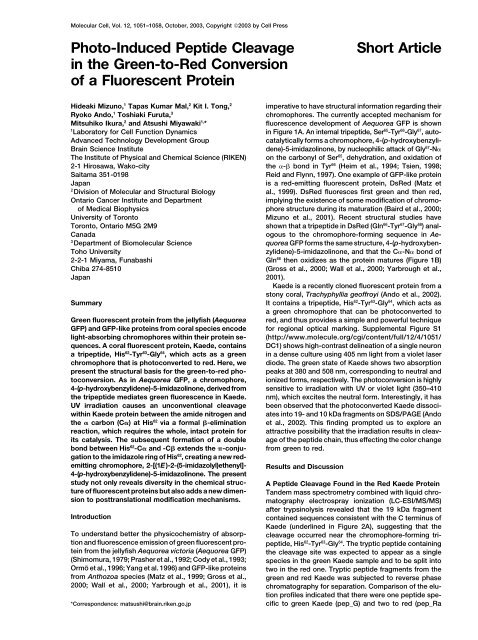
Molecular Cell, Vol. 12, 1051–1058, Oc<strong>to</strong>ber, 2003, Copyright ©2003 by Cell Press<br />
<strong>Pho<strong>to</strong></strong>-<strong>Induced</strong> <strong>Peptide</strong> <strong>Cleavage</strong><br />
<strong>in</strong> <strong>the</strong> <strong>Green</strong>-<strong>to</strong>-<strong>Red</strong> Conversion<br />
of a Fluorescent Prote<strong>in</strong><br />
Hideaki Mizuno, 1 Tapas Kumar Mal, 2 Kit I. Tong, 2<br />
Ryoko Ando, 1 Toshiaki Furuta, 3<br />
Mitsuhiko Ikura, 2 and Atsushi Miyawaki 1, *<br />
1 Labora<strong>to</strong>ry for Cell Function Dynamics<br />
Advanced Technology Development Group<br />
<strong>Short</strong> <strong>Article</strong><br />
imperative <strong>to</strong> have structural <strong>in</strong>formation regard<strong>in</strong>g <strong>the</strong>ir<br />
chromophores. The currently accepted mechanism for<br />
fluorescence development of Aequorea GFP is shown<br />
<strong>in</strong> Figure 1A. An <strong>in</strong>ternal tripeptide, Ser65-Tyr66-Gly67 , au<strong>to</strong>catalytically<br />
forms a chromophore, 4-(p-hydroxybenzyli-<br />
Bra<strong>in</strong> Science Institute dene)-5-imidazol<strong>in</strong>one, by nucleophilic attack of Gly67-N on <strong>the</strong> carbonyl of Ser65 The Institute of Physical and Chemical Science (RIKEN)<br />
, dehydration, and oxidation of<br />
2-1 Hirosawa, Wako-city <strong>the</strong> - bond <strong>in</strong> Tyr66 (Heim et al., 1994; Tsien, 1998;<br />
Saitama 351-0198<br />
Reid and Flynn, 1997). One example of GFP-like prote<strong>in</strong><br />
Japan is a red-emitt<strong>in</strong>g fluorescent prote<strong>in</strong>, Ds<strong>Red</strong> (Matz et<br />
2Division of Molecular and Structural Biology<br />
al., 1999). Ds<strong>Red</strong> fluoresces first green and <strong>the</strong>n red,<br />
Ontario Cancer Institute and Department imply<strong>in</strong>g <strong>the</strong> existence of some modification of chromo-<br />
of Medical Biophysics<br />
phore structure dur<strong>in</strong>g its maturation (Baird et al., 2000;<br />
University of Toron<strong>to</strong> Mizuno et al., 2001). Recent structural studies have<br />
shown that a tripeptide <strong>in</strong> Ds<strong>Red</strong> (Gln66-Tyr67-Gly68 Toron<strong>to</strong>, Ontario M5G 2M9<br />
) anal-<br />
Canada ogous <strong>to</strong> <strong>the</strong> chromophore-form<strong>in</strong>g sequence <strong>in</strong> Ae-<br />
3Department of Biomolecular Science<br />
quorea GFP forms <strong>the</strong> same structure, 4-(p-hydroxyben-<br />
Toho University zylidene)-5-imidazol<strong>in</strong>one, and that <strong>the</strong> C-N bond of<br />
Gln66 2-2-1 Miyama, Funabashi<br />
<strong>the</strong>n oxidizes as <strong>the</strong> prote<strong>in</strong> matures (Figure 1B)<br />
Chiba 274-8510 (Gross et al., 2000; Wall et al., 2000; Yarbrough et al.,<br />
Japan<br />
2001).<br />
Kaede is a recently cloned fluorescent prote<strong>in</strong> from a<br />
s<strong>to</strong>ny coral, Trachyphyllia geoffroyi (Ando et al., 2002).<br />
It conta<strong>in</strong>s a tripeptide, His62-Tyr63-Gly64 Summary<br />
, which acts as<br />
a green chromophore that can be pho<strong>to</strong>converted <strong>to</strong><br />
<strong>Green</strong> fluorescent prote<strong>in</strong> from <strong>the</strong> jellyfish (Aequorea red, and thus provides a simple and powerful technique<br />
GFP) and GFP-like prote<strong>in</strong>s from coral species encode for regional optical mark<strong>in</strong>g. Supplemental Figure S1<br />
light-absorb<strong>in</strong>g chromophores with<strong>in</strong> <strong>the</strong>ir prote<strong>in</strong> se- (http://www.molecule.org/cgi/content/full/12/4/1051/<br />
quences. A coral fluorescent prote<strong>in</strong>, Kaede, conta<strong>in</strong>s DC1) shows high-contrast del<strong>in</strong>eation of a s<strong>in</strong>gle neuron<br />
a tripeptide, His <strong>in</strong> a dense culture us<strong>in</strong>g 405 nm light from a violet laser<br />
62-Tyr63-Gly64 , which acts as a green<br />
chromophore that is pho<strong>to</strong>converted <strong>to</strong> red. Here, we diode. The green state of Kaede shows two absorption<br />
present <strong>the</strong> structural basis for <strong>the</strong> green-<strong>to</strong>-red pho- peaks at 380 and 508 nm, correspond<strong>in</strong>g <strong>to</strong> neutral and<br />
<strong>to</strong>conversion. As <strong>in</strong> Aequorea GFP, a chromophore, ionized forms, respectively. The pho<strong>to</strong>conversion is highly<br />
4-(p-hydroxybenzylidene)-5-imidazol<strong>in</strong>one, derived from sensitive <strong>to</strong> irradiation with UV or violet light (350–410<br />
<strong>the</strong> tripeptide mediates green fluorescence <strong>in</strong> Kaede. nm), which excites <strong>the</strong> neutral form. Interest<strong>in</strong>gly, it has<br />
UV irradiation causes an unconventional cleavage been observed that <strong>the</strong> pho<strong>to</strong>converted Kaede dissociwith<strong>in</strong><br />
Kaede prote<strong>in</strong> between <strong>the</strong> amide nitrogen and ates <strong>in</strong><strong>to</strong> 19- and 10 kDa fragments on SDS/PAGE (Ando<br />
<strong>the</strong> carbon (C) at His et al., 2002). This f<strong>in</strong>d<strong>in</strong>g prompted us <strong>to</strong> explore an<br />
62 via a formal -elim<strong>in</strong>ation<br />
reaction, which requires <strong>the</strong> whole, <strong>in</strong>tact prote<strong>in</strong> for attractive possibility that <strong>the</strong> irradiation results <strong>in</strong> cleav-<br />
its catalysis. The subsequent formation of a double age of <strong>the</strong> peptide cha<strong>in</strong>, thus effect<strong>in</strong>g <strong>the</strong> color change<br />
bond between His62-C and -C extends <strong>the</strong> -conjugation<br />
<strong>to</strong> <strong>the</strong> imidazole r<strong>in</strong>g of His<br />
from green <strong>to</strong> red.<br />
62 , creat<strong>in</strong>g a new redemitt<strong>in</strong>g<br />
chromophore, 2-[(1E)-2-(5-imidazolyl)e<strong>the</strong>nyl]-<br />
4-(p-hydroxybenzylidene)-5-imidazol<strong>in</strong>one. The present<br />
Results and Discussion<br />
study not only reveals diversity <strong>in</strong> <strong>the</strong> chemical struc- A <strong>Peptide</strong> <strong>Cleavage</strong> Found <strong>in</strong> <strong>the</strong> <strong>Red</strong> Kaede Prote<strong>in</strong><br />
ture of fluorescent prote<strong>in</strong>s but also adds a new dimen- Tandem mass spectrometry comb<strong>in</strong>ed with liquid chrosion<br />
<strong>to</strong> posttranslational modification mechanisms. ma<strong>to</strong>graphy electrospray ionization (LC-ESI/MS/MS)<br />
after tryps<strong>in</strong>olysis revealed that <strong>the</strong> 19 kDa fragment<br />
Introduction conta<strong>in</strong>ed sequences consistent with <strong>the</strong> C term<strong>in</strong>us of<br />
Kaede (underl<strong>in</strong>ed <strong>in</strong> Figure 2A), suggest<strong>in</strong>g that <strong>the</strong><br />
To understand better <strong>the</strong> physicochemistry of absorp- cleavage occurred near <strong>the</strong> chromophore-form<strong>in</strong>g trition<br />
and fluorescence emission of green fluorescent pro- peptide, His62-Tyr63-Gly64 . The tryptic peptide conta<strong>in</strong><strong>in</strong>g<br />
te<strong>in</strong> from <strong>the</strong> jellyfish Aequorea vic<strong>to</strong>ria (Aequorea GFP) <strong>the</strong> cleavage site was expected <strong>to</strong> appear as a s<strong>in</strong>gle<br />
(Shimomura, 1979; Prasher et al., 1992; Cody et al., 1993; species <strong>in</strong> <strong>the</strong> green Kaede sample and <strong>to</strong> be split <strong>in</strong><strong>to</strong><br />
Ormö et al., 1996; Yang et al. 1996) and GFP-like prote<strong>in</strong>s two <strong>in</strong> <strong>the</strong> red one. Tryptic peptide fragments from <strong>the</strong><br />
from Anthozoa species (Matz et al., 1999; Gross et al., green and red Kaede was subjected <strong>to</strong> reverse phase<br />
2000; Wall et al., 2000; Yarbrough et al., 2001), it is chroma<strong>to</strong>graphy for separation. Comparison of <strong>the</strong> elution<br />
profiles <strong>in</strong>dicated that <strong>the</strong>re were one peptide spe-<br />
*Correspondence: matsushi@bra<strong>in</strong>.riken.go.jp<br />
cific <strong>to</strong> green Kaede (pep_G) and two <strong>to</strong> red (pep_Ra
Molecular Cell<br />
1052<br />
Figure 1. A Comparison of Reported Schemes for <strong>the</strong> Au<strong>to</strong>catalytic Formation and Maturation of Chromophores <strong>in</strong> Fluorescent Prote<strong>in</strong>s<br />
(A) Aequorea GFP. (B) Ds<strong>Red</strong>. The cleavage site is <strong>in</strong>dicated by an arrowhead. -conjugation for visible-light absorption is <strong>in</strong>dicated <strong>in</strong> green<br />
or red. Neighbor<strong>in</strong>g am<strong>in</strong>o acids (s<strong>in</strong>gle-letter code) have been added.<br />
and pep_Rb) (Figure 2B). We believed that pep_G was (Figure 2D). These results strongly suggested that <strong>the</strong><br />
split <strong>in</strong><strong>to</strong> pep_Ra and pep_Rb.<br />
C-term<strong>in</strong>al end was not a carboxyl group (-COOH) gener-<br />
ated by hydrolysis but a carbamoyl group (-CONH2). pep_G: The Tryptic <strong>Peptide</strong> Conta<strong>in</strong><strong>in</strong>g<br />
This structure expla<strong>in</strong>s both <strong>the</strong> creation of <strong>the</strong> divalent<br />
<strong>the</strong> <strong>Green</strong> Chromophore cation on <strong>the</strong> ESI/MS through pro<strong>to</strong>nation of both <strong>the</strong><br />
Based on <strong>the</strong> am<strong>in</strong>o acid sequence of Kaede, <strong>the</strong> tryptic C-term<strong>in</strong>al and N-term<strong>in</strong>al am<strong>in</strong>o groups, as well as <strong>the</strong><br />
peptide bear<strong>in</strong>g His 62 -Tyr 63 -Gly 64 is predicted <strong>to</strong> conta<strong>in</strong> 1-Da mass reduction observed <strong>in</strong> <strong>the</strong> y ions. It was also<br />
concluded that <strong>the</strong> cleavage site was between His62 residues from Glu -C<br />
46 <strong>to</strong> Arg66 (a green bar <strong>in</strong> Figure 2A).<br />
The ESI/MS/MS spectrum of collision-<strong>in</strong>duced fragments<br />
of pep_G identified am<strong>in</strong>o acid sequences correspond<strong>in</strong>g<br />
<strong>to</strong> Pro<br />
and -N (Figure 3A).<br />
51-Phe61 and Asn65-Arg66 (Figure 2C), <strong>in</strong>dicat<strong>in</strong>g<br />
that pep_G is <strong>the</strong> tryptic peptide conta<strong>in</strong><strong>in</strong>g <strong>the</strong> green The Structural Basis for <strong>the</strong> <strong>Green</strong>-<strong>to</strong>-<strong>Red</strong><br />
chromophore. Because <strong>the</strong> three am<strong>in</strong>o acid residues— <strong>Pho<strong>to</strong></strong>conversion of Kaede<br />
His62 , Tyr63 , and Gly64 —were not identified <strong>in</strong> <strong>the</strong> spec- The o<strong>the</strong>r cleavage product was pep_Ra (a red bar <strong>in</strong><br />
trum, and <strong>the</strong> fragment ion conta<strong>in</strong><strong>in</strong>g His Figure 2A), which should conta<strong>in</strong> <strong>the</strong> red chromophore.<br />
62-Gly64 (y <br />
5 )<br />
exhibited a molecular mass (626.21 Da) that was 20.09 Figure 3A illustrates <strong>the</strong> events proposed <strong>to</strong> be <strong>in</strong>volved<br />
Da lower than <strong>the</strong> <strong>the</strong>oretical value (646.30 Da), we hy- <strong>in</strong> chromophore modification. The pho<strong>to</strong>cleavage <strong>in</strong>po<strong>the</strong>sized<br />
that modification of <strong>the</strong> tripeptide had oc- volves elim<strong>in</strong>ation of a carboxamide (species 4b) at<br />
His62-C and removal of a pro<strong>to</strong>n at His62 curred. The reduction of 20.09 Da suggests cyclization<br />
-C. The<br />
(loss of H2O) and oxidation (O2-mediated loss of H2) of result<strong>in</strong>g structure, 2-[(1E)-2-(5-imidazolyl)e<strong>the</strong>nyl]-4<strong>the</strong><br />
peptide <strong>to</strong> form a 4-(p-hydroxybenzylidene)-5-imida- (p-hydroxybenzylidene)-5-imidazol<strong>in</strong>one (species 5a),<br />
zol<strong>in</strong>one, as occurs <strong>in</strong> Aequorea GFP. features extended -conjugation, account<strong>in</strong>g for <strong>the</strong><br />
green-<strong>to</strong>-red conversion of Kaede. The structure is con-<br />
Site of <strong>the</strong> <strong>Pho<strong>to</strong></strong>-<strong>Induced</strong> <strong>Cleavage</strong><br />
sistent with our mass spectral data on pep_Ra. The ESI/<br />
of Kaede Prote<strong>in</strong> MS spectrum showed a peak for a monovalent ion at<br />
Next, we exam<strong>in</strong>ed <strong>the</strong> site of cleavage between pep_Ra m/z 609.09 (Figure 2F), from which <strong>the</strong> molecular<br />
and pep_Rb. Pep_Rb did not show any absorption above weight of pep_Ra was calculated <strong>to</strong> be 608.08 Da. This<br />
300 nm (results not shown), <strong>in</strong>dicat<strong>in</strong>g <strong>the</strong> absence of value is 37.22 Da lighter than <strong>the</strong> value of 645.30 Da<br />
a complete chromophore structure <strong>in</strong> this peptide. The expected for <strong>the</strong> pentapeptide His-Tyr-Gly-Asn-Arg.<br />
S<strong>in</strong>ce <strong>the</strong> region correspond<strong>in</strong>g <strong>to</strong> His62-Tyr63-Gly64 ESI/MS/MS spectrum of pep_Rb clearly identified <strong>the</strong><br />
lost<br />
am<strong>in</strong>o acid residues from Glu46 <strong>to</strong> Phe61 (Figure 2D, a 20 Da upon formation of <strong>the</strong> green chromophore, an<br />
black bar <strong>in</strong> Figure 2A). It was concluded that <strong>the</strong> Phe additional reduction by 17 Da can be attributable <strong>to</strong><br />
61<br />
located at <strong>the</strong> C term<strong>in</strong>us must <strong>the</strong>refore border on <strong>the</strong> <strong>the</strong> pho<strong>to</strong>cleavage reaction. The mass reduction can be<br />
expla<strong>in</strong>ed by an extraction of an am<strong>in</strong>o group from His62 cleavage site. With respect <strong>to</strong> <strong>the</strong> C-term<strong>in</strong>al end structure,<br />
two po<strong>in</strong>ts must be considered. First, pep_Rb was (16 Da) and liberation of a pro<strong>to</strong>n at His62-C (1 Da).<br />
detected as a divalent cation by ESI/MS (Figure 2E), To our knowledge, what we present here is <strong>the</strong> first<br />
although it conta<strong>in</strong>s no basic am<strong>in</strong>o acids, suggest<strong>in</strong>g report of a peptide cleavage <strong>in</strong> an <strong>in</strong>tact prote<strong>in</strong> at an<br />
<strong>the</strong> presence of additional am<strong>in</strong>o group <strong>in</strong> this peptide. N-C bond via a formal -elim<strong>in</strong>ation reaction (Figure<br />
Second, <strong>the</strong> y1 ,y2 ,y3 , and y4 ions all showed a loss 3B, right), while it is well accepted that peptide cleavage<br />
of 1 Da relative <strong>to</strong> <strong>the</strong>ir respective <strong>the</strong>oretical values<br />
occurs at <strong>the</strong> peptide bond, <strong>the</strong> bond between <strong>the</strong> car
Molecular Mechanism for <strong>Pho<strong>to</strong></strong>conversion<br />
1053<br />
Figure 2. Mass Spectroscopic Analysis of Tryptic <strong>Peptide</strong> Fragments from <strong>the</strong> <strong>Green</strong> and <strong>Red</strong> Kaede<br />
(A) Am<strong>in</strong>o acid sequence of Kaede. The observed sequences obta<strong>in</strong>ed by ESI-MS analysis us<strong>in</strong>g <strong>the</strong> tryptic digest from <strong>the</strong> 19 kDa fragment<br />
are underl<strong>in</strong>ed. The chromophore-form<strong>in</strong>g am<strong>in</strong>o acids are <strong>in</strong>dicated by asterisks. Open arrowheads <strong>in</strong>dicate putative tryps<strong>in</strong> digestion sites.<br />
Tryptic peptide fragments around <strong>the</strong> chromophore are <strong>in</strong>dicated by thick bars. They are pep_G (green), pep_Rb (black), and pep_Ra (red),<br />
which are written <strong>in</strong> <strong>the</strong> same colors <strong>in</strong> (B)–(F). A vertical l<strong>in</strong>e <strong>in</strong>dicates <strong>the</strong> pho<strong>to</strong>cleavage site.<br />
(B) Comparison of tryptic digests between green- and red-state Kaede by reverse phase chroma<strong>to</strong>graphy. Tryptic digests of <strong>the</strong> green- (green<br />
trace) and red- (red trace) fluoresc<strong>in</strong>g isoforms were applied on<strong>to</strong> an Inartsil ODS-3 column and eluted with a 0%–90% ace<strong>to</strong>nitrile l<strong>in</strong>ear<br />
gradient. Elution of peptides was moni<strong>to</strong>red by absorbance at 280 nm. The unique peptides <strong>in</strong> each sample (pep_G, green trace; pep_Ra and<br />
pep_Rb, red trace) are <strong>in</strong>dicated by arrowheads.<br />
(C) ESI/MS/MS spectrum of pep_G. Cyan arrowheads <strong>in</strong>dicate peaks correspond<strong>in</strong>g <strong>to</strong> C-term<strong>in</strong>al fragment ions (y ) produced through peptide<br />
bond breakage by collision-<strong>in</strong>duced dissociation.<br />
(D) ESI/MS/MS spectrum of pep_Rb. Magenta and cyan arrowheads <strong>in</strong>dicate peaks correspond<strong>in</strong>g <strong>to</strong> N- and C-term<strong>in</strong>al fragment ions (b <br />
and y , respectively). In (C) and (D), horizontal arrows with s<strong>in</strong>gle-letter codes <strong>in</strong>dicate am<strong>in</strong>o acid residues deduced from <strong>the</strong> fragment ions.<br />
At positions denoted as dotted arrows, reductions <strong>in</strong> molecular mass were observed. Intensity is multiplied by 5 times <strong>in</strong> some regions denoted<br />
as “x5.”<br />
(E) ESI/MS spectrum of pep_Rb.<br />
(F) ESI/MS spectrum of pep_Ra.<br />
Predicted am<strong>in</strong>o acid sequences of <strong>the</strong> analyzed peptides are shown at <strong>the</strong> right sides of <strong>the</strong> respective spectra. The chromophore-form<strong>in</strong>g<br />
am<strong>in</strong>o acids are <strong>in</strong>dicated by asterisks. Magenta and cyan bars under sequences <strong>in</strong>dicate deduced am<strong>in</strong>o acid residues by analyses of b <br />
and y ion series, respectively. The residues that showed reductions <strong>in</strong> molecular mass are <strong>in</strong>dicated by boldface.
Molecular Cell<br />
1054<br />
Figure 3. Molecular Mechanisms Proposed for <strong>the</strong> <strong>Green</strong>-<strong>to</strong>-<strong>Red</strong> <strong>Pho<strong>to</strong></strong>conversion<br />
(A) Scheme for <strong>the</strong> formation and pho<strong>to</strong>-<strong>in</strong>duced extension of <strong>the</strong> chromophore of Kaede. Structures derived from Phe61 , His62 , Tyr63 , and Gly64 are drawn, and <strong>the</strong> neighbor<strong>in</strong>g am<strong>in</strong>o acids (s<strong>in</strong>gle-letter code) are added. The crucial a<strong>to</strong>ms for <strong>the</strong> pho<strong>to</strong>cleavage reaction are <strong>in</strong>dicated<br />
<strong>in</strong> red <strong>in</strong> <strong>the</strong> structure of <strong>the</strong> precursor peptide (1). 4-(p-hydroxybenzylidene)-5-imidazol<strong>in</strong>one (2) is formed from 1 with <strong>the</strong> same mechanism<br />
for <strong>the</strong> chromophore formation of Aequorea GFP (Figure 1A). The chromophore <strong>in</strong> species 2 is a neutral form and nonfluorescent. Depro<strong>to</strong>nation<br />
at <strong>the</strong> hydroxyl group of Tyr63 results <strong>in</strong> a green-emitt<strong>in</strong>g species (2). The resonance structures of species 2 are <strong>in</strong> paren<strong>the</strong>ses. After excitation<br />
of <strong>the</strong> neutral form (2) by UV or violet light (h), <strong>the</strong> excited state (2*) releases pro<strong>to</strong>n <strong>to</strong> form <strong>the</strong> excited <strong>in</strong>termediate (3*) (Chat<strong>to</strong>raj et al.,<br />
1996; McAnaney et al., 2002). The p-qu<strong>in</strong>one methide-type charge density distribution has been supported by calculation for <strong>the</strong> LUMO of<br />
<strong>the</strong> neutral chromophore (Tozz<strong>in</strong>i and Nifosì, 2001). Then cleavage occurs at <strong>the</strong> N-C bond of His62 <strong>to</strong> elim<strong>in</strong>ate a carboxamide groupconta<strong>in</strong><strong>in</strong>g<br />
peptide (4b). The subsequent loss of a pro<strong>to</strong>n from His62-C gives a trans double bond between His62-C and -C, lead<strong>in</strong>g <strong>to</strong> <strong>the</strong><br />
extension of <strong>the</strong> conjugated system (5a).
Molecular Mechanism for <strong>Pho<strong>to</strong></strong>conversion<br />
1055<br />
bonyl carbon and amide nitrogen a<strong>to</strong>ms, through acid Tyr63-2,6H, as well as between His62-H and Tyr63-3,5H (Figures 4E and 4F) demonstrate that Tyr63 or protease hydrolysis (Figure 3B, left).<br />
-H and 3N<br />
of imidazol<strong>in</strong>one exist <strong>in</strong> a trans position, as occurs for<br />
Determ<strong>in</strong>ation of <strong>the</strong> Detailed Structure<br />
<strong>the</strong> chromophore of Aequorea GFP (Ormö et al., 1996;<br />
Yang et al., 1996). F<strong>in</strong>ally, an ROE between His62 of <strong>the</strong> <strong>Red</strong> Chromophore by NMR<br />
-4H and<br />
Tyr63 To validate our proposed structure of <strong>the</strong> red chromo-<br />
-3,5H (Figure 4F) supports an s-trans conformation<br />
between His62-H and His62 phore, we <strong>the</strong>n carried out extensive NMR analysis on<br />
pep_Ra. A chemically syn<strong>the</strong>sized pentapeptide, His-<br />
-1N.<br />
Tyr-Gly-Asn-Arg (pep_C) was used as a control. Comparison<br />
of <strong>the</strong> 1D<br />
Diversity of Chromophore Modification Mechanisms<br />
1H-NMR spectrum of pep_C (Figure<br />
4A) with that of pep_Ra (Figure 4B) revealed drastic<br />
changes <strong>in</strong> <strong>the</strong> chemical shifts and J-coupl<strong>in</strong>g pattern<br />
for Tyr<br />
<strong>in</strong> <strong>the</strong> GFP-like Prote<strong>in</strong> Family<br />
Both Ds<strong>Red</strong> and Kaede have -conjugation structures<br />
similar <strong>to</strong> that of Aequorea GFP, feature 4-(p-hydroxy-<br />
63 and His62 resonances. The multiplet peak corre-<br />
spond<strong>in</strong>g <strong>to</strong> Tyr<br />
benzylidene)-5-imidazol<strong>in</strong>one with<strong>in</strong> <strong>the</strong>ir chromophores,<br />
63-H of pep_C (3.01 ppm) (Figure 4A)<br />
was shifted <strong>in</strong><strong>to</strong> <strong>the</strong> aromatic/v<strong>in</strong>yl region <strong>in</strong> <strong>the</strong> pep_Ra<br />
spectrum (7.07 ppm) (Figure 4B). This downfield shift<br />
was accompanied by a characteristic change <strong>in</strong> NMR<br />
sp<strong>in</strong> properties; <strong>the</strong> peak changed from a multiplet <strong>to</strong> a<br />
s<strong>in</strong>glet. Fur<strong>the</strong>rmore, <strong>the</strong> peak correspond<strong>in</strong>g <strong>to</strong> Tyr<br />
and emit green fluorescence <strong>in</strong> <strong>the</strong>ir immature and un-<br />
pho<strong>to</strong>converted states, respectively (Gross et al., 2000;<br />
Wall et al., 2000; Yarbrough et al., 2001). The processes<br />
<strong>in</strong>volved <strong>in</strong> <strong>the</strong> development of red fluorescence, how-<br />
ever, are different. While Ds<strong>Red</strong> generates an acylim<strong>in</strong>e<br />
63-H of pep_C at 4.61 ppm <strong>in</strong> Figure 4A disappeared <strong>in</strong> <strong>the</strong><br />
spectrum of pep_Ra (Figure 4B). These observations<br />
conv<strong>in</strong>c<strong>in</strong>gly support <strong>the</strong> formation of a double bond<br />
between C and C of Tyr<br />
(-CN-CO) by an oxidation reaction <strong>to</strong> expand <strong>the</strong><br />
-conjugation structure at <strong>the</strong> 2-position of <strong>the</strong> imidazol-<br />
<strong>in</strong>one (Gross et al., 2000), Kaede becomes red specifi-<br />
cally upon UV irradiation, which causes a peptide cleav-<br />
63 <strong>in</strong> pep_Ra. The presence<br />
of a covalent bond between amide nitrogen of Gly age as a result of an elim<strong>in</strong>ation reaction. Its cleavage<br />
64<br />
and <strong>the</strong> carbonyl carbon of His site is between N and C of <strong>the</strong> His residue, which<br />
62 was verified by <strong>the</strong><br />
13 13 C-edited heteronuclear multiple bond correlation ( C-<br />
1H HMBC) spectrum (Bax and Summer, 1986) of pep_Ra<br />
(Figure 4C), which showed a correlation between Gly<br />
subsequently becomes <strong>in</strong>volved <strong>in</strong> <strong>the</strong> red chromophore.<br />
It has been reported that a fraction of Ds<strong>Red</strong> that was<br />
boiled and <strong>the</strong>n subjected <strong>to</strong> SDS/PAGE showed two<br />
64-H and 2C of <strong>the</strong> imidazol<strong>in</strong>one r<strong>in</strong>g (Figure 4G, cyan l<strong>in</strong>es).<br />
All <strong>the</strong>se results confirm <strong>the</strong> presence of a 4-(p-hydroxy-<br />
benzylidene)-5-imidazol<strong>in</strong>one chromophore structure <strong>in</strong><br />
<strong>the</strong> red chromopeptide.<br />
fragment bands of apparent masses 15 and 22 kDa<br />
(Gross et al., 2000). After <strong>the</strong> denaturation of <strong>the</strong> protective<br />
prote<strong>in</strong> shell, <strong>the</strong> CN bond <strong>in</strong> <strong>the</strong> acylim<strong>in</strong>e was<br />
thought <strong>to</strong> hydrolyze under harsh conditions. It appears<br />
The double bond formation between His that peptide cleavage does not occur <strong>in</strong> <strong>the</strong> <strong>in</strong>tact mature<br />
62-C and -C<br />
was evidenced by exceptionally large downfield shifts of Ds<strong>Red</strong>. In contrast, Kaede cleavage should occur <strong>in</strong> <strong>the</strong><br />
2.4 and 4.2 ppm for <strong>the</strong> His <strong>in</strong>tact prote<strong>in</strong> upon irradiation with UV or violet light,<br />
62-H and -H resonances,<br />
respectively (Figures 4A and 4B). In <strong>the</strong> pep_Ra spec- lead<strong>in</strong>g <strong>to</strong> formation of <strong>the</strong> structure 2-[(1E)-2-(5-imida-<br />
trum (Figure 4B), both <strong>the</strong> His zolyl)e<strong>the</strong>nyl]-4-(p-hydroxybenzylidene)-5-imidazol<strong>in</strong>one,<br />
62-H and -H resonances<br />
appeared as doublets with a J-coupl<strong>in</strong>g constant of 16.0 which accounts for <strong>the</strong> green-<strong>to</strong>-red conversion of Kaede.<br />
Hz, <strong>in</strong>dicat<strong>in</strong>g that <strong>the</strong> two pro<strong>to</strong>ns existed <strong>in</strong> a trans Ano<strong>the</strong>r GFP-like prote<strong>in</strong> absorb<strong>in</strong>g at a long waveposition<br />
(Figure 4G). Fur<strong>the</strong>rmore, <strong>the</strong> downfield shift of length is asFP595 (Lukyanov et al., 2000), whose pro-<br />
<strong>the</strong> His posed chromophore structure is different from that of<br />
62-4H peak (Figures 4A and 4B) is consistent with<br />
<strong>the</strong> expansion of -conjugation <strong>to</strong> <strong>the</strong> imidazole r<strong>in</strong>g Ds<strong>Red</strong> or Kaede. Accord<strong>in</strong>g <strong>to</strong> a study by Martynov<br />
et al. (2001), <strong>the</strong> tripeptide Met65-Tyr66-Gly67 through <strong>the</strong> C-C double bond.<br />
of asFP595<br />
To establish <strong>the</strong> preferred conformation of <strong>the</strong> red undergoes a cyclization reaction <strong>in</strong>volv<strong>in</strong>g nucleophilic<br />
attack by <strong>the</strong> Met65-N on<strong>to</strong> <strong>the</strong> Tyr66 chromophore, we carried out carbonyl, produc-<br />
1H-1H rotational nuclear<br />
Overhauser effect spectroscopy (ROESY) at three bonds <strong>in</strong>g a six-membered heterocycle r<strong>in</strong>g structure. Follow<strong>in</strong>g<br />
dehydration and oxidation of <strong>the</strong> - bond of Tyr66 with<strong>in</strong> <strong>the</strong> structure of 2-[(1E)-2-(5-imidazolyl)e<strong>the</strong>nyl]-<br />
,<br />
<strong>the</strong> peptide cleaves at <strong>the</strong> peptide bond between Cys64 4-(p-hydroxybenzylidene)-5-imidazol<strong>in</strong>one (Figure 4G).<br />
and Met65 First, a rotational nuclear Overhauser effect (ROE) was<br />
, splitt<strong>in</strong>g <strong>the</strong> prote<strong>in</strong> <strong>in</strong><strong>to</strong> 8- and 20 kDa fragobserved<br />
between His ments. Although this peptide cleavage completes <strong>the</strong><br />
62-H and Gly64-H but not between<br />
His chromophore, a hydrolysis, ra<strong>the</strong>r than elim<strong>in</strong>ation, re-<br />
62-H and Gly64-H (Figure 4D), confirm<strong>in</strong>g that<br />
His action actually cleaves <strong>the</strong> peptide bond. Fur<strong>the</strong>rmore,<br />
62-H and 1N of imidazol<strong>in</strong>one r<strong>in</strong>g are <strong>in</strong> an s-cis<br />
conformation. Second, ROEs between His62-H and <strong>the</strong> hydrolysis does not depend on illum<strong>in</strong>ation.<br />
(B) <strong>Peptide</strong> cleavage reactions. Conventional cleavage at <strong>the</strong> peptide bond, <strong>the</strong> bond between <strong>the</strong> carbonyl carbon and amide nitrogen a<strong>to</strong>ms<br />
through acid or protease hydrolysis (left). <strong>Pho<strong>to</strong></strong>-<strong>in</strong>duced peptide cleavage at an N-C bond <strong>in</strong> <strong>in</strong>tact Kaede prote<strong>in</strong> (right).<br />
(C) Provisional model for <strong>the</strong> pho<strong>to</strong>physics of <strong>the</strong> green Kaede. Direct excitation of <strong>the</strong> anionic chromophore (2) results <strong>in</strong> green fluorescence.<br />
Upon excitation of <strong>the</strong> neutral chromophore, 2* rapidly converts <strong>to</strong> 3* through <strong>the</strong> excited-state pro<strong>to</strong>n transfer (Chat<strong>to</strong>raj et al., 1996). 3* is<br />
an <strong>in</strong>termediate anionic chromophore <strong>in</strong> a nonequilibrium prote<strong>in</strong> environment, and may correspond <strong>to</strong> I* <strong>in</strong> <strong>the</strong> three-state model for <strong>the</strong><br />
pho<strong>to</strong>physics of wild-type Aequorea GFP (Chat<strong>to</strong>raj et al., 1996; McAnaney et al., 2002; Brejc et al., 1997; Weber et al., 1999). The nonfluorescence<br />
of <strong>the</strong> neutral form of Kaede is expla<strong>in</strong>ed by (1) no direct fluorescence (2*→ 2), (2) radiationless decay of 3* <strong>to</strong> 3, and (3) no conversion<br />
from 3* <strong>to</strong> 2*.
Molecular Cell<br />
1056<br />
Figure 4. Determ<strong>in</strong>ation of <strong>the</strong> Detailed Structure of <strong>the</strong> <strong>Red</strong> Chromophore by NMR<br />
(A and B) 1H NMR spectra of pep_C and pep_Ra, respectively. Downfield (9.0-6.5 ppm) and upfield (4.8-1.3 ppm) regions of <strong>the</strong> spectra are<br />
displayed <strong>in</strong> <strong>the</strong> left and right, respectively. Arrowheads <strong>in</strong>dicate peaks assigned <strong>to</strong> His-4H, His-H, and H (red arrowhead), and Tyr-H and<br />
H (green arrowhead). Arrows <strong>in</strong>dicate shifts of some peaks <strong>in</strong> pep_Ra.<br />
(C) Expanded 13C-1H HMBC spectrum with a mix<strong>in</strong>g time of 80 msec. Arrowhead <strong>in</strong>dicates spots correspond<strong>in</strong>g <strong>to</strong> a correlation between Gly-H<br />
and 2C of imidazol<strong>in</strong>one.
Molecular Mechanism for <strong>Pho<strong>to</strong></strong>conversion<br />
1057<br />
<strong>Pho<strong>to</strong></strong>physics of <strong>the</strong> <strong>Pho<strong>to</strong></strong>-<strong>Induced</strong> <strong>Peptide</strong> <strong>Cleavage</strong> col. <strong>Peptide</strong>s were purified by reverse phase chroma<strong>to</strong>graphy (In-<br />
It is <strong>in</strong>terest<strong>in</strong>g <strong>to</strong> know if His er<strong>to</strong>sil ODS-3, GL Science, Japan) and eluted with a 0 <strong>to</strong> 90%<br />
62 is replaceable with o<strong>the</strong>r<br />
am<strong>in</strong>o acids. Substitution of tyros<strong>in</strong>e, tryp<strong>to</strong>phan, aspartate,<br />
or arg<strong>in</strong><strong>in</strong>e dimmed or abolished <strong>the</strong> fluorescence.<br />
Substitution of all o<strong>the</strong>r am<strong>in</strong>o acids yielded green-emitt<strong>in</strong>g<br />
mutants that did not exhibit pho<strong>to</strong>-<strong>in</strong>duced cleav-<br />
ace<strong>to</strong>nitrile l<strong>in</strong>ear gradient <strong>in</strong> <strong>the</strong> presence of 0.1% trifluoroacetate.<br />
Pep_Ra was fur<strong>the</strong>r purified by size exclusion chroma<strong>to</strong>graphy<br />
(Superdex <strong>Peptide</strong>, Pharmacia) us<strong>in</strong>g 30% ace<strong>to</strong>nitrile:0.1% trifluoroacetate<br />
as eluent.<br />
age as well as pho<strong>to</strong>conversion (Supplemental Table S1 ESI-MS/MS Analysis of Chromopeptides<br />
[http://www.molecule.org/cgi/content/full/12/4/1051/ <strong>Peptide</strong>s were applied on<strong>to</strong> a Cadenza C18 column (Michrom BioRe-<br />
DC1]). Thus, His sources, USA) <strong>in</strong>stalled on a MAGIC 2002 HPLC system (Michrom<br />
62 is requisite for <strong>in</strong>itiat<strong>in</strong>g <strong>the</strong> -elim<strong>in</strong>ation<br />
reaction. It is possible that <strong>the</strong> imidazole of His<br />
BioResources) and eluted at a flow rate of 1 l/m<strong>in</strong> with a 2 <strong>to</strong> 90%<br />
62<br />
gets pro<strong>to</strong>nated on its 3N and participates <strong>in</strong> <strong>the</strong> reaction<br />
by supply<strong>in</strong>g a pro<strong>to</strong>n from its 1N <strong>to</strong> <strong>the</strong> carboxamide<br />
leav<strong>in</strong>g group <strong>in</strong> 4b (Figure 3A). In fact, 1N of <strong>the</strong> imidaace<strong>to</strong>nitrile<br />
l<strong>in</strong>ear gradient <strong>in</strong> <strong>the</strong> presence of 0.1% formic acid.<br />
<strong>Peptide</strong>s <strong>in</strong> <strong>the</strong> eluate were ionized by positive-mode nanoflow-LC<br />
ESI (MDS Proteomics, Denmark) at 2.4 kV of capillary voltage and<br />
<strong>in</strong>troduced <strong>in</strong><strong>to</strong> a QSTAR quadrupole-TOF mass spectrometer<br />
zole is positioned near <strong>the</strong> cleavage site as revealed by<br />
<strong>the</strong> determ<strong>in</strong>ation of <strong>the</strong> red chromophore conformation<br />
(AB/MDS Sciex, Canada).<br />
(Figure 4G). On <strong>the</strong> o<strong>the</strong>r hand, <strong>the</strong> green chromopep- NMR Analyses of Chromopeptides<br />
tide, pep_G, was not cleaved with UV irradiation (data<br />
not shown). Also, a mutation outside <strong>the</strong> chromophoreform<strong>in</strong>g<br />
region, Ala69Ser, generated a mutant that fluoresced<br />
green but did not pho<strong>to</strong>convert (data not shown).<br />
<strong>Peptide</strong>s were lyophilized from D2O and redissolved <strong>in</strong> 99.999% D2O (Isotec Inc., USA). Sodium 3-(trimethylsilyl)-propionate-2,2,3,3-d4 (Isotec Inc.) was used as a standard for calibration of NMR spectra.<br />
13C NMR spectra were acquired us<strong>in</strong>g a Bruker Advance 500 MHz<br />
spectrometer equipped with a 13C cryoprobe. O<strong>the</strong>r spectra were<br />
Therefore, <strong>the</strong> pho<strong>to</strong>-<strong>in</strong>duced peptide cleavage and re- acquired us<strong>in</strong>g a Bruker Advance 600 MHz spectrometer equipped<br />
sult<strong>in</strong>g expansion of -conjugation appear <strong>to</strong> require a<br />
strict three-dimensional structure for <strong>the</strong> catalysis. The<br />
with a QXI probe.<br />
crystal structures of <strong>the</strong> green and red Kaede will pro- Acknowledgments<br />
vide complementary <strong>in</strong>formation <strong>to</strong> fur<strong>the</strong>r our understand<strong>in</strong>g<br />
of <strong>the</strong> conversion mechanism.<br />
Also, ultra-fast time-resolved spectroscopy of Kaede<br />
We thank Dr. M. Usui, Dr. K. Otsuki, Dr. N. Hirotani, Dr. T. Suenaga,<br />
and Dr. M. I<strong>to</strong> for assistance and encouragement. We also acknowledge<br />
Dr. Roger Y. Tsien for valuable advice, and Dr. Markus Waelchli<br />
will reveal excited state dynamics <strong>in</strong> <strong>the</strong> prote<strong>in</strong> (Figure (Bruker-Biosp<strong>in</strong> Japan) for help <strong>in</strong> record<strong>in</strong>g NMR. This work was<br />
3C). These forthcom<strong>in</strong>g studies will provide an answer supported by grants from CREST of JST (Japan Science and Tech<strong>to</strong><br />
<strong>the</strong> fundamental question of why excitation of <strong>the</strong> nology), <strong>the</strong> Japanese M<strong>in</strong>istry of Education, Science and Technol-<br />
neutral form of <strong>the</strong> green chromophore causes <strong>the</strong> chem-<br />
ical reaction, while excitation of <strong>the</strong> anionic form gives<br />
fluorescence. At present, species 3* and 2* <strong>in</strong> Figure<br />
ogy, and HFSP (Human Frontier Science Program) <strong>to</strong> A.M. and by<br />
a grant from Cancer Research Society Inc/Canadian Institute of<br />
Health Research <strong>to</strong> M.I.<br />
3A are <strong>in</strong>dist<strong>in</strong>guishable. We have only a speculation<br />
that <strong>the</strong> hydrogen-bond network around <strong>the</strong> chromo-<br />
Received: June 23, 2003<br />
Revised: September 10, 2003<br />
phore is different between <strong>the</strong> two species, analogous Accepted: September 17, 2003<br />
<strong>to</strong> <strong>the</strong> three-state model for <strong>the</strong> pho<strong>to</strong>physics of wildtype<br />
Aequorea GFP (Chat<strong>to</strong>raj et al., 1996; McAnaney<br />
Published: Oc<strong>to</strong>ber 23, 2003<br />
et al., 2002; Brejc et al., 1997; Weber et al., 1999). Only References<br />
<strong>in</strong> species 3* may <strong>the</strong> network connect <strong>the</strong> phenolic<br />
hydroxyl of Tyr<br />
Ando, R., Hama, H., Yamamo<strong>to</strong>-H<strong>in</strong>o, M., Mizuno, H., and Miyawaki,<br />
63 <strong>to</strong> <strong>the</strong> imidazole r<strong>in</strong>g of His62 so that<br />
<strong>the</strong> pro<strong>to</strong>n on 1N of His<br />
A. (2002). An optical marker based on <strong>the</strong> UV-<strong>in</strong>duced green-<strong>to</strong>-red<br />
62 imidazole is efficiently released<br />
for <strong>the</strong> cleavage reaction.<br />
pho<strong>to</strong>conversion of a fluorescent prote<strong>in</strong>. Proc. Natl. Acad. Sci. USA<br />
99, 12651–12656.<br />
Baird, G.S., Zacharias, D.A., and Tsien, R.Y. (2000). Biochemistry,<br />
Experimental Procedures mutagenesis, and oligomerization of Ds<strong>Red</strong>, a red fluorescent prote<strong>in</strong><br />
from coral. Proc. Natl. Acad. Sci. USA 97, 11984–11989.<br />
Production and Purification of Kaede Prote<strong>in</strong><br />
Recomb<strong>in</strong>ant Kaede prote<strong>in</strong> was produced <strong>in</strong> E. coli and purified<br />
as described previously (Ando et al., 2002), except that all proce-<br />
dures were done <strong>in</strong> <strong>the</strong> dark. <strong>Pho<strong>to</strong></strong>conversion was performed on<br />
green Kaede by illum<strong>in</strong>ation at 365 nm us<strong>in</strong>g a UV illum<strong>in</strong>a<strong>to</strong>r <strong>in</strong> 150<br />
Bax, A., and Summer, M.F. (1986). Pro<strong>to</strong>n and carbon-13 assignments<br />
from sensitivity-enhanced detection of heteronuclear multiple-bond<br />
connectivity by 2D multiple quantum NMR. J. Am. Chem.<br />
Soc. 108, 2093–2094.<br />
mM NaCl and 10 mM MOPS (pH 7.0).<br />
Brejc, K., Sixma, T.K., Kitts, P.A., Ka<strong>in</strong>, S.R., Tsien, R.Y., Ormo, M.,<br />
and Rem<strong>in</strong>g<strong>to</strong>n, S.J. (1997). Structural basis for dual excitation and<br />
Purification of Chromopeptides after Tryps<strong>in</strong>ization<br />
pho<strong>to</strong>isomerization of <strong>the</strong> Aequorea vic<strong>to</strong>ria green fluorescent pro-<br />
Prote<strong>in</strong> was denatured and tryps<strong>in</strong>ized with sequence-grade modi- te<strong>in</strong>. Proc. Natl. Acad. Sci. USA 94, 2306–2311.<br />
fied tryps<strong>in</strong> (Promega, USA) <strong>in</strong> accordance with <strong>the</strong> supplier’s pro<strong>to</strong>- Chat<strong>to</strong>raj, M., K<strong>in</strong>g, B.A., Bublitz, G.U., and Boxer, S.G. (1996). Ultra-<br />
(D–F) Expanded 1 H- 1 H ROESY spectra of 500 msec mix<strong>in</strong>g time. Both positive (solid l<strong>in</strong>e) and negative (dotted l<strong>in</strong>e) signals are <strong>in</strong>dicated.<br />
Closed arrowheads <strong>in</strong>dicate spots correspond<strong>in</strong>g <strong>to</strong> correlations between His-H and Gly-H (D), and between His-4H and Tyr-3,5H (F). Open<br />
arrowheads <strong>in</strong>dicate spots correspond<strong>in</strong>g <strong>to</strong> correlations between His-H and Tyr-2,6H (E), and between His-H and Tyr-3,5H (F).<br />
(G) Summary of NMR analyses regard<strong>in</strong>g cis-trans isomers of <strong>the</strong> structure, 2-[(1E )-2-(5-imidazolyl)e<strong>the</strong>nyl]-4-(p-hydroxybenzylidene)-5-imidazol<strong>in</strong>one.<br />
Cyan l<strong>in</strong>es <strong>in</strong>dicate a correlation between Gly-H and 2C of imidazol<strong>in</strong>one observed on <strong>the</strong> HMBC spectrum (C). ROEs are <strong>in</strong>dicated<br />
by magenta dotted l<strong>in</strong>es (D–F). Assignments and correlations observed <strong>in</strong> NMR analyses are shown <strong>in</strong> Supplemental Data (http://www.molecule.org/cgi/content/full/12/4/1051/DC1).
Molecular Cell<br />
1058<br />
fast excited state dynamics <strong>in</strong> green fluorescent prote<strong>in</strong>: multiple<br />
states and pro<strong>to</strong>n transfer. Proc. Natl. Acad. Sci. USA 93, 8362–8367.<br />
Cody, C.W., Prasher, D.C., Westler, W.M., Prendergast, F.G., and<br />
Ward, W.W. (1993). Chemical structure of <strong>the</strong> hexapeptide chromophore<br />
of <strong>the</strong> Aequorea green-fluorescent prote<strong>in</strong>. Biochemistry<br />
32, 1212–1218.<br />
Gross, L.A., Baird, G.S., Hoffman, R.C., Baldridge, K.K., and Tsien,<br />
R.Y. (2000). The structure of <strong>the</strong> chromophore with<strong>in</strong> Ds<strong>Red</strong>, a red<br />
fluorescent prote<strong>in</strong> from coral. Proc. Natl. Acad. Sci. USA 97, 11990–<br />
11995.<br />
Heim, R., Prasher, D.C., and Tsien, R.Y. (1994). Wavelength mutations<br />
and posttranslational au<strong>to</strong>xidation of green fluorescent prote<strong>in</strong>.<br />
Proc. Natl. Acad. Sci. USA 91, 12501–12504.<br />
Lukyanov, K.A., Fradkov, A.F., Gurskaya, N.G., Matz, M.V., Labas,<br />
Y.A., Savitsky, A.P., Markelov, M.L., Zaraisky, A.G., Zhao, X., Fang,<br />
Y., et al. (2000). Natural animal coloration can be determ<strong>in</strong>ed by a<br />
nonfluorescent green fluorescent prote<strong>in</strong> homolog. J. Biol. Chem.<br />
275, 25879–25882.<br />
McAnaney, T.B., Park, E.S., Hanson, G.T., Rem<strong>in</strong>g<strong>to</strong>n, S.J., and<br />
Boxer, S.G. (2002). <strong>Green</strong> fluorescent prote<strong>in</strong> variants as ratiometric<br />
dual emission pH sensors. 2. Excited-state dynamics. Biochemistry<br />
41, 15489–15494.<br />
Martynov, V.I., Savitsky, A.P., Martynova, N.Y., Savitsky, P.A., Lukyanov,<br />
K.A., and Lukyanov, S.A. (2001). Alternative cyclization <strong>in</strong> GFPlike<br />
prote<strong>in</strong>s family. The formation and structure of <strong>the</strong> chromophore<br />
of a purple chromoprote<strong>in</strong> from Anemonia sulcata. J. Biol. Chem.<br />
276, 21012–21016.<br />
Matz, M.V., Fradkov, A.F., Labas, Y.A., Savitsky, A.P., Zaraisky, A.G.,<br />
Markelov, M.L., and Lukyanov, S.A. (1999). Fluorescent prote<strong>in</strong>s<br />
from nonbiolum<strong>in</strong>escent Anthozoa species. Nat. Biotechnol. 17,<br />
969–973.<br />
Mizuno, H., Sawano, A., Eli, P., Hama, H., and Miyawaki, A. (2001).<br />
<strong>Red</strong> fluorescent prote<strong>in</strong> from Discosoma as a fusion tag and a partner<br />
for fluorescence resonance energy transfer. Biochemistry 40,<br />
2502–2510.<br />
Ormö, M., Cubitt, A.B., Kallio, K., Gross, L.A., Tsien, R.Y., and Rem<strong>in</strong>g<strong>to</strong>n,<br />
S.J. (1996). Chemical structure of <strong>the</strong> hexapeptide chromophore<br />
of <strong>the</strong> Aequorea green-fluorescent prote<strong>in</strong>. Science 273,<br />
1392–1395.<br />
Prasher, D.C., Eckenrode, V.K., Ward, W.W., Prendergast, F.G., and<br />
Cormier, M.J. (1992). Primary structure of <strong>the</strong> Aequorea vic<strong>to</strong>ria<br />
green-fluorescent prote<strong>in</strong>. Gene 111, 229–233.<br />
Reid, B.G., and Flynn, G.C. (1997). Chromophore formation <strong>in</strong> green<br />
fluorescent prote<strong>in</strong>. Biochemistry 36, 6786–6791.<br />
Shimomura, O. (1979). Structure of <strong>the</strong> chromophore of Aequorea<br />
green fluorescent prote<strong>in</strong>. FEBS Lett. 104, 220–222.<br />
Tsien, R.Y. (1998). The green fluorescent prote<strong>in</strong>. Annu. Rev. Biochem.<br />
67, 509–544.<br />
Tozz<strong>in</strong>i, V., and Nifosì, R. (2001). Ab <strong>in</strong>itio molecular dynamics of<br />
<strong>the</strong> green fluorescent prote<strong>in</strong> (GFP) chromophore: an <strong>in</strong>sight <strong>in</strong><strong>to</strong><br />
<strong>the</strong> pho<strong>to</strong><strong>in</strong>duced dynamics of green fluorescent prote<strong>in</strong>s. J. Phys.<br />
Chem. B105, 5797–5803.<br />
Wall, M.A., Socolich, M., and Ranganathan, R. (2000). The structural<br />
basis for red fluorescence <strong>in</strong> <strong>the</strong> tetrameric GFP homolog Ds<strong>Red</strong>.<br />
Nat. Struct. Biol. 7, 1133–1138.<br />
Weber, W., Helms, V., McCammon, J.A., and Langhoff, P.W. (1999).<br />
Shedd<strong>in</strong>g light on <strong>the</strong> dark and weakly fluorescent states of green<br />
fluorescent prote<strong>in</strong>s. Proc. Natl . Acad. Sci. USA 96, 6177–6182.<br />
Yang, F., Moss, L.G., and Phillips, G.N., Jr. (1996). The molecular<br />
structure of green fluorescent prote<strong>in</strong>. Nat. Biotechnol. 14, 1246–<br />
1251.<br />
Yarbrough, D., Wachter, R.M., Kallio, K., Matz, M.V., and Rem<strong>in</strong>g<strong>to</strong>n,<br />
S.J. (2001). Ref<strong>in</strong>ed crystal structure of Ds<strong>Red</strong>, a red fluorescent<br />
prote<strong>in</strong> from coral, at 2.0-A resolution. Proc. Natl. Acad. Sci. USA<br />
98, 462–467.