The current status of laser applications in dentistry - Wiley Online ...
The current status of laser applications in dentistry - Wiley Online ...
The current status of laser applications in dentistry - Wiley Online ...

You also want an ePaper? Increase the reach of your titles
YUMPU automatically turns print PDFs into web optimized ePapers that Google loves.
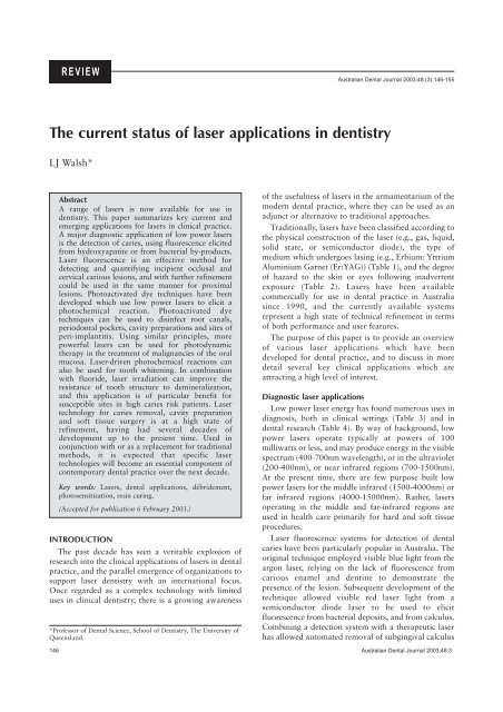
REVIEW<br />
<strong>The</strong> <strong>current</strong> <strong>status</strong> <strong>of</strong> <strong>laser</strong> <strong>applications</strong> <strong>in</strong> <strong>dentistry</strong><br />
LJ Walsh*<br />
Abstract<br />
A range <strong>of</strong> <strong>laser</strong>s is now available for use <strong>in</strong><br />
<strong>dentistry</strong>. This paper summarizes key <strong>current</strong> and<br />
emerg<strong>in</strong>g <strong>applications</strong> for <strong>laser</strong>s <strong>in</strong> cl<strong>in</strong>ical practice.<br />
A major diagnostic application <strong>of</strong> low power <strong>laser</strong>s<br />
is the detection <strong>of</strong> caries, us<strong>in</strong>g fluorescence elicited<br />
from hydroxyapatite or from bacterial by-products.<br />
Laser fluorescence is an effective method for<br />
detect<strong>in</strong>g and quantify<strong>in</strong>g <strong>in</strong>cipient occlusal and<br />
cervical carious lesions, and with further ref<strong>in</strong>ement<br />
could be used <strong>in</strong> the same manner for proximal<br />
lesions. Photoactivated dye techniques have been<br />
developed which use low power <strong>laser</strong>s to elicit a<br />
photochemical reaction. Photoactivated dye<br />
techniques can be used to dis<strong>in</strong>fect root canals,<br />
periodontal pockets, cavity preparations and sites <strong>of</strong><br />
peri-implantitis. Us<strong>in</strong>g similar pr<strong>in</strong>ciples, more<br />
powerful <strong>laser</strong>s can be used for photodynamic<br />
therapy <strong>in</strong> the treatment <strong>of</strong> malignancies <strong>of</strong> the oral<br />
mucosa. Laser-driven photochemical reactions can<br />
also be used for tooth whiten<strong>in</strong>g. In comb<strong>in</strong>ation<br />
with fluoride, <strong>laser</strong> irradiation can improve the<br />
resistance <strong>of</strong> tooth structure to dem<strong>in</strong>eralization,<br />
and this application is <strong>of</strong> particular benefit for<br />
susceptible sites <strong>in</strong> high caries risk patients. Laser<br />
technology for caries removal, cavity preparation<br />
and s<strong>of</strong>t tissue surgery is at a high state <strong>of</strong><br />
ref<strong>in</strong>ement, hav<strong>in</strong>g had several decades <strong>of</strong><br />
development up to the present time. Used <strong>in</strong><br />
conjunction with or as a replacement for traditional<br />
methods, it is expected that specific <strong>laser</strong><br />
technologies will become an essential component <strong>of</strong><br />
contemporary dental practice over the next decade.<br />
Key words: Lasers, dental <strong>applications</strong>, débridement,<br />
photosensitization, res<strong>in</strong> cur<strong>in</strong>g.<br />
(Accepted for publication 6 February 2003.)<br />
INTRODUCTION<br />
<strong>The</strong> past decade has seen a veritable explosion <strong>of</strong><br />
research <strong>in</strong>to the cl<strong>in</strong>ical <strong>applications</strong> <strong>of</strong> <strong>laser</strong>s <strong>in</strong> dental<br />
practice, and the parallel emergence <strong>of</strong> organizations to<br />
support <strong>laser</strong> <strong>dentistry</strong> with an <strong>in</strong>ternational focus.<br />
Once regarded as a complex technology with limited<br />
uses <strong>in</strong> cl<strong>in</strong>ical <strong>dentistry</strong>, there is a grow<strong>in</strong>g awareness<br />
*Pr<strong>of</strong>essor <strong>of</strong> Dental Science, School <strong>of</strong> Dentistry, <strong>The</strong> University <strong>of</strong><br />
Queensland.<br />
Australian Dental Journal 2003;48:(3):146-155<br />
<strong>of</strong> the usefulness <strong>of</strong> <strong>laser</strong>s <strong>in</strong> the armamentarium <strong>of</strong> the<br />
modern dental practice, where they can be used as an<br />
adjunct or alternative to traditional approaches.<br />
Traditionally, <strong>laser</strong>s have been classified accord<strong>in</strong>g to<br />
the physical construction <strong>of</strong> the <strong>laser</strong> (e.g., gas, liquid,<br />
solid state, or semiconductor diode), the type <strong>of</strong><br />
medium which undergoes las<strong>in</strong>g (e.g., Erbium: Yttrium<br />
Alum<strong>in</strong>ium Garnet (Er:YAG)) (Table 1), and the degree<br />
<strong>of</strong> hazard to the sk<strong>in</strong> or eyes follow<strong>in</strong>g <strong>in</strong>advertent<br />
exposure (Table 2). Lasers have been available<br />
commercially for use <strong>in</strong> dental practice <strong>in</strong> Australia<br />
s<strong>in</strong>ce 1990, and the <strong>current</strong>ly available systems<br />
represent a high state <strong>of</strong> technical ref<strong>in</strong>ement <strong>in</strong> terms<br />
<strong>of</strong> both performance and user features.<br />
<strong>The</strong> purpose <strong>of</strong> this paper is to provide an overview<br />
<strong>of</strong> various <strong>laser</strong> <strong>applications</strong> which have been<br />
developed for dental practice, and to discuss <strong>in</strong> more<br />
detail several key cl<strong>in</strong>ical <strong>applications</strong> which are<br />
attract<strong>in</strong>g a high level <strong>of</strong> <strong>in</strong>terest.<br />
Diagnostic <strong>laser</strong> <strong>applications</strong><br />
Low power <strong>laser</strong> energy has found numerous uses <strong>in</strong><br />
diagnosis, both <strong>in</strong> cl<strong>in</strong>ical sett<strong>in</strong>gs (Table 3) and <strong>in</strong><br />
dental research (Table 4). By way <strong>of</strong> background, low<br />
power <strong>laser</strong>s operate typically at powers <strong>of</strong> 100<br />
milliwatts or less, and may produce energy <strong>in</strong> the visible<br />
spectrum (400-700nm wavelength), or <strong>in</strong> the ultraviolet<br />
(200-400nm), or near <strong>in</strong>frared regions (700-1500nm).<br />
At the present time, there are few purpose built low<br />
power <strong>laser</strong>s for the middle <strong>in</strong>frared (1500-4000nm) or<br />
far <strong>in</strong>frared regions (4000-15000nm). Rather, <strong>laser</strong>s<br />
operat<strong>in</strong>g <strong>in</strong> the middle and far-<strong>in</strong>frared regions are<br />
used <strong>in</strong> health care primarily for hard and s<strong>of</strong>t tissue<br />
procedures.<br />
Laser fluorescence systems for detection <strong>of</strong> dental<br />
caries have been particularly popular <strong>in</strong> Australia. <strong>The</strong><br />
orig<strong>in</strong>al technique employed visible blue light from the<br />
argon <strong>laser</strong>, rely<strong>in</strong>g on the lack <strong>of</strong> fluorescence from<br />
carious enamel and dent<strong>in</strong>e to demonstrate the<br />
presence <strong>of</strong> the lesion. Subsequent development <strong>of</strong> the<br />
technique allowed visible red <strong>laser</strong> light from a<br />
semiconductor diode <strong>laser</strong> to be used to elicit<br />
fluorescence from bacterial deposits, and from calculus.<br />
Comb<strong>in</strong><strong>in</strong>g a detection system with a therapeutic <strong>laser</strong><br />
has allowed automated removal <strong>of</strong> subg<strong>in</strong>gival calculus<br />
146 Australian Dental Journal 2003;48:3.
Table 1. Common <strong>laser</strong> types used <strong>in</strong> <strong>dentistry</strong><br />
Laser type Construction Wavelength(s) Delivery system(s)<br />
Argon Gas <strong>laser</strong> 488, 515nm Optical fibre<br />
KTP Solid state 532nm Optical fibre<br />
Helium-neon Gas <strong>laser</strong> 633nm Optical fibre<br />
Diode Semiconductor 635, 670, 810,<br />
830, 980nm<br />
Optical fibre<br />
Nd:YAG Solid state 1064nm Optical fibre<br />
Er,Cr:YSGG Solid state 2780nm Optical fibre<br />
Er:YAG Solid state 2940nm Optical fibre,<br />
waveguide,<br />
articulated arm<br />
CO2 Gas <strong>laser</strong> 9600, 10600nm Waveguide,<br />
articulated arm<br />
from teeth and dental implants, a po<strong>in</strong>t discussed <strong>in</strong><br />
further detail below.<br />
For detection <strong>of</strong> dental caries <strong>in</strong> pits and fissures,<br />
<strong>laser</strong> fluorescence <strong>of</strong>fers greater sensitivity than<br />
conventional visual and tactile methods. 1,2 <strong>The</strong><br />
technique is also well suited to smooth surface lesions<br />
on cervical surfaces <strong>of</strong> teeth, 3,4 and to recognition <strong>of</strong><br />
caries beneath clear fissure sealants. 5 Detection <strong>of</strong><br />
proximal lesions is technically more difficult, and <strong>in</strong><br />
this sett<strong>in</strong>g, argon <strong>laser</strong>-<strong>in</strong>duced fluorescence <strong>of</strong>fers a<br />
valuable adjunct to conventional methods. 6-9 <strong>The</strong><br />
differential water content <strong>of</strong> early fissure caries and<br />
sound occlusal enamel has also led to the development<br />
<strong>of</strong> methods us<strong>in</strong>g the carbon dioxide <strong>laser</strong> to reveal<br />
such lesions, 10-12 and to modify the fissure system to<br />
<strong>in</strong>crease resistance to future carious attack. 11<br />
Two key advantages <strong>of</strong> <strong>laser</strong>-based systems are their<br />
high sensitivity, and the lack <strong>of</strong> attendant risks <strong>of</strong><br />
ioniz<strong>in</strong>g radiation. This has allowed their frequent use<br />
for monitor<strong>in</strong>g lesions <strong>of</strong> dental caries and dental<br />
erosion. 13-16 Extension <strong>of</strong> the pr<strong>in</strong>ciples <strong>of</strong> <strong>laser</strong><br />
fluorescence from the visible to the near <strong>in</strong>frared and<br />
terahertz portions <strong>of</strong> spectrum opens the possibility for<br />
more detailed analysis <strong>of</strong> the <strong>in</strong>ternal composition <strong>of</strong><br />
the tooth. 17 Moreover, the use <strong>of</strong> dyes <strong>in</strong> conjunction<br />
with <strong>laser</strong> fluorescence holds promise for us<strong>in</strong>g the<br />
method for del<strong>in</strong>eat<strong>in</strong>g cavitated from non-cavitated<br />
lesions <strong>in</strong> sites <strong>of</strong> poor cl<strong>in</strong>ical access, such as<br />
approximal surfaces. 18,19<br />
Bacterial porphyr<strong>in</strong>s <strong>in</strong> dental calculus give a strong<br />
fluorescence signal, 20 which can be used to control<br />
<strong>laser</strong>s used for scal<strong>in</strong>g. <strong>The</strong> same pr<strong>in</strong>ciple could be<br />
applied to lesions <strong>of</strong> dental caries, where a target<strong>in</strong>g<br />
<strong>laser</strong> could <strong>in</strong>duce fluorescence and provide feedback to<br />
Australian Dental Journal 2003;48:3.<br />
Table 2. Laser classification accord<strong>in</strong>g to potential<br />
hazards<br />
Class Risk Example<br />
I Fully enclosed system Nd:YAG <strong>laser</strong> weld<strong>in</strong>g system<br />
used <strong>in</strong> a dental laboratory<br />
II Visible low power <strong>laser</strong> Visible red aim<strong>in</strong>g beam <strong>of</strong> a<br />
protected by the bl<strong>in</strong>k reflex surgical <strong>laser</strong><br />
IIIa Visible <strong>laser</strong> above<br />
1 milliwatt<br />
No dental examples<br />
IIIb Higher power <strong>laser</strong> unit Low power (50 milliwatt)<br />
(up to 0.5 watts) which diode <strong>laser</strong> used for<br />
may or may not be visible.<br />
Direct view<strong>in</strong>g hazardous<br />
to the eyes<br />
biostimulation<br />
IV Damage to eyes and sk<strong>in</strong> All <strong>laser</strong>s used for oral surgery,<br />
possible. Direct or <strong>in</strong>direct whiten<strong>in</strong>g, and cavity<br />
view<strong>in</strong>g hazardous to<br />
the eyes<br />
preparation<br />
the user as to the presence <strong>of</strong> residual bacteria (i.e., the<br />
presence <strong>of</strong> <strong>in</strong>fected carious dent<strong>in</strong>e), and could also<br />
control the action <strong>of</strong> a pulsed <strong>laser</strong> to achieve<br />
automated caries removal. <strong>The</strong>re are already data on<br />
the spectral changes which occur dur<strong>in</strong>g <strong>in</strong>frared <strong>laser</strong><br />
treatment <strong>of</strong> enamel and dent<strong>in</strong>e 21,22 which could<br />
similarly be applied cl<strong>in</strong>ically when assess<strong>in</strong>g the<br />
presence or absence <strong>of</strong> carious tooth structure dur<strong>in</strong>g<br />
<strong>laser</strong>-based cavity preparation.<br />
Photoactivated dye dis<strong>in</strong>fection us<strong>in</strong>g <strong>laser</strong>s<br />
Low power <strong>laser</strong> energy <strong>in</strong> itself is not particularly<br />
lethal to bacteria, but is useful for photochemical<br />
activation <strong>of</strong> oxygen-releas<strong>in</strong>g dyes. S<strong>in</strong>glet oxygen<br />
released from the dyes causes membrane and DNA<br />
damage to micro-organisms. <strong>The</strong> photoactivated dye<br />
(PAD) technique can be undertaken with a range <strong>of</strong><br />
visible red and near <strong>in</strong>frared <strong>laser</strong>s, and systems us<strong>in</strong>g<br />
low power (100 milliwatt) visible red semiconductor<br />
diode <strong>laser</strong>s and tolonium chloride (toluid<strong>in</strong>e blue) dye<br />
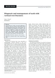
are now available commercially (Fig 1). <strong>The</strong> <strong>in</strong>itial<br />
work which demonstrated the PAD technique used<br />
helium-neon <strong>laser</strong>s. 23 However, such units have been<br />
surpassed with high efficiency diode <strong>laser</strong>s which<br />
operate at the same wavelength.<br />
<strong>The</strong> PAD technique has been shown to be effective<br />
for kill<strong>in</strong>g bacteria <strong>in</strong> complex bi<strong>of</strong>ilms, such as<br />
subg<strong>in</strong>gival plaque, which are typically resistant to the<br />
action <strong>of</strong> antimicrobial agents. 24-26 It can be used<br />
effectively <strong>in</strong> carious lesions, s<strong>in</strong>ce visible red light<br />
transmits well across dent<strong>in</strong>e, 27 and can be made<br />
species-specific by tagg<strong>in</strong>g the dye with monoclonal<br />
Table 3. Diagnostic <strong>laser</strong> <strong>applications</strong> for cl<strong>in</strong>ical practice<br />
Argon<br />
488nm<br />
Helium-neon<br />
633nm<br />
Diode<br />
633nm<br />
Diode<br />
655nm<br />
CO2 10600nm<br />
Laser fluorescence detection <strong>of</strong> dental caries ✔ ✔<br />
Laser fluorescence detection <strong>of</strong> subg<strong>in</strong>gival calculus ✔<br />
Detection <strong>of</strong> fissure caries lesions by optical changes ✔<br />
Laser doppler flowmetry to assess pulpal blood flow ✔ ✔<br />
Scann<strong>in</strong>g <strong>of</strong> phosphor plate digital radiographs ✔<br />
Scann<strong>in</strong>g <strong>of</strong> conventional radiographs for teleradiology ✔<br />
147
Table 4. Diagnostic <strong>laser</strong> <strong>applications</strong> used as research tools<br />
Nd:YAG Er:YAG Argon Helium-neon Diode<br />
1064nm 2940nm 488 and 515nm 633nm 633 and 670nm<br />
Raman spectroscopic analysis <strong>of</strong> tooth structure ✔<br />
Terahertz imag<strong>in</strong>g <strong>of</strong> <strong>in</strong>ternal tooth structure ✔<br />
Breakdown spectroscopic analysis <strong>of</strong> tooth structure ✔ ✔<br />
Confocal microscopic imag<strong>in</strong>g <strong>of</strong> s<strong>of</strong>t and hard tissues ✔<br />
Flow cytometric analysis <strong>of</strong> cells and cell sort<strong>in</strong>g ✔<br />
Pr<strong>of</strong>il<strong>in</strong>g <strong>of</strong> tooth surfaces and dental restorations ✔ ✔<br />
antibodies. 28 Photoactivated dye can be applied<br />
effectively for kill<strong>in</strong>g Gram-positive bacteria (<strong>in</strong>clud<strong>in</strong>g<br />
MRSA), Gram-negative bacteria, fungi and viruses. 29,30<br />
Major cl<strong>in</strong>ical <strong>applications</strong> <strong>of</strong> PAD <strong>in</strong>clude<br />
dis<strong>in</strong>fection <strong>of</strong> root canals, periodontal pockets, deep<br />
carious lesions, and sites <strong>of</strong> peri-implantitis. 31,32 In such<br />
locations, PAD does not give rise to deleterious thermal<br />
effects, 33 and adjacent tissues are not subjected to<br />
bystander thermal <strong>in</strong>jury. Photoactivated dye treatment<br />
does not cause sensitization and kill<strong>in</strong>g <strong>of</strong> adjacent<br />
human cells such as fibroblasts and kerat<strong>in</strong>ocytes. 34<br />
Neither the dye nor the reactive oxygen species<br />
produced from it are toxic to the patient. Tolonium<br />
chloride is used <strong>in</strong> high concentrations for screen<strong>in</strong>g<br />
patients for malignancies <strong>of</strong> the oral mucosa and<br />
oropharynx, 35,36 and does not exert toxic effects at the<br />
low concentrations used <strong>in</strong> the PAD technique.<br />
Moreover, residual reactive oxygen species are rapidly<br />
dealt with by the enzyme catalase, which is present <strong>in</strong><br />
all tissues and <strong>in</strong> the peripheral circulation, 37 and by<br />
lactoperoxidase, which is a normal component <strong>of</strong><br />
saliva.<br />
Photodynamic therapy<br />
A more powerful <strong>laser</strong>-<strong>in</strong>itiated photochemical<br />
reaction is photodynamic therapy (PDT), which has<br />
been employed <strong>in</strong> the treatment <strong>of</strong> malignancies <strong>of</strong> the<br />
oral mucosa, particularly multi-focal squamous cell<br />
carc<strong>in</strong>oma. As <strong>in</strong> PAD, <strong>laser</strong>-activation <strong>of</strong> a sensitiz<strong>in</strong>g<br />
dye <strong>in</strong> PDT generates reactive oxygen species. <strong>The</strong>se <strong>in</strong><br />
Fig 1. Laser system for photo-activated dye therapy, which uses a<br />
diode <strong>laser</strong> (635nm) and tolonium chloride dye (SaveDent, Asclepion<br />
Meditec, Fife, UK).<br />
turn directly damage cells and the associated blood<br />
vascular network, trigger<strong>in</strong>g both necrosis and<br />
apoptosis. 38<br />
Of <strong>in</strong>terest, while direct effects <strong>of</strong> PDT destroy the<br />
bulk <strong>of</strong> tumour cells, there is accumulat<strong>in</strong>g evidence<br />
that PDT activates the host immune response, and<br />
promotes anti-tumour immunity through the activation<br />
<strong>of</strong> macrophages and T lymphocytes. 37 For example,<br />
there is direct experimental evidence for photodynamic<br />
activation <strong>of</strong> the production <strong>of</strong> tumour necrosis factoralpha,<br />
39 a key cytok<strong>in</strong>e <strong>in</strong> host anti-tumour immune<br />
responses.<br />
Cl<strong>in</strong>ical studies have reported positive results for<br />
PDT treatment <strong>of</strong> carc<strong>in</strong>oma-<strong>in</strong>-situ and squamous cell<br />
carc<strong>in</strong>oma <strong>in</strong> the oral cavity, with response rates<br />
approximat<strong>in</strong>g 90 per cent. 40,41 <strong>The</strong> treated sites<br />
characteristically show erythema and oedema, followed<br />
by necrosis and frank ulceration. <strong>The</strong> ulcerated lesions<br />
typically take up to eight weeks to heal fully, and<br />
supportive analgesia is required <strong>in</strong> the first few weeks.<br />
Other than short-term photosensitivity, the treatment is<br />
tolerated well. 39<br />
Other photochemical <strong>laser</strong> effects<br />
<strong>The</strong> argon <strong>laser</strong> produces high <strong>in</strong>tensity visible blue<br />
light (488nm) which is able to <strong>in</strong>itiate photopolymerization<br />
<strong>of</strong> light-cured dental restorative<br />
materials which use camphoroqu<strong>in</strong>one as the<br />
photo<strong>in</strong>itiator. 42-44 <strong>The</strong> temperature <strong>in</strong>crease at the level<br />
<strong>of</strong> the dental pulp is much less with argon <strong>laser</strong> cur<strong>in</strong>g<br />
than when conventional quartz tungsten halogen lamp<br />
units are used. 45,46 Argon <strong>laser</strong> radiation is also able to<br />
alter the surface chemistry <strong>of</strong> both enamel and root<br />
surface dent<strong>in</strong>e, 47,48 which reduces the probability <strong>of</strong><br />
re<strong>current</strong> caries. This cl<strong>in</strong>ical benefit is arguably more<br />
important than the reduced cur<strong>in</strong>g time and improved<br />
depth <strong>of</strong> cure achieved with the argon <strong>laser</strong>.<br />
A further photochemical effect produced by high<br />
<strong>in</strong>tensity green <strong>laser</strong> light is photochemical bleach<strong>in</strong>g<br />
(Table 5). This effect relies upon specific absorption <strong>of</strong><br />
a narrow spectral range <strong>of</strong> green light (510-540nm)<br />
<strong>in</strong>to chelate compounds formed between apatites,<br />
Table 5. Laser-enhanced tooth whiten<strong>in</strong>g<br />
Argon KTP Diode CO 2<br />
515nm 532nm 810-980nm 10600nm<br />
Photochemical bleach<strong>in</strong>g ✔ ✔<br />
Photothermal bleach<strong>in</strong>g ✔ ✔<br />
148 Australian Dental Journal 2003;48:3.
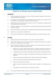
Fig 2. KTP <strong>laser</strong> photochemical bleach<strong>in</strong>g. A. Initial cl<strong>in</strong>ical<br />
appearance <strong>of</strong> the dentition <strong>in</strong> a 9-year-old patient with <strong>in</strong>tense<br />
discolouration <strong>of</strong> the <strong>in</strong>cisor teeth caused by prolonged childhood<br />
illnesses and associated medications. B. <strong>The</strong> situation immediately<br />
after three 60-m<strong>in</strong>ute appo<strong>in</strong>tments <strong>of</strong> power bleach<strong>in</strong>g us<strong>in</strong>g a<br />
conventional quartz tungsten halogen cur<strong>in</strong>g lamp and 35 per cent<br />
hydrogen peroxide gel. <strong>The</strong> <strong>in</strong>cisal enamel shade has improved<br />
somewhat, but the areas <strong>of</strong> discolouration at the g<strong>in</strong>gival third are<br />
unchanged. <strong>The</strong> arrow <strong>in</strong>dicates a residue <strong>of</strong> the protective g<strong>in</strong>gival<br />
dam. C. Cl<strong>in</strong>ical appearance immediately after one session <strong>of</strong><br />
photochemical bleach<strong>in</strong>g us<strong>in</strong>g the KTP <strong>laser</strong> and proprietary<br />
alkal<strong>in</strong>e hydrogen peroxide (Smartbleach ®) gel. <strong>The</strong> <strong>laser</strong> treatment<br />
was targeted to the g<strong>in</strong>gival third. <strong>The</strong> patient and her parents were<br />
pleased with the immediate post-operative result and did not request<br />
any additional treatment.<br />
porphyr<strong>in</strong>s, and tetracycl<strong>in</strong>e compounds. 49 <strong>The</strong> argon<br />
<strong>laser</strong> (515nm) and potassium titanyl phosphate (KTP)<br />
<strong>laser</strong> (532nm) can both be used for photochemical<br />
bleach<strong>in</strong>g, s<strong>in</strong>ce their wavelengths approximate the<br />
absorption maxima <strong>of</strong> these chelate compounds (525-<br />
530nm). 50 Argon and KTP <strong>laser</strong>s can achieve a positive<br />
result <strong>in</strong> cases which are completely unresponsive to<br />
conventional photothermal ‘power’ bleach<strong>in</strong>g (Fig 2).<br />
Laser <strong>applications</strong> <strong>in</strong> the dental laboratory<br />
<strong>The</strong>re is a range <strong>of</strong> laboratory-based <strong>laser</strong><br />
<strong>applications</strong> (Table 6). Laser holographic imag<strong>in</strong>g is a<br />
well established method for stor<strong>in</strong>g topographic<br />
<strong>in</strong>formation, such as crown preparations, occlusal<br />
tables, and facial forms. <strong>The</strong> use <strong>of</strong> two <strong>laser</strong> beams<br />
allows more complex surface detail to be mapped us<strong>in</strong>g<br />
<strong>in</strong>terferometry, 51,52 while conventional diffraction<br />
grat<strong>in</strong>gs and <strong>in</strong>terference patterns are used to generate<br />
holograms and contour pr<strong>of</strong>iles. 53-56<br />
Laser scann<strong>in</strong>g <strong>of</strong> casts can be l<strong>in</strong>ked to<br />
computerized mill<strong>in</strong>g equipment for fabrication <strong>of</strong><br />
restorations from porcela<strong>in</strong> and other materials. An<br />
alternative fabrication strategy is to s<strong>in</strong>ter ceramic<br />
materials, to create a solid restoration from a powder<br />
<strong>of</strong> alum<strong>in</strong>a or hydroxyapatite. 57 <strong>The</strong> same approach can<br />
be used to form complex shapes from dental wax and<br />
other materials which can be s<strong>in</strong>tered, such that these<br />
can then be used <strong>in</strong> conventional ‘lost wax’ cast<strong>in</strong>g. A<br />
variation on this theme is ultraviolet (helium-cadmium)<br />
<strong>laser</strong>-<strong>in</strong>itiated polymerization <strong>of</strong> liquid res<strong>in</strong> <strong>in</strong> a<br />
chamber, to create surgical templates for implant<br />
surgery and major reconstructive oral surgery. <strong>The</strong>se<br />
templates can be coupled with <strong>laser</strong>-based position<strong>in</strong>g<br />
systems for complex reconstructive and orthognathic<br />
surgical procedures.<br />
Laser procedures on dental hard tissues<br />
Cavity preparation us<strong>in</strong>g <strong>laser</strong>s has been an area <strong>of</strong><br />
major research <strong>in</strong>terest s<strong>in</strong>ce <strong>laser</strong>s were <strong>in</strong>itially<br />
developed <strong>in</strong> the early 1960s. At the present time,<br />
several <strong>laser</strong> types with similar wavelengths <strong>in</strong> the<br />
middle <strong>in</strong>frared region <strong>of</strong> the electromagnetic spectrum<br />
are used commonly for cavity preparation and caries<br />
removal. <strong>The</strong> Er:YAG, Er:YSGG and Er,Cr:YSGG <strong>laser</strong>s<br />
operate at wavelengths <strong>of</strong> 2940, 2790, and 2780nm,<br />
respectively. <strong>The</strong>se wavelengths correspond to the peak<br />
absorption range <strong>of</strong> water <strong>in</strong> the <strong>in</strong>frared spectrum<br />
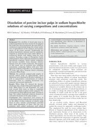
(Fig 3), although the absorption <strong>of</strong> the Er:YAG <strong>laser</strong><br />
(13,000cm -1 ) is much higher than that <strong>of</strong> the Er:YSGG<br />
(7000cm -1 ) and Er,Cr:YSGG (4000cm -1 ) 58-60 S<strong>in</strong>ce all<br />
three <strong>laser</strong>s rely on water-based absorption for cutt<strong>in</strong>g<br />
enamel and dent<strong>in</strong>e, the efficiency <strong>of</strong> ablation<br />
(measured <strong>in</strong> terms <strong>of</strong> volume and mass loss <strong>of</strong> tooth<br />
structure for identical energy parameters) is greatest for<br />
the Er:YAG <strong>laser</strong>. 58,60<br />
<strong>The</strong>se <strong>laser</strong> systems can be used for effective caries<br />
removal and cavity preparation without significant<br />
thermal effects, collateral damage to tooth structure, or<br />
patient discomfort. 61-64 Normal dental enamel conta<strong>in</strong>s<br />
sufficient water (approximately 12 per cent by volume)<br />
that a water mist spray coupled to an Er-based <strong>laser</strong><br />
Australian Dental Journal 2003;48:3. 149
Table 6. Laser <strong>applications</strong> <strong>in</strong> the dental laboratory<br />
Helium neon<br />
633nm<br />
Diode<br />
635nm<br />
Nd:YAG<br />
1064nm<br />
CO2 10600nm<br />
Helium-Cadmium<br />
300nm<br />
Scann<strong>in</strong>g <strong>of</strong> models for orthodontics or holographic storage ✔ ✔<br />
Scann<strong>in</strong>g <strong>of</strong> crown preparations for CAD-CAM ✔ ✔<br />
Weld<strong>in</strong>g <strong>of</strong> metals (Co:Cr, titanium) ✔<br />
S<strong>in</strong>ter<strong>in</strong>g <strong>of</strong> ceramics ✔<br />
CAD-s<strong>in</strong>ter<strong>in</strong>g fabrication ✔<br />
CAD-polymer fabrication <strong>of</strong> spl<strong>in</strong>ts or surgical models ✔<br />
Cutt<strong>in</strong>g <strong>of</strong> ceramics ✔<br />
system can achieve effective ablation at temperatures<br />
well below the melt<strong>in</strong>g and vapourization temperatures<br />
<strong>of</strong> enamel. 61 Er-based dental <strong>laser</strong>s can also be used to<br />
remove res<strong>in</strong> composite res<strong>in</strong> and glass-ionomer cement<br />
restorations, and to etch tooth structure (Fig 4).<br />
A characteristic operat<strong>in</strong>g feature <strong>of</strong> Er-based <strong>laser</strong><br />
systems is a popp<strong>in</strong>g sound when the <strong>laser</strong> is operat<strong>in</strong>g<br />
on dental hard tissues. Both the pitch and resonance <strong>of</strong><br />
this sound relate to the propagation <strong>of</strong> an acoustic<br />
shock wave with<strong>in</strong> the tooth, and vary accord<strong>in</strong>g to the<br />
presence or absence <strong>of</strong> caries. This feature assists the<br />
user <strong>in</strong> determ<strong>in</strong><strong>in</strong>g that caries removal is complete. 66 In<br />
contrast to the popp<strong>in</strong>g sound dur<strong>in</strong>g caries removal,<br />
one <strong>current</strong> generation Er,Cr:YSGG <strong>laser</strong> system creates<br />
a loud snapp<strong>in</strong>g sound even when not <strong>in</strong> contact with<br />
any structure <strong>in</strong> the mouth. This seem<strong>in</strong>g paradox can<br />
be expla<strong>in</strong>ed by an effect termed ‘plasma de-coupl<strong>in</strong>g’<br />
<strong>of</strong> the beam, <strong>in</strong> which <strong>in</strong>cident <strong>laser</strong> energy heats the air<br />
and water directly <strong>in</strong> front <strong>of</strong> the <strong>laser</strong> handpiece. In the<br />
Er,Cr:YSGG <strong>laser</strong>, this is done <strong>in</strong>tentionally <strong>in</strong> order to<br />
deliver energy onto the rear surface <strong>of</strong> atomized water<br />
molecules, with the aim <strong>of</strong> accelerat<strong>in</strong>g them to a higher<br />
speed (so-called ‘HydroK<strong>in</strong>etic cutt<strong>in</strong>g’). 67 Detailed<br />
studies <strong>of</strong> the cutt<strong>in</strong>g mechanisms <strong>of</strong> Er:YAG and<br />
Er,Cr:YSGG <strong>laser</strong>s have revealed that the mechanism<br />
by which enamel is removed is basically the same for<br />
both <strong>laser</strong> systems, namely explosive subsurface<br />
expansion <strong>of</strong> <strong>in</strong>terstitially trapped water. 65 <strong>The</strong> same<br />
<strong>in</strong>vestigations also failed to show Er,Cr:YSGG <strong>laser</strong><br />
cutt<strong>in</strong>g <strong>of</strong> a variety <strong>of</strong> materials which were free <strong>of</strong><br />
water, which the authors stated was ‘contradictory to<br />
the existence <strong>of</strong> the hydrok<strong>in</strong>etic phenomenon’. 65<br />
14000<br />
12000<br />
10000<br />
8000<br />
6000<br />
4000<br />
2000<br />
Er,Cr:YSGG<br />
Er:YAG<br />
0<br />
2.6 2.65 2.7 2.75 2.8 2.85 2.9 2.95 3 3.05 3.1 3.15 3.2 3.25 3.3<br />
Fig 3. <strong>The</strong> absorption curve <strong>of</strong> water <strong>in</strong> the middle <strong>in</strong>frared region.<br />
Data on the vertical axis are units <strong>of</strong> absorption, while the horizontal<br />
axis shows wavelength <strong>in</strong> micrometers. <strong>The</strong> plot shows the position<br />
<strong>of</strong> two <strong>laser</strong> wavelengths used for cavity preparation: Er,Cr:YSGG<br />
2.78 micrometers, and Er:YAG 2.94 micrometers. <strong>The</strong> figure is based<br />
on data from reference 59.<br />
An important theoretical extension to the pr<strong>in</strong>ciple<br />
<strong>of</strong> water-based <strong>laser</strong> ablation <strong>of</strong> tooth structure is the<br />
recently described effect <strong>of</strong> ‘<strong>laser</strong> abrasion’, <strong>in</strong> which<br />
Er:YAG <strong>laser</strong> energy is used to accelerate the movement<br />
<strong>of</strong> particles <strong>of</strong> sapphire 30-50 micrometers <strong>in</strong> diameter<br />
<strong>in</strong> aqueous suspension. As <strong>in</strong> air abrasion, the impact <strong>of</strong><br />
these particles causes brittle splitt<strong>in</strong>g, result<strong>in</strong>g <strong>in</strong> tooth<br />
substance removal. In the <strong>laser</strong> abrasion method, high<br />
speed photography has documented particle velocities<br />
<strong>in</strong> the range <strong>of</strong> 50-100 metres per second, which enable<br />
a rate <strong>of</strong> enamel removal ‘higher than that <strong>of</strong> high speed<br />
turb<strong>in</strong>es’ with a very low volume <strong>of</strong> abrasive particles. 68<br />
This technique could be employed with <strong>current</strong><br />
generation <strong>laser</strong>s once a suitable dispens<strong>in</strong>g system for<br />
the suspension <strong>of</strong> particles has been developed. As well<br />
as the potential <strong>of</strong> even more rapid cutt<strong>in</strong>g rates than<br />
conventional rotary <strong>in</strong>strumentation, <strong>laser</strong> abrasion<br />
<strong>of</strong>fers the promise <strong>of</strong> <strong>laser</strong>-based cutt<strong>in</strong>g <strong>of</strong> structures<br />
which are not otherwise amenable to this, such as<br />
ceramic restorations.<br />
Intensive research over the past three decades on<br />
other non-Erbium <strong>laser</strong>-based cavity preparation<br />
systems has yet to be translated to direct cl<strong>in</strong>ical<br />
application. To date, alternative <strong>laser</strong> systems,<br />
<strong>in</strong>clud<strong>in</strong>g super-pulsed CO2, Ho:YAG, Ho:YSGG,<br />
Nd:YAG, Nd:NLF, diode <strong>laser</strong>s and excimers, have not<br />
proven feasible for use for cavity preparation <strong>in</strong> general<br />
practice sett<strong>in</strong>gs.<br />
Other than caries removal, this is a range <strong>of</strong> other<br />
well established <strong>laser</strong> hard tissue procedures <strong>in</strong>clude<br />
desensitization <strong>of</strong> cervical dent<strong>in</strong>e (us<strong>in</strong>g Nd:YAG,<br />
Er:YAG, Er,Cr:YSGG CO2, KTP, and diode <strong>laser</strong>s),<br />
<strong>laser</strong> analgesia (us<strong>in</strong>g Nd:YAG, Er:YAG, and<br />
Er,Cr:YSGG <strong>laser</strong>s), <strong>laser</strong>-enhanced fluoride uptake<br />
(us<strong>in</strong>g Er:YAG, Er,Cr:YSGG, CO2, argon, and KTP<br />
<strong>laser</strong>s). Furthermore, there is a considerable range <strong>of</strong><br />
periodontal procedures (Table 7), and endodontic<br />
procedures (Table 8) which can be undertaken with<br />
<strong>laser</strong>s as an alternative to conventional approaches.<br />
S<strong>of</strong>t tissue <strong>laser</strong> procedures<br />
<strong>The</strong>re are numerous s<strong>of</strong>t tissue procedures which can<br />
be performed with <strong>laser</strong>s. 69-71 Two key features <strong>of</strong> these<br />
are reduced bleed<strong>in</strong>g <strong>in</strong>tra-operatively and less pa<strong>in</strong><br />
post-operatively compared to conventional techniques<br />
such as electrosurgery. <strong>The</strong> degree <strong>of</strong> absorption <strong>in</strong> key<br />
tissue components dictates the type <strong>of</strong> effect ga<strong>in</strong>ed by<br />
the <strong>laser</strong> on s<strong>of</strong>t tissues, and <strong>in</strong> this regard the content<br />
<strong>of</strong> water and haemoglob<strong>in</strong> <strong>in</strong> oral tissues is important<br />
150 Australian Dental Journal 2003;48:3.
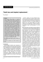
Fig 4. Restorative procedures us<strong>in</strong>g the Er:YAG <strong>laser</strong>, <strong>in</strong> anxious dental patients, without local anesthesia. <strong>The</strong> Er:YAG <strong>laser</strong> was used with a<br />
non-contact handpiece. A. Pre-operative appearance <strong>of</strong> a 22-year-old male with salivary dysfunction, and associated cervical and approximal<br />
caries. B. Areas <strong>of</strong> caries and defective res<strong>in</strong> composite have been removed. <strong>The</strong> <strong>in</strong>tense white appearance <strong>of</strong> the marg<strong>in</strong>s is typical <strong>of</strong> <strong>laser</strong> etch<strong>in</strong>g.<br />
C. <strong>The</strong> restored teeth immediately post-operatively. <strong>The</strong> etched appearance <strong>of</strong> the marg<strong>in</strong>s disappears once bond<strong>in</strong>g res<strong>in</strong> has been placed.<br />
D. 30-year-old female patient with areas <strong>of</strong> hypoplastic enamel. E. <strong>The</strong> enamel surface has been ‘peeled’ us<strong>in</strong>g a series <strong>of</strong> pulses from the <strong>laser</strong>.<br />
F. <strong>The</strong> two areas have been restored with res<strong>in</strong> composite. G. 65-year-old female patient undergo<strong>in</strong>g anti-cancer chemotherapy, with re<strong>current</strong><br />
caries at the marg<strong>in</strong>s <strong>of</strong> several restorations. H. Areas <strong>of</strong> caries and underm<strong>in</strong>ed res<strong>in</strong> composite have been removed. I. <strong>The</strong> cavity preparations<br />
have been restored.<br />
for the efficient absorption <strong>of</strong> many commonly used<br />
dental <strong>laser</strong>s. 72 Certa<strong>in</strong> procedures <strong>in</strong> patients with<br />
bleed<strong>in</strong>g disorders are better suited to <strong>laser</strong>s with<br />
greater haemostatic capabilities (Table 9). Examples <strong>of</strong><br />
simple s<strong>of</strong>t tissue procedures are presented <strong>in</strong> Fig 5.<br />
CONCLUSIONS<br />
Laser technology for caries detection, res<strong>in</strong> cur<strong>in</strong>g,<br />
cavity preparation and s<strong>of</strong>t tissue surgery is at a high<br />
state <strong>of</strong> ref<strong>in</strong>ement, hav<strong>in</strong>g had several decades <strong>of</strong><br />
development up to the present time. This is not to say<br />
that further major improvements cannot occur. Indeed,<br />
as is <strong>in</strong> the case with <strong>laser</strong> abrasion, the fusion <strong>of</strong><br />
concepts from differ<strong>in</strong>g technologies may open the<br />
door to novel techniques and treatments. <strong>The</strong> field <strong>of</strong><br />
<strong>laser</strong>-based photochemical reactions holds great<br />
promise for additional <strong>applications</strong>, particularly for<br />
target<strong>in</strong>g specific cells, pathogens or molecules. A<br />
further area <strong>of</strong> future growth is expected to be the<br />
comb<strong>in</strong>ation <strong>of</strong> diagnostic and therapeutic <strong>laser</strong><br />
techniques <strong>in</strong> the one device, for example the detection<br />
and removal <strong>of</strong> dental caries or dental calculus. For<br />
Australian Dental Journal 2003;48:3. 151
Table 7. Periodontal <strong>laser</strong> procedures<br />
Er:YAG<br />
2940nm<br />
Er,Cr:YSGG<br />
2780nm<br />
KTP<br />
532nm<br />
Argon 488<br />
and 515nm<br />
Diode<br />
810-980nm<br />
Nd:YAG<br />
1064nm<br />
Helium-neon<br />
633nm<br />
Diode 635,<br />
670 or 830nm<br />
CO2 10600nm<br />
Calculus removal<br />
Periodontal pocket<br />
✔<br />
dis<strong>in</strong>fection<br />
Photoactivated dye<br />
dis<strong>in</strong>fection <strong>of</strong><br />
✔ ✔ ✔ ✔ ✔ ✔<br />
pockets<br />
De-epithelialization to<br />
✔ ✔<br />
assist regeneration ✔ ✔ ✔<br />
Table 8. Endodontic <strong>laser</strong> procedures<br />
CO2 Erbium:YAG Er,Cr:YSGG KTP Argon 488 Diode Nd:YAG Helium-neon Diode 635,<br />
10600nm 2940nm 2780nm 532nm and 515nm 810-980nm 1064nm 633nm 670, or 830nm<br />
Direct pulp capp<strong>in</strong>g<br />
Dry<strong>in</strong>g <strong>of</strong> the<br />
✔ ✔<br />
root canal<br />
Removal <strong>of</strong><br />
✔ ✔ ✔<br />
smear layer<br />
Root canal<br />
✔ ✔<br />
dis<strong>in</strong>fection<br />
Photoactivated dye<br />
✔ ✔ ✔ ✔ ✔ ✔<br />
dis<strong>in</strong>fection <strong>of</strong> pockets ✔ ✔<br />
example, an ‘autopilot’ system for subg<strong>in</strong>gival<br />
débridement has been developed (for detailed review,<br />
see ref 73), and the potential exists to extend this<br />
concept further.<br />
<strong>The</strong>re is a large research effort <strong>in</strong>ternationally<br />
focused on develop<strong>in</strong>g new <strong>laser</strong> <strong>applications</strong> for dental<br />
practice, and each year several large meet<strong>in</strong>gs are held<br />
which br<strong>in</strong>g together this research. Examples <strong>in</strong>clude<br />
the International Society for Lasers <strong>in</strong> Dentistry (ISLD),<br />
the European Society for Oral Laser Applications<br />
(ESOLA), and the Academy <strong>of</strong> Laser Dentistry (ALD).<br />
With the further development <strong>of</strong> <strong>laser</strong> <strong>dentistry</strong> as an<br />
area <strong>of</strong> cl<strong>in</strong>ical pursuit, there will be considerable<br />
opportunity for cl<strong>in</strong>icians to become <strong>in</strong>volved <strong>in</strong> these<br />
research meet<strong>in</strong>gs and <strong>in</strong> specific research projects. <strong>The</strong><br />
Australasian region has played a substantial role <strong>in</strong> the<br />
development <strong>of</strong> hard tissue <strong>laser</strong> <strong>applications</strong>, 74,75 and<br />
this level <strong>of</strong> <strong>in</strong>volvement is expected to cont<strong>in</strong>ue <strong>in</strong> the<br />
future, as various research groups exam<strong>in</strong>e uses for<br />
<strong>laser</strong>s <strong>in</strong> conjunction with or as a replacement for<br />
traditional methods.<br />
<strong>The</strong>re is little argument that over recent years the use<br />
<strong>of</strong> <strong>laser</strong>s <strong>in</strong> <strong>dentistry</strong> <strong>in</strong> Australia has moved beyond<br />
Table 9. Surgical <strong>laser</strong> <strong>applications</strong><br />
academic centres and specialist units <strong>in</strong>to the<br />
ma<strong>in</strong>stream <strong>of</strong> general practice. Look<strong>in</strong>g to the future,<br />
it is expected that specific <strong>laser</strong> technologies will<br />
become an essential component <strong>of</strong> contemporary dental<br />
practice over the next decade.<br />
ACKNOWLEDGMENTS<br />
I thank the dental practitioners who have referred<br />
patients to the Laser Cl<strong>in</strong>ic over the past 12 years, and<br />
the numerous staff and students who have contributed<br />
to the dental <strong>laser</strong> research programme at the<br />
University <strong>of</strong> Queensland.<br />
REFERENCES<br />
1. Shi XQ, Welander U, Angmar-Månsson B. Occlusal caries<br />
detection with KaVo DIAGNOdent and radiography: an <strong>in</strong> vitro<br />
comparison. Caries Res 2000;34:151-158.<br />
2. Lussi A, Megert B, Longbottom C, Reich E, Francescut P. Cl<strong>in</strong>ical<br />
performance <strong>of</strong> a <strong>laser</strong> fluorescence device for detection <strong>of</strong><br />
occlusal caries lesions. Eur J Oral Sci 2001;109:14-19.<br />
3. Shi XQ, Tranaeus S, Angmar-Månsson B. Validation <strong>of</strong><br />
DIAGNOdent for quantification <strong>of</strong> smooth-surface caries: an <strong>in</strong><br />
vitro study. Acta Odontol Scand 2001;59:74-78.<br />
4. Shi XQ, Tranaeus S, Angmar-Månsson B. Comparison <strong>of</strong> QLF<br />
and DIAGNOdent for quantification <strong>of</strong> smooth surface caries.<br />
Caries Res 2001;35:21-26.<br />
Er:YAG 2940nm<br />
(least haemostasis)<br />
Er,Cr:YSGG<br />
2780nm<br />
CO2 10600nm<br />
KTP<br />
532nm<br />
Diode<br />
810-980nm<br />
Argon 488<br />
and 515nm<br />
Nd:YAG 1064nm<br />
(most haemostatis)<br />
M<strong>in</strong>or s<strong>of</strong>t<br />
tissue surgery<br />
Major s<strong>of</strong>t<br />
✔ ✔ ✔ ✔ ✔ ✔<br />
tissue surgery<br />
Surgical treatment<br />
<strong>of</strong> large<br />
✔<br />
vascular lesions ✔<br />
Bone cutt<strong>in</strong>g<br />
Implant exposure<br />
✔ ✔<br />
with bone removal ✔ ✔ ✔<br />
152 Australian Dental Journal 2003;48:3.
Fig 5. S<strong>of</strong>t tissue procedures us<strong>in</strong>g middle and far <strong>in</strong>frared <strong>laser</strong>s. A. Pre-operative cl<strong>in</strong>ical appearance <strong>of</strong> a 22-year-old female with marked g<strong>in</strong>gival<br />
overgrowth from nifedip<strong>in</strong>e and cyclospor<strong>in</strong>. <strong>The</strong> patient has received a kidney transplant, is immuno-suppressed, and has a bleed<strong>in</strong>g tendency.<br />
B. the immediate post-operative appearance follow<strong>in</strong>g g<strong>in</strong>givoplasty with the carbon dioxide <strong>laser</strong>. Complete haemostasis is ma<strong>in</strong>ta<strong>in</strong>ed dur<strong>in</strong>g the<br />
procedure. C. Initial cl<strong>in</strong>ical appearance <strong>of</strong> a 16-year-old female patient with marked g<strong>in</strong>gival overgrowth, which has obscured the orthodontic<br />
brackets and caused the cessation <strong>of</strong> fixed orthodontic treatment. D. Immediate post-operative view <strong>of</strong> quadrant 1 follow<strong>in</strong>g g<strong>in</strong>gival recontour<strong>in</strong>g<br />
with the carbon dioxide <strong>laser</strong>. E. Immediate post-operative view <strong>of</strong> quadrant 2 follow<strong>in</strong>g recontour<strong>in</strong>g with the Er:YAG <strong>laser</strong> <strong>in</strong> contact mode.<br />
Note the different appearance <strong>of</strong> the tissues compared to quadrant 1. Both segments were treated at the same appo<strong>in</strong>tment. F and G. Cl<strong>in</strong>ical<br />
appearance <strong>of</strong> the two sites two weeks follow<strong>in</strong>g surgery. <strong>The</strong> tissue contours are identical to those determ<strong>in</strong>ed at the time <strong>of</strong> surgery.<br />
5. Takamori K, Hokari N, Okumura Y, Watanabe S. Detection <strong>of</strong><br />
occlusal caries under sealants by use <strong>of</strong> a <strong>laser</strong> fluorescence<br />
system. J Cl<strong>in</strong> Laser Med Surg 2001;19:267-271.<br />
6. Bjelkhagen H, Sundström F, Angmar-Månsson B, Ryden H. Early<br />
detection <strong>of</strong> enamel caries by the lum<strong>in</strong>escence excited by visible<br />
<strong>laser</strong> light. Swed Dent J 1982;6:1-7.<br />
7. Angmar-Månsson B, ten Bosch JJ. Optical methods for the<br />
detection and quantification <strong>of</strong> caries. Adv Dent Res 1987;1:14-<br />
20.<br />
8. Hafström-Bjorkman U, Sundström F, Angmar-Månsson B. Initial<br />
caries diagnosis <strong>in</strong> rat molars, us<strong>in</strong>g <strong>laser</strong> fluorescence. Acta<br />
Odontol Scand 1991;49:27-33.<br />
9. Hafström-Bjorkman U, Sundström F, de Jossel<strong>in</strong> de Jong E,<br />
Oliveby A, Angmar-Månsson B. Comparison <strong>of</strong> <strong>laser</strong> fluorescence<br />
and longitud<strong>in</strong>al microradiography for quantitative assessment <strong>of</strong><br />
<strong>in</strong> vitro enamel caries. Caries Res 1992;26:241-247.<br />
10. Longbottom C, Pitts NB. CO2 <strong>laser</strong> and the diagnosis <strong>of</strong> occlusal<br />
caries: <strong>in</strong> vitro study. J Dent 1993;21:234-239.<br />
Australian Dental Journal 2003;48:3. 153
11. Walsh LJ, Perham S. Enamel fusion us<strong>in</strong>g a carbon dioxide <strong>laser</strong>:<br />
a technique for seal<strong>in</strong>g pits and fissures. Cl<strong>in</strong> Prev Dent<br />
1991;13:16-20.<br />
12. Benedetto MD, Antonson DE. Use <strong>of</strong> CO2 <strong>laser</strong> for visible<br />
detection <strong>of</strong> enamel fissure caries. Qu<strong>in</strong>tessence Int 1988;19:187-<br />
190.<br />
13. Angmar-Månsson B, al-Khateeb S, Tranaeus S. Monitor<strong>in</strong>g the<br />
caries process. Optical methods for cl<strong>in</strong>ical diagnosis and<br />
quantification <strong>of</strong> enamel caries. Eur J Oral Sci 1996;104<br />
(4(Pt 2)):480-485.<br />
14. Ando M, Hall AF, Eckert GJ, Schemehorn BR, Analoui M,<br />
Stookey GK. Relative ability <strong>of</strong> <strong>laser</strong> fluorescence techniques to<br />
quantitate early m<strong>in</strong>eral loss <strong>in</strong> vitro. Caries Res 1997;31:125-<br />
131.<br />
15. al-Khateeb S, ten Cate JM, Angmar-Månsson B, et al.<br />
Quantification <strong>of</strong> formation and rem<strong>in</strong>eralization <strong>of</strong> artificial<br />
enamel lesions with a new portable fluorescence device. Adv<br />
Dent Res 1997;11:502-506.<br />
16. al-Khateeb S, Forsberg CM, de Jossel<strong>in</strong> de Jong E, Angmar-<br />
Månsson B. A longitud<strong>in</strong>al <strong>laser</strong> fluorescence study <strong>of</strong> white spot<br />
lesions <strong>in</strong> orthodontic patients. Am J Orthod Dent<strong>of</strong>acial Orthop<br />
1998;113:595-602.<br />
17. Konig K, Flemm<strong>in</strong>g G, Hibst R. Laser-<strong>in</strong>duced aut<strong>of</strong>luorescence<br />
spectroscopy <strong>of</strong> dental caries. Cell Mol Biol (Noisy-le-grand)<br />
1998;44:1293-1300.<br />
18. Eggertsson H, Analoui M, van der Veen M, Gonzalez-Cabezas C,<br />
Eckert G, Stookey G. Detection <strong>of</strong> early <strong>in</strong>terproximal caries <strong>in</strong><br />
vitro us<strong>in</strong>g <strong>laser</strong> fluorescence, dye-enhanced <strong>laser</strong> fluorescence<br />
and direct visual exam<strong>in</strong>ation. Caries Res 1999;33:227-233.<br />
19. Featherstone JD. Caries detection and prevention with <strong>laser</strong><br />
energy. Dent Cl<strong>in</strong> North Am 2000;44:955-969.<br />
20. Folwaczny M, Heym R, Mehl A, Hickel R. Subg<strong>in</strong>gival calculus<br />
detection with fluorescence <strong>in</strong>duced by 655 nm InGaAsP diode<br />
<strong>laser</strong> radiation. J Periodontol 2002;73:597-601.<br />
21. Am<strong>in</strong>zadeh A, Shahabi S, Walsh LJ. Raman spectroscopic studies<br />
<strong>of</strong> CO2 <strong>laser</strong>-irradiated human dental enamel. Spectrochim Acta<br />
A Mol Biomol Spectrosc 1999;55:1303-1308.<br />
22. Sasaki KM, Aoki A, Masuno H, Ich<strong>in</strong>ose S, Yamada S, Ishikawa<br />
I. Compositional analysis <strong>of</strong> root cementum and dent<strong>in</strong> after<br />
Er:YAG <strong>laser</strong> irradiation compared with CO2 lased and <strong>in</strong>tact<br />
roots us<strong>in</strong>g Fourier transformed <strong>in</strong>frared spectroscopy. J<br />
Periodontal Res 2002;37:50-59.<br />
23. Wilson M, Dobson J, Harvey W. Sensitization <strong>of</strong> oral bacteria to<br />
kill<strong>in</strong>g by low-power <strong>laser</strong> radiation. Curr Microbiol<br />
1992;25:77-81.<br />
24. Dobson J, Wilson M. Sensitization <strong>of</strong> oral bacteria <strong>in</strong> bi<strong>of</strong>ilms to<br />
kill<strong>in</strong>g by light from a low-power <strong>laser</strong>. Arch Oral Biol<br />
1992;37:883-887.<br />
25. Sarker S, Wilson M. Lethal photosensitization <strong>of</strong> bacteria <strong>in</strong><br />
subg<strong>in</strong>gival plaque from patients with chronic periodontitis. J<br />
Periodontal Res 1993;28:204-210.<br />
26. Wilson M. Bacterial effect <strong>of</strong> <strong>laser</strong> light and its potential use <strong>in</strong><br />
the treatment <strong>of</strong> plaque-related diseases. Int Dent J 1994;44:181-<br />
189.<br />
27. Burns T, Wilson M, Pearson GJ. Effect <strong>of</strong> dent<strong>in</strong>e and collagen on<br />
the lethal photosensitization <strong>of</strong> Streptococcus mutans. Caries Res<br />
1995;29:192-197.<br />
28. Bhatti M, MacRobert A, Henderson B, Shepherd P, Cridland J,<br />
Wilson M. Antibody-targeted lethal photosensitization <strong>of</strong><br />
Porphyromonas g<strong>in</strong>givalis. Antimicrob Agents Chemother<br />
2000;44:2615-2618.<br />
29. O’Neill JF, Hope CK, Wilson M. Oral bacteria <strong>in</strong> multi-species<br />
bi<strong>of</strong>ilms can be killed by red light <strong>in</strong> the presence <strong>of</strong> toluid<strong>in</strong>e<br />
blue. Lasers Surg Med 2002;31:86-90.<br />
30. Seal GJ, Ng YL, Spratt D, Bhatti M, Gulabivala K. An <strong>in</strong> vitro<br />
comparison <strong>of</strong> the bactericidal efficacy <strong>of</strong> lethal<br />
photosensitization or sodium hyphochlorite irrigation on<br />
Streptococcus <strong>in</strong>termedius bi<strong>of</strong>ilm <strong>in</strong> root canals. Int Endodont J<br />
2002;35:268-274.<br />
31. Walsh LJ. <strong>The</strong> <strong>current</strong> <strong>status</strong> <strong>of</strong> low level <strong>laser</strong> therapy <strong>in</strong><br />
<strong>dentistry</strong>. Part 2. Hard tissue <strong>applications</strong>. Aust Dent J<br />
1997;42:302-306.<br />
32. Dortbudak O, Haas R, Bernhart T, Mailath-Pokorny G. Lethal<br />
photosensitization for decontam<strong>in</strong>ation <strong>of</strong> implant surfaces <strong>in</strong> the<br />
treatment <strong>of</strong> peri-implantitis. Cl<strong>in</strong> Oral Implants Res<br />
2001;12:104-108.<br />
33. Str<strong>in</strong>ger GJ, Bird PS, Walsh LJ. Lethal <strong>laser</strong> photosensitization <strong>of</strong><br />
Streptococcus mutans with a visible red diode <strong>laser</strong>. Aust Dent J<br />
2000;45(Suppl):S22.<br />
34. Soukos N, Wilson M, Burns T, Speight PM. Photodynamic effects<br />
<strong>of</strong> toluid<strong>in</strong>e blue on human oral kerat<strong>in</strong>ocytes and fibroblasts<br />
and Streptococcus sanguis evaluated <strong>in</strong> vitro. Lasers Surg Med<br />
1996;18:253-259.<br />
35. Epste<strong>in</strong> JB, Oakley C, Millner A, Emerton S, van der Meij E, Le<br />
N. <strong>The</strong> utility <strong>of</strong> toluid<strong>in</strong>e blue application as a diagnostic aid <strong>in</strong><br />
patients previously treated for upper oropharyngeal carc<strong>in</strong>oma.<br />
Oral Surg Oral Med Oral Pathol Oral Radiol Endod<br />
1997;83:537-547.<br />
36. Feaver GP, Morrison T, Humphris G. A study to determ<strong>in</strong>e the<br />
acceptability <strong>in</strong> patients and dentists <strong>of</strong> toluid<strong>in</strong>e blue <strong>in</strong><br />
screen<strong>in</strong>g for oral cancer. Prim Dent Care 1999;6:45-50.<br />
37. Walsh LJ. Safety issues relat<strong>in</strong>g to the use <strong>of</strong> hydrogen peroxide<br />
<strong>in</strong> <strong>dentistry</strong>. Aust Dent J 2000;45:257-269.<br />
38. Dougherty TJ. An update on photodynamic therapy <strong>applications</strong>.<br />
J Cl<strong>in</strong> Laser Med Surg 2002;20:3-7.<br />
39. Vowels BR, Cass<strong>in</strong> M, Boufal MH, Walsh LJ, Rook AH.<br />
Extracorporeal photophoresis <strong>in</strong>duces the production <strong>of</strong> tumor<br />
necrosis factor-alpha by monocytes: implications for the<br />
treatment <strong>of</strong> cutaneous T-cell lymphoma and systemic sclerosis. J<br />
Invest Dermatol 1992;98:686-692.<br />
40. Fan KFM, Hopper C, Speight PM, Buonaccorsi GA, Bown SG.<br />
Photodynamic therapy us<strong>in</strong>g mTHPC for malignant disease <strong>in</strong><br />
the oral cavity. Int J Cancer 1997;73:25-32.<br />
41. Biel MA. Photodynamic therapy and the treatment <strong>of</strong> head and<br />
neck neoplasia. Laryngoscope 1998;108:1259-1268.<br />
42. Tarle Z, Meniga A, Ristic M, Sutalo J, Pichler G. Polymerization<br />
<strong>of</strong> composites us<strong>in</strong>g pulsed <strong>laser</strong>. Eur J Oral Sci 1995;103:394-<br />
398.<br />
43. Bouschlicher MR, Vargas MA, Boyer DB. Effect <strong>of</strong> composite<br />
type, light <strong>in</strong>tensity, configuration factor and <strong>laser</strong><br />
polymerization on polymerization contraction forces. Am J Dent<br />
1997;10:88-96.<br />
44. Flem<strong>in</strong>g MG, Maillet WA. Photopolymerization <strong>of</strong> composite<br />
res<strong>in</strong> us<strong>in</strong>g the argon <strong>laser</strong>. J Can Dent Assoc 1999;65:447-450.<br />
45. Anic I, Pavelic B, Peric B, Matsumoto K. In vitro pulp chamber<br />
temperature rises associated with the argon <strong>laser</strong> polymerization<br />
<strong>of</strong> composite res<strong>in</strong>. Lasers Surg Med 1996;19:438-444.<br />
46. Cobb DS, Dederich DN, Gardner TV. In vitro temperature<br />
change at the dent<strong>in</strong>/pulpal <strong>in</strong>terface by us<strong>in</strong>g conventional<br />
visible light versus argon <strong>laser</strong>. Lasers Surg Med 2000;26:386-<br />
397.<br />
47. Hicks MJ, Flaitz CM, Westerman GH, Blankenau RJ, Powell GL,<br />
Berg JH. Caries-like lesion <strong>in</strong>itiation and progression around<br />
<strong>laser</strong>-cured sealants. Am J Dent 1993;6:176-180.<br />
48. Westerman G, Hicks J, Flaitz C. Argon <strong>laser</strong> cur<strong>in</strong>g <strong>of</strong> fluoridereleas<strong>in</strong>g<br />
pit and fissure sealant: <strong>in</strong> vitro caries development.<br />
ASDC J Dent Child 2000;67:385-390.<br />
49. L<strong>in</strong> LC, Pitts DL, Burgess LW. An <strong>in</strong>vestigation <strong>in</strong>to the feasibility<br />
<strong>of</strong> photobleach<strong>in</strong>g tetracycl<strong>in</strong>e-sta<strong>in</strong>ed teeth. J Endodont<br />
1988;14:293-299.<br />
50. Davies AK, Cundall RB, Dandiker Y, Sifk<strong>in</strong> MA. Photooxidation<br />
<strong>of</strong> tetracycl<strong>in</strong>e adsorbed onto hydroxyapatite <strong>in</strong><br />
relation to the light-<strong>in</strong>duced sta<strong>in</strong><strong>in</strong>g <strong>of</strong> teeth. J Dent Res<br />
1985;64:936-939.<br />
51. Fogleman EA, Kelly MT, Grubbs WT. Laser <strong>in</strong>terferometric<br />
method for measur<strong>in</strong>g l<strong>in</strong>ear polymerization shr<strong>in</strong>kage <strong>in</strong> light<br />
cured dental restoratives. Dent Mater 2002;18:324-330.<br />
52. Ros<strong>in</strong> M, Splieth CH, Hessler M, Gartner CH, Kordass B,<br />
Kocher T. Quantification <strong>of</strong> g<strong>in</strong>gival edema us<strong>in</strong>g a new 3-D <strong>laser</strong><br />
scann<strong>in</strong>g method. J Cl<strong>in</strong> Periodontol 2002;29:240-246.<br />
53. Wakabayashi K, Sohmura T, Takahashi J, et al. Development <strong>of</strong><br />
the computerized dental cast form analyz<strong>in</strong>g system – three<br />
dimensional diagnosis <strong>of</strong> dental arch form and the <strong>in</strong>vestigation<br />
<strong>of</strong> measur<strong>in</strong>g condition. Dent Mater J 1997;16:180-190.<br />
154 Australian Dental Journal 2003;48:3.
54. Ayoub AF, Wray D, Moos KF, et al. Three-dimensional model<strong>in</strong>g<br />
for modern diagnosis and plann<strong>in</strong>g <strong>in</strong> maxill<strong>of</strong>acial surgery. Int J<br />
Adult Orthodon Orthognath Surg 1996;11:225-233.<br />
55. Motohashi N, Kuroda T. A 3D computer-aided design system<br />
applied to diagnosis and treatment plann<strong>in</strong>g <strong>in</strong> orthodontics and<br />
orthognathic surgery. Eur J Orthod 1999;21:263-274.<br />
56. Ryden H, Bjelkhagen H, Soder PO. <strong>The</strong> use <strong>of</strong> <strong>laser</strong> beams for<br />
measur<strong>in</strong>g tooth mobility and tooth movements. J Periodontol<br />
1975;46:421-425.<br />
57. Walsh LJ. Burgeon<strong>in</strong>g technology: future directions <strong>in</strong> oral<br />
health. In: Dental Perspectives. An overview <strong>of</strong> cl<strong>in</strong>ical issues<br />
fac<strong>in</strong>g community <strong>dentistry</strong>. <strong>The</strong> Rowland Company: Issue 2,<br />
1998;6-8.<br />
58. Stock K, Hibst R, Keller U. Comparison <strong>of</strong> Er:YAG and<br />
Er:YSGG <strong>laser</strong> ablation <strong>of</strong> dental hard tissues. SPIE<br />
2000;3192:88-95.<br />
59. Weber MJ. Handbook <strong>of</strong> optical materials. Boca Taon, Florida:<br />
CRC Press, 2002;375-377.<br />
60. Belikov AV, Er<strong>of</strong>eev AV, Shumil<strong>in</strong> VV, Tkachuk AM.<br />
Comparative study <strong>of</strong> the 3 micron <strong>laser</strong> action on different hard<br />
tissue samples us<strong>in</strong>g free runn<strong>in</strong>g pulsed Er-doped YAG, YSGG,<br />
YAP and YLF <strong>laser</strong>s. SPIE 1993;2080:60-67.<br />
61. Hibst R, Keller U. Experimental studies <strong>of</strong> the application <strong>of</strong> the<br />
Er:YAG <strong>laser</strong> on dental hard substances: I. Measurement <strong>of</strong> the<br />
ablation rate. Lasers Surg Med 1989;9:338-344.<br />
62. Hibst R, Keller U. Experimental studies <strong>of</strong> the application <strong>of</strong> the<br />
Er:YAG <strong>laser</strong> on dental hard substances: II. Light microscopic<br />
and SEM <strong>in</strong>vestigations. Lasers Surg Med 1989;9:345-351.<br />
63. Walsh JT, Flotte TJ, Deutsch TF. Er:YAG <strong>laser</strong> ablation <strong>of</strong> tissue:<br />
effect <strong>of</strong> pulse duration and tissue type on thermal damage.<br />
Lasers Surg Med 1989;9:314-326.<br />
64. Walsh JT, Deutsch TF. Er: YAG <strong>laser</strong> ablation <strong>of</strong> tissue:<br />
measurement <strong>of</strong> ablation rates. Lasers Surg Med 1989;9:327-<br />
337.<br />
65. Freiberg RJ, Cozean C. Pulse erbium <strong>laser</strong> ablation <strong>of</strong> hard dental<br />
tissue: the effects <strong>of</strong> atomised water spray vs water surface film.<br />
SPIE 2002;4610:74-84.<br />
66. Clark J, Symons AL, Diklic S, Walsh LJ. Effectiveness <strong>of</strong><br />
diagnos<strong>in</strong>g residual caries with various methods dur<strong>in</strong>g cavity<br />
preparation us<strong>in</strong>g conventional methods, chemo-mechanical<br />
caries removal, and Er:YAG <strong>laser</strong>. Aust Dent J 2001;46<br />
(Suppl):S20.<br />
67. Riziou I, Kimmel A. Atomized fluid particles for<br />
electromagnetically <strong>in</strong>duced cutt<strong>in</strong>g. US Patent 5,741,247. 1998.<br />
68. Altschuler GB, Belikov AV, S<strong>in</strong>elnik YA. A <strong>laser</strong>-abrasive method<br />
for the cutt<strong>in</strong>g <strong>of</strong> neamle and dent<strong>in</strong>e. Lasers Surg Med<br />
2001;28:435-444.<br />
69. Walsh LJ. S<strong>of</strong>t tissue management <strong>in</strong> periodontics us<strong>in</strong>g a carbon<br />
dioxide surgical <strong>laser</strong>. Periodontol 1992;13:13-19.<br />
70. Walsh LJ. <strong>The</strong> use <strong>of</strong> <strong>laser</strong>s <strong>in</strong> implantology: an overview. J Oral<br />
Implantol 1992;18:335-340.<br />
71. Walsh LJ. <strong>The</strong> cl<strong>in</strong>ical challenge <strong>of</strong> <strong>laser</strong> use <strong>in</strong> periodontics.<br />
Periodontol 1996;17:66-72.<br />
72. Walsh LJ. Dental <strong>laser</strong>s: Some basic pr<strong>in</strong>ciples. Postgrad Dent<br />
1994;4:26-29.<br />
73. Walsh LJ. Emerg<strong>in</strong>g <strong>applications</strong> for <strong>in</strong>frared <strong>laser</strong>s <strong>in</strong><br />
implantology. Periodontol 2002;23:8-15.<br />
74. Cernav<strong>in</strong> I. Laser <strong>dentistry</strong> – revolution <strong>of</strong> dental treatment <strong>in</strong> the<br />
new millennium. ADA News Bullet<strong>in</strong> 2002;304:8-9.<br />
75. Walsh LJ. Laser <strong>dentistry</strong>: 14 years <strong>of</strong> laboratory and cl<strong>in</strong>ical<br />
research at <strong>The</strong> University <strong>of</strong> Queensland. ADAQ News<br />
2002;477:12-13.<br />
Address for correspondence/repr<strong>in</strong>ts:<br />
Pr<strong>of</strong>essor Laurence J Walsh<br />
School <strong>of</strong> Dentistry<br />
<strong>The</strong> University <strong>of</strong> Queensland<br />
200 Turbot Street<br />
Brisbane, Queensland 4000<br />
Email: l.walsh@uq.edu.au<br />
Australian Dental Journal 2003;48:3. 155