design and development of riluzole loaded chitosan nanoparticles ...
design and development of riluzole loaded chitosan nanoparticles ...
design and development of riluzole loaded chitosan nanoparticles ...

You also want an ePaper? Increase the reach of your titles
YUMPU automatically turns print PDFs into web optimized ePapers that Google loves.
Academic Sciences<br />
Research Article<br />
DESIGN AND DEVELOPMENT OF RILUZOLE LOADED CHITOSAN NANOPARTICLES BY<br />
EMULSIFICATION CROSSLINKING<br />
YATEENDRA SHANMUKHAPUVVADA* 1 , SAIKISHORE VANKAYALAPATI 2<br />
1 M.pharm Bapatla College <strong>of</strong> Pharmacy, Bapatla, Andhra Pradesh, 2 Associate pr<strong>of</strong>essor Bapatla College <strong>of</strong> Pharmacy, Bapatla, Andhra<br />
Pradesh, India.<br />
ABSTRACT<br />
Received: 16 July 2012, Revised <strong>and</strong> Accepted: 06 Sep 2012<br />
The major difficulty in treating most <strong>of</strong> the central nervous disorder was <strong>design</strong>ing a formulation that overcoming the blood brain barrier <strong>and</strong><br />
reaching the intended targeted site. In that case nanopariculate drug delivery is a successful dosage form to break the impasse In present study<br />
<strong>nanoparticles</strong> with hydrophilic polymers Chitosan <strong>nanoparticles</strong> were prepared using emulsification crossliking method. The formulation is<br />
evaluated for its particle size, zeta potential, drug entrapment efficiency, <strong>and</strong> invitro drug release pr<strong>of</strong>iles. The particle average particles size <strong>of</strong> all<br />
formulation were found to be 348, 492, 418, 382, 608 nm respectively. The zeta potential <strong>of</strong> different formulations gave 17.3±0.5, 18.4±0.4,<br />
22.6±0.4, 23.9±0.8, 27.6±0.9. The practical evaluation <strong>of</strong> these formulation conclude formulation having 3:1.5 ratio was the best formulation<br />
based up on its release kinetics having 86.5% <strong>and</strong> 55% <strong>of</strong> entrapment efficiency. This formulation was exceptionally have prolonged period <strong>of</strong> drug<br />
releasing capability thus providing a sustained release drug delivery <strong>of</strong> <strong>riluzole</strong>.<br />
Keywords: Riluzole, Chitosan, Nano particles, Emulsification, Characterization.<br />
INTRODUCTION<br />
Amyotropic lateral sclerosis is a neurological disorder which<br />
majorly affects the motor neurons <strong>of</strong> the upper <strong>and</strong> lower limbs. The<br />
disease is characterized by wasting <strong>of</strong> muscle <strong>and</strong> loss <strong>of</strong> muscle.<br />
Pathology mechanisms drawn in advancement <strong>of</strong> ALS have been<br />
inter correlated to the glutamatergic neurotransmitter system, with<br />
wastage <strong>of</strong> motor neurons triggered in the course <strong>of</strong> superfluous<br />
opening <strong>of</strong> glutamate receptors at the synaptic cleft 1 .<br />
Riluzole is the only drug licensed for symptomatic ALS treatment<br />
<strong>and</strong> is proposed to delay disease progression.Riluzole is a potent<br />
neuroprotective agent that intervenes at several sites in the<br />
process <strong>of</strong> signal transmission by the messenger glutamate. It is<br />
known that it reduces the release <strong>of</strong> glutamate into the synaptic<br />
gap <strong>and</strong> thus glutamate mediated activation <strong>of</strong> glutamate<br />
receptors <strong>and</strong> protects dopamine neurons 2 . In most <strong>of</strong> the cases<br />
the drug reaches the targeted site at very low concentrations <strong>of</strong><br />
the total concentration. Bioavailability <strong>of</strong> the drug is affected by<br />
the physiochemical properties <strong>of</strong> the drug. In case <strong>of</strong> <strong>riluzole</strong> 60<br />
% <strong>of</strong> the drug is metabolized in the liver giving inactive<br />
metabolites.<br />
The moderate efficacy <strong>of</strong> Riluzole may be due to low bioavailability,<br />
lack <strong>of</strong> multifunctionality, Riluzole is approximately 90% absorbed<br />
following an oral dose but only 30-60% reaches the target site. This<br />
can be explained by the fact that this agent primarily undergoes<br />
rapid chemical degradation into its inactive metabolites (e.g.<br />
Riluzole-glucuronide) in the liver 3 .<br />
Hence, developing <strong>and</strong> <strong>design</strong>ing a drug delivery system which<br />
enhances the therapeutic outcome <strong>of</strong> a drug at the same time<br />
reducing the unwanted toxic effects in-vivo is indeed purposeful<br />
task.<br />
Drug delivery to central nervous system is a foremosthurdle in<br />
treating multiple cerebral diseases like, Amyotropic lateral sclerosis<br />
(Motor Neuron Disease) Alzheimer’s, brain tumours, Parkinsons<br />
diseases. The blood brain barrier (BBB) is an obstinatehindrance for<br />
very good number <strong>of</strong> drugs including antibiotics, antineoplastic,<br />
antiepileptics, agents <strong>and</strong> a variety <strong>of</strong> central nervous system (CNS)<br />
active drugs.<br />
Nanoparticulate drug delivery is specifically chosen to deliver such<br />
drugs to brain by penetrating BBB <strong>and</strong> these may provide a<br />
momentousstratagem to make way through thetough barrier 4 .<br />
In addition, due to the exceptionalphysiochemical properties<br />
<strong>chitosan</strong> a cationic hydrophilic polymer could reach overwhelming<br />
International Journal <strong>of</strong> Pharmacy <strong>and</strong> Pharmaceutical Sciences<br />
ISSN- 0975-1491 Vol 4, Issue 4, 2012<br />
the blood–brain barrier (BBB) by endocytosis or transcytosis<br />
mechanism that occurs in the endothelial cells. When these<br />
<strong>nanoparticles</strong> reach the bloodstream, they may avoid the<br />
macrophage uptake <strong>of</strong> the mononuclear phagocyte system owing to<br />
their small size 5 .<br />
Chitosan is the second most abundant natural polymer after<br />
cellulose obtained by deacetylation <strong>of</strong> chitin. Chitosan possess some<br />
ideal properties <strong>of</strong> polymeric carriers for <strong>nanoparticles</strong> such as<br />
biocompatible, biodegradable,nontoxic <strong>and</strong> inexpensive. These<br />
properties make <strong>chitosan</strong> a very attractive material as drug delivery<br />
carriers.<br />
Chitosan <strong>nanoparticles</strong> are prepared by the emulsificationbased on<br />
the interaction between the negative groups <strong>of</strong> sodium<br />
tripolyphosphate (TPP) <strong>and</strong> Rendering positively charged amino (-<br />
NH2) <strong>and</strong> hydroxyl (-OH) groups, CS enables a high degree <strong>of</strong><br />
chemical modification.<br />
MATERIALS AND METHODS<br />
Chitosan gratis sample was obtained from India Sea foods,<br />
Chochi.Viscosity was apparently 304mpaswith 89.79%degree <strong>of</strong><br />
deacetylation, <strong>and</strong> molecular weight was about 1500 k Daltons.<br />
Trypolyphosphate was obtained from S.D.fines Mumbai. Gratis<br />
sample <strong>of</strong> Riluzole was obtained from the Hetero drugs, Hyderabad.<br />
Buffer chemicals were all reagent grade obtained from Sigma<br />
Aldrich Hyderabad.<br />
Preparation <strong>of</strong> <strong>nanoparticles</strong><br />
<strong>chitosan</strong> <strong>nanoparticles</strong> were prepared by emulsification crosslinking<br />
method. gels were prepared by dissolving various concentrations <strong>of</strong><br />
Chitosan( 1-4mg/ml) 2%(W/V)containing 200 mg <strong>of</strong> Riluzole using<br />
ethanol as a cosolventglacial acetic acid with magnetic stirrer until<br />
homogenous gel like solution is obtained. Half the quantity Acetone<br />
equivalent to gel <strong>of</strong> was added into 15 ml <strong>of</strong> arachis oil <strong>and</strong><br />
emulsified with magnetic stirrer. This emulsion is continuously<br />
stirred for half an hour for rapid formation <strong>of</strong> <strong>nanoparticles</strong> in the oil<br />
phase due to evaporation <strong>of</strong> acetone. To crosslink <strong>and</strong> separate the<br />
<strong>nanoparticles</strong> trypolyphosphate is added to the system. This is done<br />
by slowly adding with a micropipette 6 .<br />
The stirring is continued for about 1hr. The resultant <strong>nanoparticles</strong><br />
suspensions are centrifuged at 20000x g for 30 min using REMI C24<br />
centrifuge 7 . Excess <strong>of</strong> water is added to draw the <strong>nanoparticles</strong> into<br />
the aqueous phase. Out <strong>of</strong> which 5 formulations are found to be the<br />
better ones to investigate further <strong>and</strong> to reproduce a consistent<br />
formulation as depicted in the table 1.
Table 1: Selected ratios <strong>of</strong> <strong>chitosan</strong>- trypolyphosphate<br />
formulations<br />
S. No. Batch<br />
code<br />
Amount<br />
<strong>of</strong> drug<br />
(mg/ml)<br />
1 F1 10 2<br />
2 F2 10 2<br />
3 F3 10 3<br />
4 F4 10 4<br />
5 F5 10 5<br />
Conc.<strong>of</strong><br />
<strong>chitosan</strong><br />
(mg/ml)<br />
Characterization <strong>of</strong> prepared Chitosan Nanoparticles<br />
Determination <strong>of</strong> particle size <strong>and</strong> morphology<br />
The <strong>chitosan</strong> <strong>nanoparticles</strong> were observed with JOEL model JSM<br />
6610LV (Detector- Everhart thornley). It was photographed using<br />
scanning electron gun operated with accelerating voltage<br />
30KV with a pre-centered tungsten hairpin filament<br />
are characterized simultaneously8 <strong>nanoparticles</strong> were observed with JOEL model JSM-<br />
It was photographed using<br />
scanning electron gun operated with accelerating voltage <strong>of</strong> 0.3filamentshape<br />
<strong>and</strong> size<br />
.<br />
Surface charge determination<br />
The zeta potential <strong>of</strong> the <strong>chitosan</strong> <strong>nanoparticles</strong> are measured by<br />
using a Zetasizer® 3000 (Malvern Instruments,) at 90° scattering<br />
angle recorded for 90 seconds. The sample is distributed in the<br />
proper suspending nding medium, specifically an aqueous solution <strong>of</strong> NaCl<br />
(0.9% w/v), filtered (0.2 μm) double-distilled distilled water 9 .<br />
Percentage yield<br />
The <strong>nanoparticles</strong> production yield is calculated by<br />
analysis. Fixed volumes <strong>of</strong> <strong>nanoparticles</strong><br />
centrifuged (16,000×g, 30 min, 15 ºC) <strong>and</strong> sediments are dried. The<br />
process yield is calculated as follows 10 The <strong>nanoparticles</strong> production yield is calculated by gravimetric<br />
. Fixed volumes <strong>of</strong> <strong>nanoparticles</strong> suspensions are<br />
centrifuged (16,000×g, 30 min, 15 ºC) <strong>and</strong> sediments are dried. The<br />
.<br />
<strong>nanoparticles</strong> weight<br />
percentage yeild = × 100<br />
total solidweight<br />
Percentage entrapment efficiency:<br />
Conc. <strong>of</strong> cross<br />
linking<br />
agent.(mg/ml)<br />
1.0<br />
1.5<br />
1.5<br />
2<br />
1<br />
The entrapment efficiency <strong>of</strong> the <strong>nanoparticles</strong> is<br />
analyzedgravimetric analysis (mass balance). The drug trapped in<br />
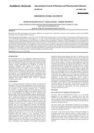
Fig. 1, 2: Chitosan <strong>nanoparticles</strong> prepared by emulsification crosslinking technique.<br />
It was observed that the formulation with lower polymer <strong>and</strong><br />
crosslinking agent no formation <strong>of</strong> particulate system was found.<br />
The formulations at particular ratio a proportional increase in the<br />
concentration increased size <strong>of</strong> the <strong>nanoparticles</strong>. It was found that<br />
the concentration at very higher ratio large particle size is observed<br />
due to high viscosity <strong>of</strong> the gel <strong>and</strong> no distrib distributing room in the<br />
emulsion, above these concentrations resulted in formation <strong>of</strong><br />
aggregates.<br />
Yateendra et al.<br />
Int J Pharm arm Pharm Sci, Vol 4, Issue 4, 244-248<br />
<strong>chitosan</strong> <strong>nanoparticles</strong> <strong>and</strong> the free drug, unentrapped drug is<br />
estimated to know total amount <strong>of</strong> the drug entrapped.<br />
The drug present in the formulated is extracted from the<br />
formulation <strong>and</strong> then analyzed for the drug content. A known<br />
volume <strong>of</strong> the nanosuspension is filtered through 0.22µm whatmann<br />
filter paper. To the sediment obtained 2% <strong>of</strong> sodium citrate is added<br />
<strong>and</strong> centrifuged at 1000rpm for 15 min, to damage the <strong>chitosan</strong><br />
crosslinks. Add 4% <strong>of</strong> acetic acid solution <strong>and</strong> till the sediment forms<br />
a clear solution. 5ml <strong>of</strong> methanol is taken into <strong>chitosan</strong> solution <strong>and</strong><br />
vortexed for 10 min, colloidal solution is estimated under UV<br />
spectrophotometer at 265 nm.<br />
The supernatant t is also estimated as well to know the amount <strong>of</strong> the<br />
drug unentrapped.1ml <strong>of</strong> the methanol is added to 1ml <strong>of</strong><br />
supernatant solution <strong>and</strong> filtered through 0.22µm filter <strong>and</strong><br />
absorbance at 265nm is noted under UV spectrophotometer.<br />
In vitro release study<br />
A franz diffusion cell is used to monitor Riluzole release from the<br />
<strong>nanoparticles</strong>. The receptor phase is 2:10 ratios <strong>of</strong> phosphate<br />
buffered saline (PBS, pH 7.4) <strong>and</strong> methanol respectively<br />
thermostatically maintained at 37°C, with each release experiment<br />
run in triplicate. Dialysis membrane) with molecular weight cut <strong>of</strong>f<br />
12,000 to 14000 Daltons is used to separate receptor <strong>and</strong> donor<br />
phases. The latter consisted suspension <strong>of</strong> <strong>nanoparticles</strong> containing<br />
Riluzole100 mg mixed for 5 seconds to aid re re-suspension in<br />
Phosphate osphate Buffer Solution. Samples (1ml) from the receptor phase<br />
are taken at time intervals <strong>and</strong> an equivalent volume <strong>of</strong> Phosphate<br />
Buffer Solution replaced into the receiver compartment. Diffusion <strong>of</strong><br />
Riluzole into the receptor phase is evaluated<br />
spectrophotometrically.<br />
RESULTS AND DISCUSSION<br />
Determination <strong>of</strong> particle size <strong>and</strong> morphology<br />
Morphology study <strong>of</strong> the <strong>nanoparticles</strong>prepared by emulsification<br />
crosslinking technique were found be spherical with good structural<br />
composition having a definite nite boundary as shown in the fig 1. The<br />
average particle sizes were found to 348, 492, 418, 382, 608 nm for<br />
the formulations F1, F2, F3, F4, F5 respectively.<br />
Determination <strong>of</strong> surface charge<br />
The <strong>nanoparticles</strong> prepared are maintained at ambient pH <strong>and</strong><br />
temperature to prevent the degradation <strong>of</strong> the formulation.<br />
the crosslinking agent being proton rich have influence on the zeta<br />
potential11 The <strong>nanoparticles</strong> prepared are maintained at ambient pH <strong>and</strong><br />
temperature to prevent the degradation <strong>of</strong> the formulation. However<br />
rosslinking agent being proton rich have influence on the zeta<br />
. (Table 2)<br />
It was clear from the data at pH range <strong>of</strong> 4.8 4.8-5.5 increase in TPP<br />
concentration has equivalenteffect on zeta potential. The<br />
245
esponsible molecule for this effect is tripolyphosphate which is<br />
proton rich. The more the ions are exhausted to neutralize the more<br />
the amino groups present in the <strong>chitosan</strong> <strong>and</strong> these free ions on the<br />
surface are responsible for the surface charge.<br />
Table 2: Corresponding zeta potentials <strong>of</strong> various formulations<br />
Formulation Zeta potential mV<br />
F1 17.3±0.5<br />
F2 18.4±0.4<br />
F3 22.6±0.4<br />
F4 23.9±0.8<br />
F5 27.6±0.9<br />
Percentage yield<br />
The entrapment efficacy <strong>of</strong> the drug in to the formulations is<br />
analysed by the gravemetric (mass balance analysis) which shows<br />
the results <strong>of</strong> each formulations. Formulation with higher <strong>chitosan</strong><br />
<strong>and</strong> Tpp concentrations was found to have more entrapment<br />
efficacy entrapping 55% <strong>of</strong> the drug for F3. While F4 is next best<br />
Yateendra et al.<br />
Int J Pharm Pharm Sci, Vol 4, Issue 4, 244-248<br />
formulation with 48% entrapment efficacy.Where F1, F2 <strong>and</strong> F5 are<br />
not much efficient in entrapping not more than 36%, 39%, <strong>and</strong>42 %<br />
respectively.<br />
Chitosan unique molecular geometry plays an important role in the<br />
drug entrapment efficacy. Most <strong>of</strong> the drug having positive charge is<br />
more suitable for the loading <strong>of</strong> drug into the <strong>chitosan</strong> network. The<br />
cationic drug like <strong>riluzole</strong> is barely difficult task to have good<br />
entrapment. Interestingly this major hurdle is solved by the method<br />
<strong>of</strong> preparation. Where the <strong>chitosan</strong> gel solution, is presented into<br />
immiscible oil phase consequently cross-linked by a crosslinking<br />
agent.<br />
In vitro release study<br />
In vitro studies <strong>of</strong> optimized <strong>chitosan</strong> nano formulations were<br />
carried out for release pattern across cellophane membrane. The<br />
release patterns <strong>of</strong> F1, F2, F3,F4, <strong>and</strong> F5 are<br />
78.79,81.61,86.20,83.99,84.35 <strong>and</strong> 84.35 respectively (table<br />
3)(graph 2). It was quite evident from the release pr<strong>of</strong>ile that show<br />
extended the drug release through <strong>nanoparticles</strong> based upon which<br />
F3 was found to be the best formulation.<br />
Table 3: Drug release pr<strong>of</strong>iles, first order kinetics, peppas plot, n values <strong>of</strong> selected formulations<br />
Formulations Percentage <strong>of</strong> drug<br />
released (%)<br />
first order kinetics Peppas plot n values<br />
F1 78.79 0.9787 0.9777 0.9058<br />
F2 81.61 0.9750 0.9772 0.8896<br />
F3 86.20 0.9841 0.9282 0.8641<br />
F4 83.99 0.9781 0.9786 0.8606<br />
F5 84.35 0.9756 0.9746 0.8695<br />
Study <strong>of</strong> Release kinetics<br />
In order to define <strong>and</strong> correlate the release kinetics <strong>of</strong> all five<br />
formulations the release kinetics were done. The corresponding<br />
dissolution data were fitted in suited kinetic dissolution models<br />
(Table 3) (fig 3&4)(graph 3&4). The equation, which is used to<br />
describe drug release mechanism, is:<br />
/ = <br />
Where, m t / m 8 is the portion <strong>of</strong> drug release‘t’ is the release time<br />
‘k’ is the constant. K dictates the properties <strong>of</strong> the macromolecular<br />
polymer system. ‘n’ is the release exponent indicative <strong>of</strong> the<br />
mechanism <strong>of</strong> release. The values <strong>of</strong> n <strong>and</strong> r 2 for coated batch was<br />
0.521<strong>and</strong> 0.680.Since the values <strong>of</strong> slope (n) lies in between 0.5 <strong>and</strong><br />
1 it was concluded that the mechanism by which drug is being<br />
released is a non-Fickian (anomalous) solute diffusion mechanism,<br />
that is, drug release during dissolution test may be controlled by all<br />
diffusion, erosion <strong>and</strong> swelling mechanism. 12<br />
CONCLUSION<br />
Graph 1: St<strong>and</strong>ard calibration curve for <strong>riluzole</strong><br />
Emulsification cross linking technique was employed to formulate<br />
the <strong>nanoparticles</strong> using <strong>chitosan</strong> as polymer <strong>and</strong> trypolyphosphate<br />
as cross linking agent. The <strong>nanoparticles</strong> produce were found to be<br />
nano range with acceptable physical chemical nature. Based on<br />
percentage yield, drug entrapment efficiency, particle size<br />
morphology, zeta potential <strong>and</strong> in vitro release, formulation F3 was<br />
found to be the optimal formulation. Thus <strong>nanoparticles</strong> <strong>of</strong> Riluzole<br />
F3 with polymer crosslinking agent ratio 3:1.5 were found to be<br />
spherical, discrete <strong>and</strong> able to sustain the drug release effectively.<br />
246
Yateendra et al.<br />
Int J Pharm Pharm Sci, Vol 4, Issue 4, 244-248<br />
Graph 2: Drug release pr<strong>of</strong>ile <strong>of</strong> some selected formulations prepared by emulsification crosslinking method.<br />
Graph 3: First oder release kinetics <strong>of</strong> some selected formulations<br />
Graph 4: Peppas plot <strong>of</strong> some slected formulations<br />
247
REFERENCES<br />
1. Cheah B.C, Vucic S, Krishnan A.V, Kiernan M.C: Riluzole,<br />
Neuroprotection <strong>and</strong> Amyotrophic Lateral Sclerosis. Current<br />
Medicinal Chemistry 2010; (1)7: 1942-1959.<br />
2. Cifra A, Nani F, Nistri A: Riluzole is a potent drug to protect<br />
neonatal rat hypoglossal motoneurons in vitro from<br />
excitotoxicity due to glutamate uptake block. European Journal<br />
<strong>of</strong> Neuroscience 2011; 33(5): 899- 913.<br />
3. Bensimon G, Lacomblez L, Meininger V: A Controlled Trial <strong>of</strong><br />
Riluzole in Amyotrophic Lateral Sclerosis. N Engl J Med 1994;<br />
330:585-591.<br />
4. Chien-Liang GL, Lynn Bristol A, Lin Jin, Dykes-Hoberg M,<br />
Thomas Crawford, Lora Clawson, <strong>and</strong> Jeffrey D : Rothstein<br />
Aberrant RNA Processing in a Neurodegenerative Disease- the<br />
Cause for Absent EAAT2, a Glutamate Transporter, in<br />
Amyotrophic Lateral Sclerosis. Neuron1998; Vol. 20: 589–602.<br />
5. Bondì ML, Craparo EF, Giammona G, Drago F: Brain-targeted<br />
solid lipid <strong>nanoparticles</strong> containing Riluzole - preparation,<br />
characterization <strong>and</strong> biodistribution. Nanomedicine 2010;<br />
5(1):25–32.<br />
6. Dhanya KP, santhi K, Dhanaraj SA <strong>and</strong> Sajeeth CI: Formulation<br />
<strong>and</strong> evaluation <strong>of</strong> <strong>chitosan</strong> nanospheres as a carrier for the<br />
Yateendra et al.<br />
Int J Pharm Pharm Sci, Vol 4, Issue 4, 244-248<br />
targeted delivery <strong>of</strong> lamivudine to the brain. International<br />
Journal <strong>of</strong> Comprehensive Pharmacy 2011; 5 (13).<br />
7. Simar PK, Rekha Rao, Afzal Hussain, Sarita Khatkar:<br />
Preparation <strong>and</strong> Characterization <strong>of</strong> rivastigmine Loaded<br />
Chitosan Nanoparticles. J. Pharm Sci. & Res 2011; 3(5): 1227-<br />
1232.<br />
8. Calvo P, Remunan-Lopez C, Vila-Jato JL, Alonso MJ: Novel<br />
hydrophilic Chitosan–Polyethylene Oxide <strong>nanoparticles</strong> as<br />
protein carriers. J. Appl. Polym. Sci 1997; 63: 125–132.<br />
9. Sangeetha S, Harish G, Malay Samanta K: Chitosan-Based<br />
nanospheres as Drug Delivery Systems for Cytarabine.<br />
International Journal <strong>of</strong> pharma <strong>and</strong> Bio Sciences 2010; 1(2).<br />
10. Adlin Jino Nesalin J, Gowthamarajan K, Somashekhara C N:<br />
Formulation <strong>and</strong> Evaluation <strong>of</strong> Nanoparticles Containing<br />
Flutamide. International Journal <strong>of</strong> Chem tech Research 2009;<br />
1(4): 1331-1334.<br />
11. Liu H, Changyou Gao: Preparation <strong>and</strong> properties <strong>of</strong> ionically<br />
cross-linked <strong>chitosan</strong> <strong>nanoparticles</strong>. Polym. Adv. Technol 2009;<br />
20: 613–619.<br />
12. Sijumon K, Sajan J,Twan L: Underst<strong>and</strong>ing the mechanism <strong>of</strong><br />
ionic gelation for synthesis <strong>of</strong> <strong>chitosan</strong> <strong>nanoparticles</strong> using<br />
qualitative techniques. Asian Journal <strong>of</strong> Pharmaceutics 2010;<br />
4(2): 148-153.<br />
248