fourth joint meeting of dutch and german
fourth joint meeting of dutch and german
fourth joint meeting of dutch and german

Create successful ePaper yourself
Turn your PDF publications into a flip-book with our unique Google optimized e-Paper software.
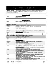
FOURTH JOINT MEETING OF DUTCH AND GERMAN<br />
PLANT VIROLOGISTS<br />
WICC, Wageningen, March 10 <strong>and</strong> 11, 2005<br />
A binational scientific <strong>meeting</strong> organised under auspices <strong>of</strong> the<br />
Nederl<strong>and</strong>se Kring voor Plantevirologie<br />
<strong>and</strong> the<br />
DPG Arbeitskreis Viruskrankheiten der Pflanzen<br />
This symposium has been sponsored by<br />
Naktuinbouw - Roel<strong>of</strong>arendsveen<br />
Bloembollenkeuringsdienst - Lisse<br />
Nunhems Netherl<strong>and</strong>s BV - Haelen<br />
De Ruiter Seeds – Bergschenhoek<br />
Rijk Zwaan BV – De Lier<br />
Plantenziektenkundige Dienst LNV – Wageningen<br />
Prime Diagnostics - Wageningen
FOURTH JOINT MEETING OF DUTCH AND GERMAN PLANT VIROLOGISTS<br />
THURSDAY March 10<br />
08.00 - 08.50 Registration<br />
08.50 - 09.00 Welcome address<br />
WICC, Wageningen, March 10 <strong>and</strong> 11, 2005<br />
Programme<br />
Session: Plant virus genome organisation <strong>and</strong> expression (Chair: Rob Goldbach)<br />
09.00 - 09.20<br />
John Bol: Coat protein enhances translational efficiency <strong>of</strong> Alfalfa mosaic virus RNAs<br />
<strong>and</strong> interacts with elF4F<br />
09.20 - 09.40<br />
Holger Jeske: Recombination-replication-repair interface <strong>of</strong> geminiviruses<br />
09.40 - 10.00<br />
Renate Koenig: BNYVV-like particles formed by coat protein expressed from a<br />
potexvirus-based vector construct<br />
10.00 - 10.20<br />
Richard Kormelink: TSWV transcriptase in vitro prefers cap donors with multiple base<br />
complementarity to the viral template<br />
10.20 - 10.50 C<strong>of</strong>fee/Tea<br />
Session: Virus - Plant Interactions (Chair: Willi Jelkmann)<br />
10.50 - 11.10<br />
Tatjana Kleinow: Interactions <strong>of</strong> movement protein BC1 <strong>and</strong> nuclear shuttle protein BV1 <strong>of</strong><br />
Abutilon mosaic geminivirus<br />
11.10 - 11.30<br />
Jan van Lent: Interaction between Cowpea mosaic virus movement tubules <strong>and</strong> the<br />
plasma membrane<br />
11.30 - 11.50<br />
Conny Heinze: Symptom variants <strong>of</strong> Cucumber mosaic virus (CMV) are based on more<br />
than a single mutation<br />
11.50 - 12.10<br />
Ilona Rolfes: GFP-labeling <strong>of</strong> mite transmitted Brome streak mosaic virus (BStMV)<br />
12.10 - 12.30<br />
Marjolein Snippe: TSWV particle assembly: in vivo interactions between the structural<br />
nucleoprotein <strong>and</strong> G1 spike protein<br />
12.30 - 13.30 Lunch (<strong>and</strong> poster set-up)<br />
1
Continuation Session: Virus - Plant Interactions (Chair: Willi Jelkmann)<br />
13.30 - 13.50<br />
Christina Wege: Transgenic AbMV DNA B supports mechanical transmission but does not<br />
release phloem restriction<br />
Session: New Diagnostic Tools (Chair: Harm Huttinga)<br />
13.50 - 14.10<br />
Nicolette Klijn: Future challenges in the innovation <strong>of</strong> detection <strong>and</strong> identification<br />
techniques in plant virology<br />
14.10 - 14.30<br />
Frank Rabenstein: Serological <strong>and</strong> molecular differentiation <strong>of</strong> Soil-borne cereal mosaic<br />
virus <strong>and</strong> Soil-borne wheat mosaic virus – two furoviruses occurring on<br />
wheat <strong>and</strong> rye in Germany<br />
14.30 - 15.00 Tea<br />
Session: Host responses: Resistance <strong>and</strong> RNAi (Chair: Holger Jeske)<br />
15.00 - 15.20<br />
Artur Pfitzner: The allelic resistance genes Tm-2 <strong>and</strong> Tm-22 against Tomato mosaic virus<br />
differ only in 4 amino acids but recognise two independent domains <strong>of</strong> the<br />
ToMV MP<br />
15.20 - 15.40<br />
Hans Hemmes: Functional analysis <strong>of</strong> RNAi suppressors <strong>of</strong> negative str<strong>and</strong> plant viruses<br />
15.40 - 16.00<br />
Konstanze Dietrich: Occurrence <strong>of</strong> short interfering RNA in virus-infected Cassava (Manihot<br />
esculenta) varieties with differential resistance/susceptibility against<br />
geminivirus infections<br />
16.00 - 16.20<br />
Simone Ribeiro: The use <strong>of</strong> RNA silencing for resistance to geminiviruses<br />
16.20 - 16.40<br />
Jörg Schubert: Attempts for genetic improvement <strong>of</strong> resistance <strong>of</strong> barley to Barley yellow dwarf<br />
virus<br />
16.40 - 17.00<br />
Gabi Krczal: Single-chain antibodies against a plant viral RNA-dependent RNA<br />
polymerase confer virus resistance<br />
17.30 - 19.00 Posters <strong>and</strong> Drinks<br />
19.30 - Dinner<br />
2
FRIDAY March 11<br />
08.30 - 09.00 AK - VK business <strong>meeting</strong><br />
Session: New, Emerging <strong>and</strong> Quarantine viruses I (Chair: John Bol)<br />
09.00 - 09.20<br />
Inas Farouk: Translocation <strong>and</strong> immunolocalisation <strong>of</strong> Watermelon chlorotic stunt virus,<br />
WmCSV, in its vector Bemisia tabaci (Genn.)<br />
09.20 - 09.40<br />
Vincent Bijman: Recent developments in Augusta disease <strong>of</strong> tulip in the Netherl<strong>and</strong>s<br />
09.40 - 10.00<br />
Arjen Werkman: Identification <strong>of</strong> viroids occuring in tomato <strong>and</strong> potato, <strong>and</strong> consequences<br />
for testing<br />
10.00 - 10.20<br />
Sharif Barends: NeRNV, a new tymovirus with a genomic RNA having a histidylatable<br />
tobamovirus-like 3’end<br />
10.20 - 10.50 C<strong>of</strong>fee/Tea<br />
Session: New, Emerging <strong>and</strong> Quarantine Viruses (Chair: Guenter Adam)<br />
10.50 - 11.10<br />
Joe Vetten: Identification <strong>and</strong> detection <strong>of</strong> a closterovirus from carrot in Germany<br />
11.10 - 11.30<br />
Ko Verhoeven: A new strain <strong>of</strong> Cowpea mild mottle virus infects French bean in Spain <strong>and</strong><br />
Morocco<br />
11.30 - 11.50<br />
Hans-Peter A new bunyaviral-type plant virus is associated with the ringspot disease <strong>of</strong><br />
Muehlbach: European mountain ash (Sorbus aucuparia L.)<br />
11.50 - 12.10<br />
Dennis Knierim: Sequence analysis <strong>of</strong> a serogroup IV tospovirus isolated from Lycopersicum<br />
esculentum in Thail<strong>and</strong><br />
12.10 - 12.30<br />
René van der Vlugt: Natural variation in Pepino mosaic virus<br />
12.30 Closing Remarks<br />
12.45 - 13.45 Lunch<br />
Departure<br />
3
Poster presentations (Thursday, 10 th March, 17.30-19.00 hr)<br />
1. Evidence for the occurrence <strong>of</strong> three nanovirus species infecting faba bean in Ethiopia<br />
A Abraham<br />
2. Investigations on Citrus tristeza virus (CTV) <strong>and</strong> its occurrence in citrus orchards in arid <strong>and</strong><br />
semi arid zones <strong>of</strong> Sudan<br />
M Abubaker<br />
3. Differentiation <strong>of</strong> Cucumber mosaic virus isolates by hybridisation to oligonucleotides in a<br />
microarray format<br />
G Adam<br />
4. Characterisation <strong>of</strong> Cherry leaf roll virus (CLRV) isolates from different host plants<br />
S von Bargen<br />
5. Molecular characterization <strong>of</strong> Vicia cryptic virus<br />
R Blawid<br />
6. A new potyvirus causing flower breaking in Begonia semperflorens<br />
I Bouwen<br />
7. High frequency silencing <strong>of</strong> multiple targets using a single inverted repeat construct<br />
E Bucher<br />
8. Construction <strong>and</strong> agroinfection <strong>of</strong> a Potato virus M full-length clone<br />
S Flatken<br />
9. Characterisation <strong>of</strong> proteins involved in translocation <strong>of</strong> the geminivirus Watermelon chlorotic<br />
stunt virus, WmCSV, in its vector Bemisia tabaci (Genn.)<br />
A Gadelseed<br />
10. Biological <strong>and</strong> molecular characteristics <strong>of</strong> different Cherry leaf roll virus (CLRV) isolates<br />
J Gentkow<br />
11. Large-scale application <strong>of</strong> real-time RT-PCR for testing Potato spindle tuber viroid in potato<br />
CCC Jansen<br />
12. Multiple infections <strong>of</strong> commercial Poinsettia<br />
H Jeske<br />
13. TMV coat protein: Expression, in vivo self assembly <strong>and</strong> mutagenesis<br />
A Kadri<br />
14. Investigation <strong>of</strong> development <strong>of</strong> infection by soil-borne viruses in cereals<br />
U Kastirr<br />
15. Dutch-German cooperations in evaluating species demarcation criteria for tombus- <strong>and</strong><br />
tymoviruses<br />
R Koenig<br />
16. Nuclei instabilities correlated with active rolling circle replication (RCR) <strong>and</strong> recombination<br />
dependent replication (RDR) <strong>of</strong> Abutilon mosaic virus<br />
B-J Koo<br />
17. The nucleoprotein gene <strong>of</strong> Tomato spotted wilt virus as a tag protein fusion for easy<br />
purification<br />
<strong>and</strong> enhanced production <strong>of</strong> recombinant protein in plants<br />
C Lacorte<br />
4
18. Changes in the spectrum <strong>of</strong> PVY strain groups <strong>and</strong> their involvement in potato <strong>and</strong> tobacco<br />
diseases<br />
K Lindner<br />
19. Identification <strong>and</strong> characterization <strong>of</strong> Tospoviruses from Iran<br />
A Mehraban<br />
20. Detection <strong>and</strong> Identification <strong>of</strong> a novel potexvirus infecting allium by paramagnetic beads<br />
ssRNA isolation <strong>and</strong> one tube RT-PCR assay with a new potexvirus genus primer set<br />
R Miglino<br />
21. Characterization <strong>and</strong> nucleotide sequence <strong>of</strong> Grapevine leafroll associated virus-7 (GLRaV-7)<br />
C Mikona<br />
22. The spread <strong>of</strong> Rice yellow mottle virus in irrigated rice crops<br />
D Peters<br />
23. Identification <strong>of</strong> viruses in ornamental Allium species <strong>and</strong> control strategies<br />
KTK Pham<br />
24. Molecular <strong>and</strong> biological characterization <strong>of</strong> Beet mild yellowing virus <strong>and</strong> determination <strong>of</strong> the<br />
host plant spectrum by agroinfection<br />
D Stephan<br />
25. Epidemiological developments in Potato virus Y<br />
M Verbeek<br />
26. Expression analysis <strong>of</strong> potyviral 6K1 in Nicotiana benthamiana<br />
A Waltermann<br />
27. Alfalfa mosaic virus detected in true seed <strong>of</strong> Solanum kurtzianum during post-entry quarantine<br />
testing<br />
AW Werkman<br />
28. Investigating the mechanism(s) <strong>of</strong> cross-protection between strains <strong>of</strong> Cucumber mosaic virus<br />
H Ziebell<br />
5
ABSTRACTS<br />
Oral Presentations<br />
Coat protein enhances translational efficiency <strong>of</strong> Alfalfa mosaic virus RNAs <strong>and</strong> interacts<br />
with eIF4F<br />
JF Bol 1 , IM Krab 1 <strong>and</strong> DR Gallie 2 .<br />
1 Institute <strong>of</strong> Biology, Gorlaeus Laboratories, Leiden University <strong>and</strong> 2 Department <strong>of</strong> Biochemistry,<br />
University <strong>of</strong> California, Riverside, California 92521<br />
E-mail: j.bol@chem.leidenuniv.nl.<br />
Translation <strong>of</strong> eukaryotic mRNAs is strongly enhanced by the formation <strong>of</strong> a closed loop structure<br />
due to the interaction between the poly(A)-binding protein bound to the 3'-poly(A) tail <strong>and</strong> the<br />
eIF4G subunit <strong>of</strong> the eIF4F complex bound to the 5' cap structure. The three plus-str<strong>and</strong> genomic<br />
RNAs <strong>of</strong> Alfalfa mosaic virus (AMV) <strong>and</strong> the subgenomic messenger for viral coat protein (CP)<br />
contain a 5'-cap structure but no 3'-poly(A) tail. Binding <strong>of</strong> CP to the 3'-end <strong>of</strong> AMV RNAs is<br />
required for efficient translation <strong>of</strong> the viral RNAs <strong>and</strong> to initiate infection in plant cells. To study the<br />
role <strong>of</strong> CP in translation, plant protoplasts were transfected with luciferase (Luc) transcripts with 3'terminal<br />
sequences consisting <strong>of</strong> the 3'-untranslated region <strong>of</strong> AMV RNA 3 (Luc-AMV), a poly(A)<br />
tail <strong>of</strong> 50 residues (Luc-poly(A)), or a short vector-derived sequence (Luc-control). Pre-incubation <strong>of</strong><br />
the transcripts with CP had no effect on Luc-expression from Luc-poly(A) or Luc-control but<br />
strongly stimulated Luc-expression from Luc-AMV. From time-course experiments it was calculated<br />
that CP-binding increased the half-life <strong>of</strong> Luc-AMV by 20% <strong>and</strong> enhanced its translational efficiency<br />
about 40-fold. The stimulation <strong>of</strong> translation by CP was cap-dependent <strong>and</strong> could be reproduced in<br />
yeast cells. GST-pull-down assays revealed the binding <strong>of</strong> AMV CP to initiation factor complexes<br />
eIF4F <strong>and</strong> eIFiso4F from wheat germ. By Far-Western blotting it was shown that this binding<br />
occurred through an interaction <strong>of</strong> CP with the eIF4G <strong>and</strong> eIFiso4G subunits <strong>of</strong> eIF4F <strong>and</strong><br />
eIFiso4F, respectively. The results support the hypothesis that the role <strong>of</strong> CP in translation <strong>of</strong> viral<br />
RNAs mimics the role <strong>of</strong> the poly(A) binding protein in translation <strong>of</strong> cellular mRNAs.<br />
Recombination-replication-repair interface <strong>of</strong> geminiviruses<br />
H Jeske, B Alberter, E Bejarano, J Castillo, G Morilla, AM Rezaian, W Preiß <strong>and</strong> C Wege<br />
Institute <strong>of</strong> Biology, Department <strong>of</strong> Plant Molecular Biology <strong>and</strong> Virology, University <strong>of</strong> Stuttgart<br />
E-mail: holger.jeske@bio.uni-stuttgart.de<br />
One major driving force <strong>of</strong> geminivirus evolution is recombination. They may exchange genomic<br />
components (pseudorecombination) as a prerequisite for true molecular recombination <strong>and</strong> may<br />
acquire thus new DNA components like DNAs B, beta DNAs, satellite DNAs or even DNAs <strong>of</strong><br />
different viruses as it has been shown for nanovirus DNA circles. Using an optimized twodimensional<br />
gel electrophoresis in combination with hybridization <strong>and</strong> electron microscopy we have<br />
discovered that the recombinational flexibility is reasoned by a recombination-dependent replication<br />
mode (RDR) that is widespread at least among begomoviruses <strong>and</strong> curtoviruses but does also<br />
occur for satellites, beta DNAs <strong>and</strong> artificial episomes. Compared to complementary str<strong>and</strong><br />
synthesis (CSR) <strong>and</strong> rolling circle replication (RCR), which both were found together with RDR, the<br />
latter mechanism allows the virus to repair every broken or unfinished DNA intermediate as far as<br />
homologous templates are available. Using virus-specific two colour detection, we found out the<br />
the frequency by which different geminiviruses enter the same nucleus was surprisingly high in<br />
tomatoes <strong>and</strong> N. benthamiana, so that the chance to recombine two viruses is extremely high.<br />
Moreover, weeds are ideal cradles for new geminiviruses since they are reservoirs <strong>and</strong> collect<br />
several virus species over years, as exemplified for Sida micrantha-associated geminiviruses.<br />
Surveying different combinations <strong>of</strong> geminiviral <strong>and</strong> satellite DNAs two strategies can be<br />
discriminated between Old World <strong>and</strong> New World begomoviruses. The first were rather promicous<br />
to transreplicate other DNAs, even without a cognate Rep-binding sequence, whereas the latter<br />
need the interaction <strong>of</strong> Rep <strong>and</strong> Rep-binding sequences <strong>and</strong> consequently DNAs B were acquired<br />
by shuffling <strong>of</strong> the regulatory region (common region CR) by homologous recombination. The<br />
consequences <strong>of</strong> these findings for efficient resistance breeding <strong>and</strong> epidemiology will be<br />
discussed.<br />
6
BNYVV-like particles formed by coat protein expressed from a potexvirus-based vector<br />
construct<br />
R Koenig, S Loss <strong>and</strong> D-E Lesemann<br />
c/o Biologische Bundesanstalt, Institut für Pflanzenvirologie, Mikrobiologie und biologische<br />
Sicherheit, Braunschweig, Germany<br />
E-mail: r.koenig@bba.de<br />
We have recently determined the nucleotide (nt) sequences <strong>of</strong> the genomic RNAs <strong>of</strong> three<br />
potexviruses which had been isolated long ago from different species in the Cactaceae <strong>and</strong> were<br />
originally designated as different strains, i.e. BS, CC10 <strong>and</strong> K11, <strong>of</strong> Cactus virus X. Because<br />
serological <strong>and</strong> molecular studies revealed only rather distant relationships between these viruses,<br />
we have proposed to change their taxonomic status <strong>and</strong> rename them to Zygocactus virus X<br />
(ZVX), Opuntia virus X (OVX) <strong>and</strong> Schlumbergera virus X (ScVX), respectively. The full length<br />
cDNA sequence <strong>of</strong> ZVX RNA was cloned into the 35S promoter-containing vector p35Stupa kindly<br />
provided by E. Maiß, Hannover. The cDNA clones obtained consistently produced symptomless<br />
local <strong>and</strong> occasionally also systemic infections in C. quinoa. In order to enable insertion <strong>and</strong><br />
expression <strong>of</strong> foreign genes an AscI <strong>and</strong> a SpeI site were introduced downstream <strong>of</strong> the coat<br />
protein (cp) promoter <strong>and</strong> directly upstream <strong>of</strong> the cp gene. Most <strong>of</strong> the ZVX cp gene except for its<br />
56 3’-terminal nucleotides were replaced by the corresponding sequence <strong>of</strong> the ScVX cp gene <strong>and</strong><br />
by additional 45 nts upstream <strong>of</strong> the ScVX cp gene which presumably contain the ScVX cp<br />
promoter. The expression <strong>of</strong> foreign genes which are inserted between the AscI/SpeI sites is thus<br />
directed by the ZVX cp promoter whereas the expression <strong>of</strong> the ScVX cp gene is directed by its<br />
own promoter. Clones, into which the cp gene <strong>of</strong> Beet necrotic yellow vein virus (BNYVV) was<br />
inserted, produced local infections in C. quinoa <strong>and</strong> sugarbeet. In the former, BNYVV cp was<br />
readily detected by means <strong>of</strong> ELISA. Immunoelectron microscopy revealed the presence <strong>of</strong> viruslike<br />
particles which were strongly decorated by antibodies to BNYVV. This particle formation is<br />
surprising, because in natural infections it is assumed that the cp readthrough protein is necessary<br />
to initiate particle assembly.<br />
TSWV transcriptase in vitro prefers cap donors with multiple base complementarity to the<br />
viral template<br />
IC van Knippenberg, M Lamine, P Verbruggen, M Nischang, R Goldbach <strong>and</strong> R Kormelink<br />
Laboratory <strong>of</strong> Virology, Wageningen University<br />
E-mail: richard.kormelink@wur.nl<br />
Transcription <strong>of</strong> segmented negative str<strong>and</strong> RNA viruses is initiated by cap snatching, i.e. a<br />
mechanism in which host mRNAs are cleaved generally at 10-20 nt from their 5' capped end <strong>and</strong><br />
the resulting capped leaders used to prime transcription <strong>of</strong> the viral genome. This mechanism has<br />
first been decribed for Influenza A virus in the early 80’s <strong>and</strong> since reported for several other<br />
viruses. For Tomato spotted wilt virus (TSWV), type species <strong>of</strong> the plant-infecting Tospovirus<br />
genus within the Bunyaviridae, the occurrence <strong>of</strong> cap-snatching was reported in 1992 <strong>and</strong> following<br />
studies have shown that cap donors require a single base complementarity to the ultimate or<br />
penultimate viral template sequence. More recently, the occurrence in vitro <strong>of</strong> "re-snatching" <strong>of</strong> viral<br />
mRNAs, i.e. the use <strong>of</strong> viral mRNAs as cap donors, has been demonstrated for TSWV. To estimate<br />
the relative occurrence <strong>of</strong> re-snatching compared to snatching <strong>of</strong> host mRNAs, the use <strong>of</strong> cap<br />
donors with either single, double or multiple complementarity to the viral template was analysed in<br />
pair-wise competition in TSWV in vitro transcription assays. These analyses have shown a strong<br />
preference for multiple-basepairing donors. Similar experiments are currently performed for<br />
Influenza A virus transcription initiation. Furthermore, a (first) series <strong>of</strong> cap-donor mutants has been<br />
made to analyse the requirements <strong>of</strong> cap-donor leader sequences upstream the baseparing<br />
residues for TSWV as well as Influenza A virus transcription initiation.<br />
7
Interactions <strong>of</strong> movement protein BC1 <strong>and</strong> nuclear shuttle protein BV1 <strong>of</strong> Abutilon mosaic<br />
geminivirus<br />
T Kleinow 1 , S Frischmuth 1 , S Zhang 1 , S Hehnle 1 , A Aberle 1 , C Wege 1 , D Hülser 2 <strong>and</strong> H Jeske 1<br />
1) Department <strong>of</strong> Plant Molecular Biology <strong>and</strong> Virology, Biological Institute, University <strong>of</strong> Stuttgart<br />
<strong>and</strong> 2) Department <strong>of</strong> Biophysics, Biological Institute, University <strong>of</strong> Stuttgart<br />
E-mail: tatjana.kleinow@bio.uni-stuttgart.de<br />
Bipartite geminiviruses such as Abutilon mosaic virus encodes two proteins responsible for virus<br />
movement <strong>and</strong> pathogenic properties, the nuclear shuttle protein (NSP; syn. BV1) <strong>and</strong> the<br />
movement protein (MP; syn. BC1). Transient expression <strong>of</strong> green fluorescent protein fusions <strong>of</strong> MP<br />
<strong>and</strong> NSP in host plant cells has shown that BV1 localizes to nuclei <strong>and</strong> BC1 to the cell periphery or<br />
close to nuclei. By electron microscopy techniques NSP expressed in fission yeast was detected in<br />
nuclei whereas MP was localized to protoplasmic leaflets <strong>of</strong> plasma membranes. Upon coexpression<br />
in plants or yeast, BC1 redirected BV1 from nuclei to cell periphery <strong>and</strong> in sink leaves <strong>of</strong><br />
host plants, additionally to adjacent cells. BC1 deletion mutants lacking the membrane-binding<br />
domain indicated a homo-oligomerization <strong>of</strong> its C-terminus by yeast two-hybrid <strong>and</strong> Cyto-Trap<br />
analysis. In-vitro assembly <strong>of</strong> double-str<strong>and</strong>ed super coiled DNA with NSP <strong>and</strong> MP into<br />
conspicuous structures was confirmed using electron microscopy <strong>and</strong> provided evidence for<br />
cooperative interaction <strong>of</strong> MP, NSP <strong>and</strong> DNA. These results support a model, in which NSP<br />
transports viral DNA to the cell periphery, <strong>and</strong> BC1 acts as a membrane adaptor for NSP-DNA<br />
complexes to facilitate cooperative cell-to-cell movement within plants. By use <strong>of</strong> two-hybrid<br />
system, we identified BC1-interacting plant factors, which are currently under investigation.<br />
Interaction between Cowpea mosaic virus movement tubules <strong>and</strong> the plasma membrane<br />
JWM van Lent 1 , J Pouwels 2 , M Rolloos 1,2 , CM Carvalho 1 , J Wellink 2 <strong>and</strong> RW Goldbach 1<br />
1 2<br />
Laboratory <strong>of</strong> Virology, Wageningen University <strong>and</strong> Laboratory <strong>of</strong> Molecular Biology, Wageningen<br />
University<br />
E-mail: jan.vanlent@wur.nl.<br />
Cowpea mosaic virus (CPMV) virions traverse the cell wall through nanotubules that are<br />
assembled from the viral movement protein (MP). In planta these tubules are found exclusively in<br />
the plasmodesmal canal. In isolated plant protoplasts, devoid <strong>of</strong> cell walls <strong>and</strong> plasmodesmata, a<br />
similar polarity <strong>of</strong> tubule assembly is observed. The MP anchors somehow at the plasma<br />
membrane (PM) <strong>and</strong> from there an outward growing tubule is assembled tightly encased with PM<br />
as also occurs within the plasmodesma. Transport tubules are not only formed in protoplasts <strong>of</strong><br />
host plants, but also in those isolated from non-host plants or even animal cells that express the<br />
MP. Obviously, MP interacts with the PM or with PM-associated proteins <strong>and</strong> such proteins should<br />
be conserved in plant <strong>and</strong> animal cells.<br />
This intriguing interaction between MP <strong>and</strong> PM was further analyzed by MP-mutant analysis, timelapse<br />
microscopy <strong>and</strong> FRET/FRAP studies. It has been shown before that prior to tubule formation,<br />
MP (-GFP) accumulates in so-called peripheral punctate spots at the PM <strong>and</strong> it was speculated<br />
that these were the nucleation sites for tubule assembly. Time-lapse fluorescent microscopy was<br />
performed on protoplasts expressing MP-GFP <strong>and</strong> showed that the punctate spots are dynamic<br />
structures that are immobilized at the PM <strong>and</strong> that tubules indeed arise from (a sub-population <strong>of</strong>)<br />
these spots.<br />
To investigate the interaction <strong>of</strong> tubules with the surrounding PM, the diffusion herein <strong>of</strong> small <strong>and</strong><br />
large fluorescent PM-associated proteins was examined by fluorescence recovery after<br />
photobleaching (FRAP). These studies indicate that tubules made by CPMV MP do not interact<br />
directly with the PM, but most likely via a PM intrinsic protein (PIP) or otherwise associated host<br />
protein. Using affinity chromatography it was found that purified MP a.o. binds to aquaporin present<br />
in the plasmamembrane. Transient expression <strong>of</strong> MP-YFP <strong>and</strong> Arabidopsis aquaporin P1;4-CFP<br />
revealed that these proteins colocalize in punctuate spots on the surface <strong>of</strong> cowpea protoplasts.<br />
Acceptor photobleaching experiments indicate that they also physically interact at these sites,<br />
suggesting that aquaporin is used by the virus as a primer for the tubule formation at the<br />
plasmamembrane.<br />
8
Symptom variants <strong>of</strong> Cucumber mosaic virus (CMV) are based on more than a single<br />
mutation<br />
D Zhang, P Willingmann, G Adam <strong>and</strong> C Heinze<br />
Biocentre Klein-Flottbek <strong>and</strong> Botanical Garden, University <strong>of</strong> Hamburg<br />
E-mail: cheinze@iangbot.uni-hamburg.de<br />
In Asia chili is <strong>of</strong> high importance for the daily nutrition <strong>and</strong> grown as local varieties from small<br />
holders. Beside other pathogens chili is susceptible to Cucumber mosaic virus (CMV), a (+) ssRNA<br />
virus with a tripartite genome. Commercial chili varieties with resistance to this disease were not<br />
available <strong>and</strong> therefore resistant varieties have been developed from the Asian Vegetable<br />
Research <strong>and</strong> Development Center (AVRDC, Taiwan). Although some lines showed resistance to<br />
many strains, originating in several Asian countries, it was overcome at least by two isolates,<br />
CMV-AN from India <strong>and</strong> CMV-KS44 from Thail<strong>and</strong>. The responsible genetic determinant was<br />
located on RNA 2 by reassortment studies. Chimeric constructs <strong>of</strong> full-length RNA 2 cDNA clones<br />
based on isolate Fny comprising the complete gene silencing suppressor (2b) gene <strong>of</strong> resistance<br />
breaking isolate CMV-AN <strong>and</strong> partial sequences <strong>of</strong> 2a gene <strong>and</strong> 3´ non coding region were able to<br />
overcome the resistance in concert with RNA 1 <strong>and</strong> RNA 3 from the non resistance breaking<br />
isolate CMV-P3613. These data suggested that the gene silencing suppressor 2b <strong>and</strong>/or the 2a<br />
protein is responsible for this biological behaviour.<br />
Within the 2a- <strong>and</strong> 2b-genes two striking mutations between resistant <strong>and</strong> non-resistant isolates<br />
were present. However, site directed mutagenesis did not confirm the significance for these<br />
mutations but suggest that other factors are involved for symptom expression. Additional<br />
reassortants between different combinations <strong>of</strong> isolates indicated that also RNA 1 <strong>and</strong>/or RNA 3<br />
may play a role in symptom determination, <strong>and</strong> - in some cases - in concert with RNA 2. The<br />
symptom expression <strong>of</strong> reassortants <strong>and</strong> genetically modified constructs in other host systems<br />
were not predictable. These results show that symptom expression is a very complex process<br />
depending on synergistic interactions. The results derived from single mutation experiments should<br />
therefore not be generalized.<br />
GFP-labelling <strong>of</strong> mite transmitted Brome streak mosaic virus (BStMV)<br />
I Rolfes <strong>and</strong> E Maiss<br />
Institute <strong>of</strong> Plant Diseases <strong>and</strong> Plant Protection, University <strong>of</strong> Hannover<br />
E-mail: rolfes@ipp.uni-hannover.de<br />
Brome streak mosaic virus (BStMV) infects Poaceae <strong>and</strong> is transmitted by eriophyid mites like the<br />
other members <strong>of</strong> the genus Tritimovirus: Wheat streak mosaic virus (WSMV) <strong>and</strong> Oat necrotic<br />
mottle virus (ONMV). For construction <strong>of</strong> a BStMV full-length cDNA clone (pBStMV-FL) viral RNA<br />
was extracted from a French BStMV isolate (11-Cal; BStMV-wt) <strong>and</strong> cDNA fragments were<br />
amplified by RT-PCR. The fragments were cloned in succession under control <strong>of</strong> an enhanced<br />
Cauliflower mosaic virus (CAMV) 35S-promoter <strong>and</strong> its 3'-polyadenylation signal, resulting in<br />
pBStMV-FL. This plasmid was transferred into plants by particle bombardment. The virus deriving<br />
from pBStMV-FL was able to infect the plant systemically <strong>and</strong> was detected immunologically.<br />
Comparison <strong>of</strong> the host spectrum <strong>of</strong> BStMV-FL <strong>and</strong> BStMV-wt revealed no differences in terms <strong>of</strong><br />
infected plants <strong>and</strong> symptom severity. Six species were identified as host plants, at which Bromus<br />
mollis, Hordeum vulgare <strong>and</strong> Triticum aestivum showed obvious virus symptoms. BStMV-FL was<br />
labelled with a green fluorescent protein (GFP) at two different positions in the genome. For this<br />
purpose the gfp gene was integrated in-between P1 <strong>and</strong> helper component protease (pBStMV-<br />
P1/GFP/HC-Pro) <strong>and</strong> in-between nuclear inclusion body b <strong>and</strong> coat protein (pBStMV-NIb/GFP/CP).<br />
Four weeks after particle bombardment virus symptoms were observed. BStMV-CP <strong>and</strong> GFP were<br />
detected by electroblot immunoassays. Fluorescence <strong>of</strong> GFP was visualized with a confocal-laser<br />
scanning microscope. Therefore, correct proteolytic processing <strong>of</strong> GFP occurred in BStMV-<br />
P1/GFP/HC-Pro as well as in BStMV-NIb/GFP/CP <strong>and</strong> the released GFPs were functional. In<br />
addition, eriophyid mites were collected from the field <strong>and</strong> used successfully to transmit BStMV.<br />
Altogether these findings enable further studies on infection, replication <strong>and</strong> transport <strong>of</strong> BStMV-FL<br />
<strong>and</strong> mutants there<strong>of</strong> in plants as well as determination <strong>of</strong> proteins <strong>and</strong> motifs involved in vector<br />
transmission.<br />
9
TSWV particle asssembly: in vivo interactions between the structural nucleoprotein <strong>and</strong> G1<br />
spike protein<br />
M Snippe 1 , JW Borst 2 , R Goldbach 1 <strong>and</strong> R Kormelink 1<br />
1 2<br />
Laboratory <strong>of</strong> Virology, Wageningen University <strong>and</strong> Laboratory <strong>of</strong> Biochemistry, Wageningen<br />
University<br />
E-mail: marjolein.snippe@wur.nl<br />
TSWV (Tomato spotted wilt virus) is a member <strong>of</strong> the Bunyaviridae family. Virus particles are<br />
spherical <strong>and</strong> membrane bound, containing spike proteins that consist <strong>of</strong> two glycoproteins, G1 <strong>and</strong><br />
G2. The core contains ribonucleoproteins (RNPs) that consists <strong>of</strong> genomic RNA tightly associated<br />
with the nucleoprotein (N) <strong>and</strong> small amounts <strong>of</strong> the viral RNA-dependent RNA polymerase.<br />
Enveloped virus particles arise as a result <strong>of</strong> RNP envelopment with membranes from the Golgi<br />
apparatus containing G1 <strong>and</strong> G2. To investigate in vivo interactions between the structural proteins<br />
involved in virus assembly, fluorescence techniques (FRET: fluorescence resonance energy<br />
transfer <strong>and</strong> FLIM: fluorescence lifetime imaging microscopy) were employed. To this end, N, G1<br />
<strong>and</strong> G2 were fused at their N or C-terminus to CFP or YFP. Upon co-expression <strong>of</strong> N-YFP <strong>and</strong> N-<br />
CFP in mammalian cells, N dimerisation was observed in peri-nuclear aggregates as well as<br />
throughout the cytoplasm. The peri-nuclear localisation <strong>of</strong> N oligomers required actin filaments <strong>and</strong><br />
microtubules, as demonstrated with the use <strong>of</strong> inhibitors, which suggested the possible involvement<br />
<strong>of</strong> these cellular elements in TSWV particle formation. Upon co-expression <strong>of</strong> N-YFP with G2-CFP<br />
no FRET was observed, whereas co-expression <strong>of</strong> N-YFP <strong>and</strong> G1-CFP did show FRET.<br />
Furthermore, G1 was observed to show an altered localisation pattern in the presence <strong>of</strong> N, i.e.<br />
upon single expression <strong>of</strong> G1 only ER localisation was observed whereas a peri-nuclear<br />
accumulation partly overlapping with that <strong>of</strong> N was observed in the presence <strong>of</strong> N. Altogether,<br />
these results suggest that the actual envelopment <strong>of</strong> TSWV RNPs’ could be triggered by an<br />
interaction between N <strong>and</strong> the cytoplasmic tail <strong>of</strong> G1.<br />
Transgenic AbMV DNA B supports mechanical transmission but does not release phloem<br />
restriction<br />
D Pohl, H Jeske <strong>and</strong> C Wege<br />
Institute <strong>of</strong> Biology Department <strong>of</strong> Molecular Biology <strong>and</strong> Virology <strong>of</strong> Plants, Universität Stuttgart<br />
E-mail: christina.wege@bio.uni-stuttgart.de<br />
Strict phloem limitation <strong>of</strong> the bipartite Abutilon mosaic virus (AbMV) in different host plants is<br />
accompanied by a lack <strong>of</strong> mechanical transmissibility. In order to analyse the functionality <strong>and</strong> thus<br />
contribution <strong>of</strong> movement proteins to tissue restriction, Nicotiana benthamiana plants were<br />
transformed with AbMV DNA B dimers <strong>and</strong> challenged by agroinoculation <strong>of</strong> AbMV DNA A. Several<br />
independent plant lines (generations T0 to T3) were capable for replicating <strong>and</strong> systemically<br />
spreading complete bipartite virus genomes, including free DNA B copies. In contrast to reports on<br />
other begomoviruses, transgenically expressed AbMV BC1 protein was not able to induce<br />
pathogenic symptoms permanently. Different transgenic lines containing functional DNA B copies<br />
<strong>and</strong> expressing BC1 protein indeed developed abnormal leaf phenotypes transiently, but later on<br />
recovered <strong>and</strong> were indistinguishable from wildtype plants. Upon AbMV infection, symptoms were<br />
the same in transgenic <strong>and</strong> wildtype plants, as was the amount <strong>of</strong> viral DNA accumulating in leaf<br />
tissues. Macroscopic <strong>and</strong> microscopic in situ hybridisation revealed that AbMV remained phloemlimited<br />
in the DNA B-transgenic plants. Nevertheless, mechanical transmission <strong>of</strong> the virus had<br />
changed: About one fifth <strong>of</strong> transgenic plants treated with sap from systemically infected wildtype<br />
N. benthamiana developed full AbMV infections. Thus AbMV DNA B genes BV1 <strong>and</strong> BC1 can<br />
complement for mechanical transmission in inoculated leaves, but in systemically infected leaves<br />
fail to support viral invasion <strong>of</strong> non-phloem cells. Since in mixed infections with CMV, AbMV has<br />
been shown to escape from the phloem, some other (perhaps gene silencing-) mechanism may be<br />
responsible for AbMV tissue limitation.<br />
10
Future challenges in the innovation <strong>of</strong> detection <strong>and</strong> identification techniques in plant<br />
virology<br />
N Klijn <strong>and</strong> JW Roenhorst<br />
Plant Protection Service, Wageningen<br />
E-mail: n.klijn@minlnv.nl<br />
Since the end <strong>of</strong> the eighties the strategy <strong>of</strong> the Dutch government focussed on the reduction <strong>of</strong> the<br />
use <strong>of</strong> pesticides. As a result less capacity <strong>and</strong> budget was available for research in the field <strong>of</strong><br />
plant health. This led to a dramatic loss <strong>of</strong> expertise, especially in the field <strong>of</strong> virology. A similar<br />
tendency could be observed internationally. Since healthy seeds <strong>and</strong> planting material are <strong>of</strong><br />
crucial importance to manage virus diseases, this loss eventually will result in increasing problems<br />
caused by viruses. The consequences <strong>of</strong> the lack <strong>of</strong> diagnostic methods for potentially harmful<br />
viruses <strong>and</strong> the loss <strong>of</strong> knowledge <strong>and</strong> expertise to develop such methods, has to be brought into<br />
attention <strong>of</strong> the responsible decision makers. Therefore coherent research proposals have to be<br />
drafted <strong>and</strong> submitted to national <strong>and</strong> international governments <strong>and</strong> organisations. It should be<br />
stressed that is necessary to maintain both knowledge <strong>and</strong> practical expertise in the field <strong>of</strong> plant<br />
virology. New detection <strong>and</strong> identification methods have to be developed based on reference<br />
collection material that, in combination with the classical methods, enable to study the biological<br />
<strong>and</strong> epidemiological aspects <strong>of</strong> viruses. Such approach will provide a solid base, which is essential<br />
to manage actual <strong>and</strong> future virus problems by prevention <strong>and</strong> control.<br />
Serological <strong>and</strong> molecular differentiation <strong>of</strong> Soil-borne cereal mosaic virus <strong>and</strong> Soil-borne<br />
wheat mosaic virus - two furoviruses occurring on wheat <strong>and</strong> rye in Germany<br />
F Rabenstein, H Mühlheim, M Wesemann, U Kastirr <strong>and</strong> T Kühne<br />
Federal Center for Breeding Research on Cultivated Plants, Institute <strong>of</strong> Resistance Research <strong>and</strong><br />
Pathogen Diagnostics, Aschersleben<br />
E-mail: f.rabenstein@bafz.de<br />
Rod-shaped virus particles approx. 20 nm in diameter were observed by electron microscopy in<br />
leaves <strong>of</strong> rye <strong>and</strong> wheat varieties grown in different regions in Germany. Virus isolates were<br />
transmitted through infected soil to wheat, rye, <strong>and</strong> triticale <strong>and</strong> by mechanical inoculations to<br />
Chenopodium quinoa. By use <strong>of</strong> polyclonal antisera to Soil-borne cereal mosaic virus (SBCMV)<br />
(BBA Braunschweig) <strong>and</strong> Soil-borne wheat mosaic virus (SBWMV) (Univ. <strong>of</strong> Nebraska, USA) it was<br />
neither possible to discriminate the isolates in ELISA <strong>and</strong> Western blots (WB) nor on the basis <strong>of</strong><br />
their symptoms on C. quinoa. SBWMV (ATCC type strain) <strong>and</strong> the German isolates ‘Eilte’,<br />
‘Eickeloh’ <strong>and</strong> ‘Heddesheim’ were propagated on rye variety ‘Nikita’ for virus purification <strong>and</strong><br />
antiserum production. Altogether 8 polyclonal antisera (PAS) were raised in rabbits. PAS-45 <strong>and</strong><br />
PAS-69 produced to the type SBWMV <strong>and</strong> to isolate ‘Heddesheim’ showed no or only very weak<br />
cross-reactions with the other isolates. By means <strong>of</strong> specific primer combinations these two<br />
isolates were verified in immunocapture RT-PCR as SBWMV whereas all the other isolates were<br />
assigned to SBCMV. Two <strong>of</strong> the four PAS produced to SBCMV reacted in DAS-ELISA, WB <strong>and</strong><br />
tissue print immunoassay (TPIA) mainly with the homologous virus. However, at high virus<br />
concentrations a specific discrimination between SBWMV <strong>and</strong> SBCMV with PAS was doubtful,<br />
particularly by their application in TPIA. Therefore, monoclonal antibodies (MAbs) were produced to<br />
both furoviruses. For SBWMV detection a DAS-ELISA based on MAb 4G4 (IgG3, ) was<br />
developed. For specific discrimination <strong>of</strong> all SBCMV isolates MAb 4G11 (IgG2a, ) can be<br />
recommended for use in TAS-ELISA. The virus species specific MAbs allow in TPIA an excellent<br />
discrimination between both furoviruses <strong>and</strong> can be applied in large-scale resistance trials. By<br />
sequences comparison we assume that the coat protein motifs ATHAY <strong>and</strong> SIHPF could form<br />
specific epitopes for SBWMV <strong>and</strong> SBCMV, respectively.<br />
11
The allelic resistance genes Tm-2 <strong>and</strong> Tm-22 against Tomato mosaic virus differ only in 4<br />
amino acids but recognize two independent domains <strong>of</strong> the ToMV MP<br />
A Gerhardts, M Strasser, M Silber und AJP Pfitzner<br />
Institute <strong>of</strong> Genetics, Department <strong>of</strong> General Virology, University <strong>of</strong> Hohenheim, Stuttgart<br />
E-mail: pfitzner@uni-hohenheim.de<br />
Transposon insertion experiments in tomato plants led to the isolation <strong>of</strong> the Tm-2 resistance gene<br />
against ToMV.. Sequence analysis revealed that Tm-2 codes for a 861 aa protein which belongs to<br />
the CC-NBS-LRR type <strong>of</strong> resistance genes. Surprisingly, Tm-2 contains only 4 amino acid<br />
exchanges located in two different domains <strong>of</strong> the protein in comparison to the allelic Tm-22,<br />
although sequence analysis <strong>of</strong> diverse resistance breaking virus strains proved that both resistance<br />
genes recognize different parts <strong>of</strong> the ToMV movement protein. Further investigations on the<br />
molecular interaction <strong>of</strong> these resistance genes with the ToMV MP indicated that Tm-2 <strong>and</strong> Tm-22<br />
contain two distinct domains for the interaction with the avirulence gene product.<br />
Functional analysis <strong>of</strong> RNAi suppressors <strong>of</strong> negative str<strong>and</strong> plant viruses<br />
JC Hemmes, EC Bucher, I Cahyani, E Buurman, RW Goldbach <strong>and</strong> M Prins<br />
Laboratory <strong>of</strong> Virology, Wageningen University<br />
E-mail: hans.hemmes@wur.nl<br />
In recent studies we have shown that the NSs protein <strong>of</strong> Tomato spotted wilt tospovirus (TSWV) as<br />
well as the NS3 protein <strong>of</strong> Rice hoja blanca tenuivirus (RHBV) suppress RNA silencing in plants.<br />
To further analyse the function <strong>of</strong> suppressor NS3 in N. benthamiana in RNA silencing, expression<br />
constructs <strong>of</strong> these proteins were produced resulting in the production <strong>of</strong> MBP tagged proteins in<br />
bacteria <strong>and</strong> plants (via A. tumefaciens infiltration). The addition <strong>of</strong> the fusion partner, included to<br />
facilitate pull down experiments, was shown not to interfere with the RNA silencing activity <strong>of</strong> the<br />
proteins. In subsequent experiments deletion mutants were generated. Two conserved domains <strong>of</strong><br />
the in NS3 protein were deleted. Both NS3 mutant proteins were proven to be expressed, while the<br />
RNA silencing suppression activity in plants was lost. Analysis <strong>of</strong> mRNA <strong>and</strong> siRNA levels<br />
compared to NS3 confirmed these observations <strong>and</strong> will be discussed. A series <strong>of</strong> point <strong>and</strong><br />
deletion mutations in the NSs protein was also produced <strong>and</strong> analysis will be presented. To further<br />
elucidate the biochemical function, in vitro RNAi experiments were performed <strong>and</strong> the function <strong>of</strong><br />
bacterial purified suppressors examined.<br />
Both TSWV <strong>and</strong> RHBV are transmitted by insects in the natural situation, <strong>and</strong> moreover are<br />
replicating in their insect vector. To further analyse the possible function <strong>of</strong> the RNAi suppressor<br />
proteins in insects, we set up RNAi (suppression) experiments in Drosophila S2 cells using eGFP<br />
as reporter. Using this system we were capable to show that S2 cells can efficiently silence the<br />
GFP transgene using specific siRNAs, hence RNAi is fully functional. Upon addition <strong>of</strong> the RNAi<br />
suppressor proteins, this effect was completely reversible indicating that NSs <strong>and</strong> NS3 are efficient<br />
RNAi suppressors in both plants <strong>and</strong> insects.<br />
Occurrence <strong>of</strong> short interfering RNA in virus-infected Cassava (Manihot esculenta) varieties<br />
with differential resistance /susceptibility against geminivirus infections<br />
K Dietrich <strong>and</strong> S Winter<br />
DSMZ Plant Virus Division, Braunschweig<br />
E-mail: k.dietrich@bba.de<br />
Geminiviruses are single str<strong>and</strong>ed DNA viruses that replicate in nuclei <strong>of</strong> infected cells, cause<br />
serious disease in cassava. It has been shown that these viruses despite their missing dsRNA<br />
phase within their replication can trigger post-transcriptional gene silencing (PTGS) in infected<br />
plants. We have therefore studied the accumulation <strong>of</strong> short interfering RNAs (siRNAs), in resistant<br />
<strong>and</strong> susceptible cassava varieties to correlate the appearance <strong>of</strong> these RNA species with recovery<br />
from infection <strong>and</strong> level <strong>of</strong> resistance. Northern analysis <strong>of</strong> RNA extractions prepared from virus<br />
infected cassava, hybridized with viral DNA-A <strong>and</strong> DNA-B specific probes revealed presence <strong>of</strong> two<br />
classes <strong>of</strong> virus specific siRNA. All cassava varieties infected with different virus species/strains<br />
revealed siRNA accumulation in symptomatic tissues, however hybridization analysis with genomic<br />
component DNA-A or DNA-B specific probes indicated that for certain viruses the primary target <strong>of</strong><br />
PTGS is within DNA-A while for others DNA-B is targeted. Still, high levels <strong>of</strong> siRNA accumulation<br />
12
were only found in symptomatic cassava leaves, hence correlated with symptom severity <strong>and</strong> virus<br />
concentration. In the resistant cassava varieties TME 4, TME 3 <strong>and</strong> TMS 96/1089A that recover<br />
from begomovirus infection, siRNA was found only in low concentrations in non-symptomatic plant<br />
parts. The recovery in resistant cassava varieties did not depend on the virus species or strain as<br />
reported earlier, but rather reflected the resistance status <strong>of</strong> the plant.<br />
The use <strong>of</strong> RNA silencing for resistance to geminiviruses<br />
SG Ribeiro 1,2 <strong>and</strong> M Prins 1<br />
1 Laboratory <strong>of</strong> Virology, Wageningen University<br />
2 Embrapa Recursos Genéticos e Biotecnologia, Brasília<br />
E-mail: simone.ribeiro@wur.nl<br />
In recent years, whitefly-transmitted geminiviruses (Genus Begomovirus) have become one <strong>of</strong> the<br />
major constraints to tomato production world wide. To evaluate the use <strong>of</strong> RNA silencing for the<br />
control <strong>of</strong> geminiviruses, we have created a construct containing coding <strong>and</strong> non-coding (promoter)<br />
sequences derived from Tomato chlorotic mottle virus (ToCMoV-[BR]), a major problem for tomato<br />
cultivation in Brazil. A ~1500 bp fragment containing the 5’ end <strong>of</strong> rep gene, the nested AC4 gene,<br />
the entire common (promoter) region <strong>and</strong> the 5’ end <strong>of</strong> the cp gene <strong>and</strong> nested AC5 gene, was<br />
PCR amplified <strong>and</strong> cloned in a GATEWAY donor vector. Subsequent recombination resulted in the<br />
fragment being cloned into a GATEWAY a binary vector downstream <strong>of</strong> the 35S promoter in an<br />
inverted repeat array, spaced by an intron. Forty eight transgenic Nicotiana benthamiana plant<br />
lines harboring the construct were produced <strong>and</strong> ten were challenged with ToCMoV-[BR]. When<br />
the transgenic lines were mechanically inoculated twice, in a ten days interval, most transgenic<br />
lines showed delayed onset <strong>of</strong> symptoms <strong>of</strong> at least 8 days, but at 45 days post inoculation, viral<br />
DNA could be detected in most plants. In two lines (19.3 <strong>and</strong> 24.2) 50% <strong>and</strong> 40% <strong>of</strong> the plants<br />
were symptomless <strong>and</strong> in 20% <strong>and</strong> 30%, respectively, the virus could not be detected. Northern<br />
blot analysis <strong>of</strong> siRNAs showed the presence <strong>of</strong> transgene-specific siRNAs <strong>and</strong> an increase upon<br />
virus inoculation. Interestingly this occurred in both resistant <strong>and</strong> susceptible plants. We are<br />
currently investigating to what extent post-transcriptional gene silencing (viral mRNA degradation)<br />
or transcriptional gene silencing (viral DNA methylation) are responsible for the observed<br />
resistance.<br />
Attempts for genetic improvement <strong>of</strong> resistance <strong>of</strong> barley to Barley yellow dwarf virus<br />
VW Fomitcheva 1 , J Schubert 1 , A Habekuss 2 , I Saalbach 3 , U Conrad 3 <strong>and</strong> J Kumlehn 3<br />
Federal Centre for Breeding Research on Cultivated Plants, 1 Institute <strong>of</strong> Resistance Research <strong>and</strong><br />
Pathogen Diagnostics <strong>and</strong> 2 Institute <strong>of</strong> Epidemiology <strong>and</strong> Resistance, Aschersleben <strong>and</strong> 3 Institute<br />
<strong>of</strong> Plant Genetics <strong>and</strong> Crop Plant Research, 0 Gatersleben<br />
E-mail: j.schubert@bafz.de<br />
In several years barley suffers from dwarfing caused by Barley yellow dwarf virus (BYDV). In<br />
Germany, PAV is the most spread serotype <strong>of</strong> this luteovirus. Except tolerance no strongly acting<br />
resistance is available <strong>and</strong> only a limited number <strong>of</strong> reports exists that resistance was improved<br />
genetically applying the principle <strong>of</strong> pathogen derived resistance.<br />
We tried to use two approaches to improve resistance genetically – expression <strong>of</strong> RNAi <strong>and</strong> single<br />
chain variable fragment antibodies (scFv). The RNAi-constructs have been based on inverted<br />
repeats <strong>of</strong> the 3‘-NTR <strong>of</strong> BYDV-PAV, divided by an intron. The scFv used in these experiments was<br />
directed against the GDD domain <strong>of</strong> viral RNA-dependent RNA polymerases.<br />
Among approximately 100 barley clones (‘Golden Promise’) expressing the RNAi only one was<br />
identified which showed a stable symptom tolerance still in T3. Resistant genotypes could not be<br />
identified.<br />
scFv induced in barley symptom tolerance too, even in T1 generation. The problem consisted in its<br />
instability in planta – only those clones showed a stable expression which have been based on<br />
constructs with a Suc promoter <strong>and</strong> where the scFv was targeted to the ER (lepB leader, KDEL<br />
ER-retention signal). The same holds true for Nicotiana benthamiana which revealed delayed virus<br />
symptoms after infection with Potato leafroll virus <strong>and</strong> Potato virus Y. At the moment homozygous<br />
N. benthamiana plants are produced to test stability <strong>of</strong> the scFv-induced resistance.<br />
13
Single-chain antibodies against a plant viral RNA-dependent RNA polymerase confer virus<br />
resistance<br />
K Boonrod 1 , D Galetzka 1,2 , PD Nagy 3 , U Conrad 4 <strong>and</strong> G Krczal 1<br />
1 Centrum Grüne Gentechnik, RLP AgroScience GmbH, Neustadt, 2 Klinikum der Johannes<br />
Gutenberg Universität Mainz, Institut für Humangenetik, Mainz, 3 Department <strong>of</strong> Plant Pathology,<br />
University <strong>of</strong> Kentucky, Lexington, USA <strong>and</strong> 4 Institute <strong>of</strong> Plant Genetics <strong>and</strong> Crop Research<br />
Gatersleben (IPK), Gatersleben<br />
E-mail: gabi.krczal@dlr.rlp.de<br />
Crop loss due to viral diseases is still a major problem for agriculture today. We present a strategy<br />
to achieve virus resistance based on the expression <strong>of</strong> single-chain Fv fragments (scFvs) against a<br />
conserved domain in a plant viral RNA-dependent RNA polymerase (RdRp), a key enzyme in virus<br />
replication. The selected scFvs inhibited complementary RNA synthesis <strong>of</strong> different plant virus<br />
RdRps in vitro <strong>and</strong> virus replication in planta. Moreover, the scFvs also bound to the RdRp <strong>of</strong> the<br />
distantly related hepatitis C virus. T1 <strong>and</strong> T2 progeny <strong>of</strong> transgenic lines <strong>of</strong> Nicotiana benthamiana<br />
expressing different scFvs either in the cytosol or in the endoplasmic reticulum showed varying<br />
degrees <strong>of</strong> resistance against four plant viruses from different genera, three <strong>of</strong> which belong to the<br />
Tombusviridae family. Virus resistance based on antibodies to RdRps adds another tool to the<br />
repertoire for combating plant viruses.<br />
Translocation <strong>and</strong> immunolocalisation <strong>of</strong> Watermelon chlorotic stunt virus, WmCSV, in its<br />
vector Bemisia tabaci (Genn.)<br />
I Fahmy Farouk 1 , D-E Lesemann 2 <strong>and</strong> S Winter 1<br />
1 2<br />
DSMZ Plant Virus Division, Braunschweig <strong>and</strong> BBA, Dept. <strong>of</strong> Plant Virology, Microbiology <strong>and</strong><br />
Biosafety, Braunschweig<br />
E-mail: s.winter@bba.de<br />
To investigate the mechanisms <strong>of</strong> begomovirus transmission by Bemisia tabaci, acquisition <strong>and</strong><br />
translocation <strong>of</strong> Watermelon chlorotic stunt virus, WmCSV, in the insect was studied. A whitefly<br />
transmissible WmCSV isolate from Sudan <strong>and</strong> 4 non transmissible mutants carrying a single amino<br />
acid mutation in the core region <strong>of</strong> the capsid protein were used for the translocation experiments.<br />
B. tabaci fed on virus infected watermelon plants were transferred to non host plants for virus<br />
discharge <strong>and</strong> subsequently used to infect watermelons. Transmission was taken as evidence for a<br />
functional interaction between virions, the gut membrane <strong>and</strong> the primary or accessory salivary<br />
gl<strong>and</strong>s. Fresh, dissected organs from viruliferous whiteflies feeding on wild type or mutant virus<br />
were examined by PCR to determine the presence <strong>of</strong> virus. WmCSV was detected in the midgut,<br />
hemocoel <strong>and</strong> salivary gl<strong>and</strong>s <strong>of</strong> B. tabaci for the transmissible <strong>and</strong> the non transmissible virus<br />
mutants. However, in Trialeurodes vaporariorum (a non vector <strong>of</strong> geminiviruses), wild-type<br />
WmCSV was detected in the midgut only, thus suggesting that the virus was not capable <strong>of</strong><br />
crossing the gut wall. In contrast, in B. tabaci, the accessory <strong>and</strong>/or the primary salivary gl<strong>and</strong>s<br />
apparently present the significant epithelial barrier to virus transmission. Hence, for the<br />
begomovirus WmCSV <strong>and</strong> its whitefly vector, a pathway similar to the luteovirus / aphid<br />
translocation is expected. In immunolocalization studies with organs excised from viruliferous<br />
insects carrying wild type or non transmissible virus mutants, a specific labelling with WmCSV<br />
antiserum was obtained at the microvilli region <strong>of</strong> the epithelial cells <strong>of</strong> the gut wall food canal,<br />
indicating for putative virus adsorption sites or, for a putative receptor-mediated delivery to the<br />
hemoceol. Respective experiments carried out with primary <strong>and</strong> accessory salivary gl<strong>and</strong>s so far<br />
did not indicate for specific adhesion/entry sites into these organs.<br />
14
Recent developments in Augusta disease <strong>of</strong> tulip in the Netherl<strong>and</strong>s<br />
VP Bijman, AFIM Derks <strong>and</strong> GJ Blom-Barnhoorn<br />
Applied Plant Research, section Flower Bulbs, Lisse<br />
E-mail: vincent.bijman@WUR.nl<br />
Augusta disease in tulips is caused by Tobacco necrosis virus (TNV). The virus is transmitted by<br />
zoospores <strong>of</strong> Olpidium brassicae. The host range <strong>of</strong> this fungus includes many commonly found<br />
weeds such as annual blue grass, annual sowthistle, chickweed, d<strong>and</strong>elion, shepherd’s purse <strong>and</strong><br />
others. Some <strong>of</strong> these Olpidium brassicae hosts are also hosts <strong>of</strong> TNV.<br />
Damage caused by Augusta disease can be severe in the Netherl<strong>and</strong>s. Disease incidence<br />
occurred up to 60% in fields in some years <strong>and</strong> up to 20 % in flower forcing. There has been a shift<br />
in tulip bulb production on s<strong>and</strong>y soils towards production on heavy clays. Since Olpidium<br />
brassicae is spread by water <strong>and</strong> clay soils are more sensitive to st<strong>and</strong>ing water, the occurrence<br />
<strong>and</strong> establishment <strong>of</strong> the disease is enforced. As a common practice in certain areas, bulbs are<br />
grown on soils, which have been in pasture for a period <strong>of</strong> circa 6 years. These pastures are a<br />
favourable environment to enhance the spread <strong>of</strong> Olpidium brassicae.<br />
The influence <strong>of</strong> soil type, plant debris <strong>and</strong> planting time are examined. The addition <strong>of</strong><br />
Pseudomonas to the bulb dipping bath with fungicides leads to a significant decrease <strong>of</strong> Augusta.<br />
Preliminary results suggest that incorporating Sarepta mustard in the soil might prevent the<br />
occurrence <strong>of</strong> Augusta disease. Several other crops are currently under investigation.<br />
The fungus Olpidium brassicae can be detected with the aid <strong>of</strong> PCR in soils <strong>and</strong> in closed water<br />
supply systems. These methods will be applied in epidemiological studies.<br />
Identification <strong>of</strong> viroids occurring in tomato <strong>and</strong> potato, <strong>and</strong> consequences for testing<br />
AW Werkman, JThJ Verhoeven, CCC Jansen <strong>and</strong> JW Roenhorst<br />
Plantenziektenkundige Dienst, Wageningen<br />
E-mail: a.w.werkman@minlnv.nl<br />
Over the last decades, viroids have been occasionally detected in samples <strong>of</strong> tomato <strong>and</strong> potato at<br />
the Plant Protection Service in the Netherl<strong>and</strong>s. In tomato the viroid infections concerned<br />
commercial greenhouses both in the Netherl<strong>and</strong>s <strong>and</strong> abroad. In potato all viroid infections have<br />
been detected during post-entry quarantine testing <strong>of</strong> imported breeding material. The viroids found<br />
in tomato were identified as Citrus exocortis viroid (CEVd), Columnea latent viroid (CLVd), Potato<br />
spindle tuber viroid (PSTVd) <strong>and</strong> Tomato chlorotic dwarf viroid. All viroids from potato were<br />
identified as PSTVd.<br />
The biological characteristics <strong>of</strong> CEVd, CLVd <strong>and</strong> PSTVd from tomato were studied by mechanical<br />
inoculation <strong>of</strong> young plants <strong>of</strong> tomato <strong>and</strong> potato under greenhouse conditions. Inoculation <strong>of</strong><br />
tomato resulted in growth reduction as well as distortion <strong>and</strong> chlorosis <strong>of</strong> younger leaves. In potato<br />
only tubers showed symptoms. Planting these tubers in the field resulted in severely stunted plants,<br />
tubers <strong>of</strong> poor quality <strong>and</strong> significant lower yields. The severity <strong>of</strong> the symptoms <strong>and</strong> yield losses<br />
differed between the viroid isolates tested.<br />
Since PSTVd has a quarantine status in the European Union, the question arose whether the<br />
status <strong>of</strong> the other viroids should be reconsidered. Therefore, the risks <strong>of</strong> CLVd, appearing the<br />
most harmful viroid, will be evaluated <strong>and</strong> used for national <strong>and</strong> international discussions about the<br />
potential risks <strong>and</strong> future measures on these viroids.<br />
With regard to testing, it is advised to use a broad-spectrum method (e.g. R-PAGE) for initial<br />
screening, <strong>and</strong> subsequent sequence analysis for identification. In cases <strong>of</strong> outbreaks, when the<br />
identity <strong>of</strong> the viroid isolate is known, <strong>and</strong> large numbers <strong>of</strong> samples have to be tested molecular<br />
methods (e.g. real-time RT-PCR) are more suitable.<br />
15
NeRNV, a new tymovirus with a genomic RNA having a histidylatable tobamovirus-like 3’end<br />
S Barends 1 , R Koenig 2 , AP Gultyaev 3 , D-E Lesemann 2 , HJ Vetten 2 , S Loss 2 <strong>and</strong> CWA Pleij 1<br />
1 Leiden Institute <strong>of</strong> Chemistry, Leiden University, 2 Biologische Bundesanstalt für L<strong>and</strong>- und<br />
Forstwirtschaft, Institut für Pflanzenvirologie, Mikrobiologie und biologische Sicherheit,<br />
Braunschweig <strong>and</strong> 3 Institute <strong>of</strong> Biology, Leiden University<br />
E-mail: barends@chem.leidenuniv.nl.<br />
The complete nucleotide sequence has been determined for the genomic RNA <strong>of</strong> the new Nemesia<br />
ring necrosis virus (NeRNV) which is widely spread in various ornamental plant species belonging<br />
to the Scrophulariaceae <strong>and</strong> Verbenaceae. Judged from its gene content, the folding properties <strong>of</strong><br />
its 5’untranslated region <strong>and</strong> from in vitro translation experiments, NeRNV RNA is a typical<br />
tymovirus RNA. Its 3’end, however, differs greatly from those <strong>of</strong> the valine-specific tymoviral RNAs<br />
which have been analysed previously. It can be folded into an upstream pseudoknot domain (UPD)<br />
<strong>and</strong> a histidine-specific tRNA-like structure (TLS), a combination which so far has been found only<br />
for tobamoviral RNAs. The identity elements found in NeRNV RNA for the recognition by yeast<br />
histidyl-tRNA synthetase are more similar to those <strong>of</strong> yeast tRNA His than the ones found in Tobacco<br />
mosaic virus (TMV) RNA. As a result NeRNV RNA can be charged with histidine even more<br />
efficiently than TMV RNA.<br />
Identification <strong>and</strong> detection <strong>of</strong> a closterovirus from carrot in Germany<br />
W Menzel, D-E Lesemann <strong>and</strong> HJ Vetten<br />
Federal Research Center for Agriculture <strong>and</strong> Forestry, Institute for Plant Virology, Microbiology <strong>and</strong><br />
Biosafety, Braunschweig<br />
E-mail:h.j.vetten@bba.de<br />
Following dsRNA isolation from a stunted <strong>and</strong> chlorotic carrot plant growing in a seed propagation<br />
plot near Bingenheim, Hessen, Germany, a complex pattern <strong>of</strong> dsRNA b<strong>and</strong>s was obtained. Since<br />
one <strong>of</strong> the dsRNAs had a notably high molecular weight, it was used as starting material for<br />
r<strong>and</strong>om RT-PCR, cloning <strong>and</strong> sequencing. Several clones were obtained which shared sequence<br />
similarity with viruses <strong>of</strong> the genus Closterovirus. A stretch <strong>of</strong> about 12,000 nucleotides comprising<br />
the complete 3’ half <strong>of</strong> the genome <strong>of</strong> the carrot closterovirus (CCV) was sequenced <strong>and</strong> analysed.<br />
Based on the hsp70h gene, which is commonly used for illustrating relationships in the family<br />
Closteroviridae, CCV is most closely related to Beet yellow stunt virus <strong>and</strong> Beet yellows virus, with<br />
which it shares hsp70h amino acid sequence similarities <strong>of</strong> 49% und 48% respectively. Moreover,<br />
CCV has a genomic organisation characteristic <strong>of</strong> the genus Closterovirus <strong>and</strong>, thus, can be confidently<br />
assigned to the genus Closterovirus <strong>of</strong> the family Closteroviridae. In attempts to develop a<br />
serological detection method for CCV, the major coat protein (CP) gene <strong>of</strong> CCV was expressed in<br />
E. coli <strong>and</strong> the resulting protein preparation was used for polyclonal antibody (PAb) production in a<br />
rabbit. These PAbs allowed visualisation <strong>of</strong> numerous closterovirus-like particles by<br />
immunosorbent electron microscopy. CCV particles had a normal length <strong>of</strong> 1600 nm <strong>and</strong> a width <strong>of</strong><br />
12 nm. In Western-blot experiments, the PAbs reacted with a protein b<strong>and</strong> <strong>of</strong> about 25 kDa which<br />
was only present in CCV-infected but not in non-infected carrot plants. PAbs also permitted<br />
sensitive detection <strong>of</strong> CCV in field carrot samples by DAS-ELISA. A reference sample <strong>of</strong> Carrot<br />
yellow leaf virus (CYLV) from the Netherl<strong>and</strong>s also gave a strong Western-blot reaction with the<br />
PAbs to CCV CP. Therefore, we regard CCV as a German isolate <strong>of</strong> CYLV, the only known<br />
closterovirus infecting Apiaceae.<br />
A new strain <strong>of</strong> Cowpea mild mottle virus infects French bean in Spain <strong>and</strong> Morocco<br />
JThJ Verhoeven 1 , JW Roenhorst 1 , CCC Jansen 1 , R Miglino 2 , AR van Schadewijk 2 , E Segundo 3 , IM<br />
Cuadrado 3 <strong>and</strong> D Janssen 3<br />
1 Plant Protection Service, Wageningen, 2 Bulb Inspection Service, Lisse <strong>and</strong> 3 Dirección General de<br />
Investigación y Formación Agraria, El Ejido, Spain<br />
E-mail: j.th.j.verhoeven@minlnv.nl<br />
In Spain recently two new virus diseases in French bean (Phaseolus vulgaris) have been reported,<br />
caused by Southern bean mosaic virus ( SBMV, Verhoeven et al., 2003) <strong>and</strong> bean yellow disorder<br />
virus BnYDV, Segundo et al., 2004), respectively. During autumn 2003, another disease turned up.<br />
Pods <strong>of</strong> infected plants showed necrotic spots at the time <strong>of</strong> harvesting, while obvious leaf<br />
16
symptoms were not observed. The symptoms did not correlate with SBMV, BnYDV or any other<br />
virus from Spain. In addition, at the same time similar symptoms were observed in Morocco.<br />
From the symptomatic pods a virus could be transmitted mechanically to Arachis hypogaea,<br />
Nicotiana occidentalis-P1, Pisum sativum ‘Kelvedon Wonder’, Phaseolus vulgaris ‘Dubbele Witte<br />
zonder draad’ <strong>and</strong> Vigna unguiculata ‘Black Eye’. In addition, it was transmitted to French bean by<br />
the tobacco whitefly (Bemisia tabaci). Using electron microscopy slightly flexuous filamentous<br />
particles with a length <strong>of</strong> about. 615 nm typical for a carlavirus were observed. In DAS-ELISA both<br />
infected plants <strong>and</strong> viruliferous whiteflies reacted positive with an antiserum to Cowpea mild mottle<br />
virus (CPMMV). The involvement <strong>of</strong> a carlavirus was further substantiated by RT-PCR with<br />
carlavirus-specific primers (Badge et al., 1996) yielding amplicons <strong>of</strong> the expected size. By using<br />
different primers part <strong>of</strong> the coat protein gene was sequenced for both a Spanish <strong>and</strong> a Moroccan<br />
isolate. Sequence analysis <strong>and</strong> comparison <strong>of</strong> deduced amino acid sequences with gene bank data<br />
revealed that both isolates are closely related (identity 99.4%) <strong>and</strong> show highest identity with the<br />
two sequenced cowpea isolates <strong>of</strong> CPMMV in the NCBI Genbank. The amino acid sequence<br />
identities with these cowpea isolates, however, varied between 91 <strong>and</strong> 95%. Therefore, it is<br />
proposed to consider the bean isolates from Spain <strong>and</strong> Morocco as a separate strain <strong>of</strong> CPMMV.<br />
To correlate the identified virus to the symptoms in French bean, five isolates were mechanically<br />
inoculated onto four bean varieties. In two varieties necrotic spots appeared on the pods for all<br />
isolates, while obvious leaf symptoms did not occur in any combination. These results indicate that<br />
this new strain <strong>of</strong> CPMMV caused the necrotic spots on the pods <strong>of</strong> the originally infected plants.<br />
A new bunyaviral-type plant virus is associated with the ringspot disease <strong>of</strong> European<br />
mountain ash (Sorbus aucuparia L.)<br />
HP Muehlbach, N Mielke, W Benthack <strong>and</strong> N Schlatermund<br />
Biocenter Klein Flottbek <strong>and</strong> Botanical Garden, University <strong>of</strong> Hamburg<br />
E-mail: muehlbach@botanik.uni-hamburg.de<br />
European mountain ash trees (Sorbus aucuparia L.) in Germany suffer from ringspot <strong>and</strong> mottling<br />
symptoms on leaves <strong>and</strong> from a gradual decay in general. We could isolate double str<strong>and</strong>ed RNA<br />
(dsRNA) from symptomatic tissue, which indicates RNA virus infection. Cloning <strong>and</strong> sequencing <strong>of</strong><br />
putative viral RNAs allowed the characterization <strong>of</strong> a new virus associated with the mountain ash<br />
disease.<br />
Fractions <strong>of</strong> dsRNA were extracted by column chromatography. A characteristic pattern <strong>of</strong> dsRNA<br />
<strong>of</strong> approximately 7 kb, 2.3 kb, 1.5 kb, <strong>and</strong> 1.3 kb, respectively, was found in leaf samples <strong>of</strong><br />
symptomatic mountain ash trees from various sites in Germany. No dsRNA was detected in<br />
asymptomatic trees.<br />
By r<strong>and</strong>om primed reverse transcription, DOP-PCR (degenerate oligonucleotide primed PCR),<br />
cloning <strong>and</strong> sequencing, dsRNA-specific cDNA fragments were obtained. Using 5´-RACE<br />
analyses, modified by biotin labelling <strong>and</strong> magnetic separation, longer cDNAs could be enriched.<br />
With our cloning strategy a cDNA <strong>of</strong> 7.0 kb in length was obtained first. The corresponding RNA<br />
harbours one ORF with homology to the RNA dependent RNA polymerase (RdRP) <strong>of</strong> members <strong>of</strong><br />
the family Bunyaviridae. It shows all conserved sequence motifs <strong>of</strong> the bunyaviral RdRP <strong>and</strong> also<br />
the typical terminus sequences at its 5´- <strong>and</strong> 3´-end. Primers derived from the terminus sequences<br />
allowed the subsequent identification <strong>of</strong> three further RNAs <strong>of</strong> 2.3, 1.6 <strong>and</strong> 1.3 kb. The<br />
corresponding ORFs encode a putative glycoprotein precursor, a putative nucleocapsid protein,<br />
<strong>and</strong> a protein <strong>of</strong> unknown function. In situ hybridization studies using digoxigenin labelled<br />
riboprobes for the viral RNA 1 <strong>and</strong> RNA 3 showed a scattered pattern <strong>of</strong> virus accumulation in the<br />
mesophyll <strong>of</strong> mountain ash leaves.<br />
The dsRNA pattern, the sequence information <strong>and</strong> the present image <strong>of</strong> the viral genome<br />
organisation strongly indicate that a new plant RNA virus with similarity to the Bunyaviridae is<br />
associated with the mountain ash ringspot disease.<br />
17
Sequence analysis <strong>of</strong> a serogroup IV tospovirus isolated from Lycospericum esculentum in<br />
Thail<strong>and</strong><br />
D Knierim, R Blawid <strong>and</strong> E Maiss<br />
University <strong>of</strong> Hannover, Institute <strong>of</strong> Plant Diseases <strong>and</strong> Plant Protection, Hannover<br />
E-mail: knierim@ipp.uni-hannover.de<br />
Tomato plants (Lycopersicum esculentum) cultivated at the Asian Institute <strong>of</strong> Technology (AIT) in<br />
Thail<strong>and</strong> showed symptoms typical for a tosopvirus infection. Serological tests revealed no reaction<br />
with an antiserum specific for tospoviruses <strong>of</strong> serogroup I, II <strong>and</strong> III (TospoBroadRange; LOEWE ®<br />
No.07507). However, antibodies specific for tospoviruses <strong>of</strong> serogroup IV (AGDIA ® SRA 61500)<br />
strongly reacted with saps <strong>of</strong> infected plants. The main objective was to determine the nucleotide<br />
sequences <strong>of</strong> the serogroup IV tospovirus. The tospovirus was mechanically transmitted to<br />
Nicotiana benthamiana <strong>and</strong> Lycopersicum esculentum cv. Lizzy plants. Total RNA was extracted<br />
<strong>and</strong> RT-PCR using primer J13 (CCCGGATCCAGAGCAAT), with conserved eight terminal<br />
nucleotides found in all tospovirus RNA termini (underlined) was used together with primers<br />
designed from conserved regions <strong>of</strong> serogroup IV tospoviruses. Six PCR fragments from the RNAends<br />
were generated, cloned <strong>and</strong> sequenced. Additional amplification <strong>and</strong> cloning steps were<br />
performed with primers located in the determined regions to complete the sequences. From the M-<br />
<strong>and</strong> L-RNA PCR-fragments <strong>of</strong> about 4000 nt were amplified with the Phusion TM high-fidelity DNA<br />
polymerase (Finnzymes). The L-RNA comprises <strong>of</strong> 8912 nt <strong>and</strong> codes for the RNA-dependent<br />
RNA-polymerase. Two ORFs are located on the M-RNA (4823 nt) encoding the Nsm protein <strong>and</strong><br />
the viral glycoprotein precursors (G1/G2) separated by an intergenic region <strong>of</strong> 433 nt. ORFs coding<br />
for NSs- <strong>and</strong> N-protein were identified on the S-RNA. However, the sequence <strong>of</strong> the S-segment<br />
intergenic region has still to be confirmed. The N-protein showed with 92,7% the highest aminoacid<br />
sequence similarity to the recently described Capsicum chlorosis virus (CaCV, AY036058). A<br />
sequence similarity <strong>of</strong> 84,4% was found to Groundnut bud necrosis virus (GBNV, NC_003619) <strong>and</strong><br />
<strong>of</strong> 85,5 % to Watermelon silver mottle virus (WSMoV, NC_003843). According to these findings we<br />
consider the new tomato infecting tospovirus from Thail<strong>and</strong> as an isolate <strong>of</strong> CaCV, tentatively<br />
named as CaCV-AIT.<br />
Natural variation in Pepino mosaic virus<br />
RAA van der Vlugt 1 , C Cuperus 1 , A Dullemans 1 <strong>and</strong> CCMM Stijger 2<br />
1 2<br />
Plant Research International BV, Wageningen <strong>and</strong> Applied Plant Research, Naaldwijk<br />
E-mail: rene.v<strong>and</strong>erVlugt@wur.nl<br />
In 1999 Pepino mosaic virus (PepMV), a member <strong>of</strong> the potexvirus genus, was diagnosed in Dutch<br />
glasshouse tomatoes. Since then the virus has established itself in Europe <strong>and</strong> many other<br />
countries worldwide. Comparison with the original PepMV, first <strong>and</strong> last described in 1980 from<br />
pepino plants (Solunum muricatum) from Peru, showed reproducible biological <strong>and</strong> molecular<br />
differences. These differences lead to the conclusion that the isolates from tomato should be<br />
considered a distinct strain <strong>of</strong> PepMV.<br />
Most tomato isolates appear very similar both in biological <strong>and</strong> molecular properties. However<br />
differences in symptom development <strong>and</strong> severity on PepMV infected tomato plants in different<br />
European countries <strong>and</strong> different growing conditions have been reported. This has led to some<br />
controversy over the phytosanitary status <strong>of</strong> the virus.<br />
Since 1999 the virus has become widespread in the Netherl<strong>and</strong>s as in some other European<br />
countries. This is partly due to the easy spread through infected fruits that contain high<br />
concentrations <strong>of</strong> the mechanically transmitted virus. Generally the virus causes only mild<br />
symptoms under Dutch conditions. However, more serious symptoms on fruits <strong>and</strong> plants have<br />
been reported sporadically. To investigate the variability <strong>of</strong> the virus <strong>and</strong> possible correlations with<br />
presence or absence <strong>of</strong> symptoms a large number <strong>of</strong> isolates from different greenhouses were<br />
collected <strong>and</strong> investigated. Detailed comparisons between these isolates, the original tomato <strong>and</strong><br />
pepino isolates <strong>and</strong> other deviant PepMV isolates will be discussed.<br />
18
Poster presentations (Thursday 10 th March 17.30 – 19.00 h)<br />
Evidence for the occurrence <strong>of</strong> three nanovirus species infecting faba bean in Ethiopia<br />
A Abraham 1,2,3 , M Varrelmann 2 <strong>and</strong> HJ Vetten 1<br />
1<br />
Federal Research Center for Agriculture & Forestry, Institute for Plant Virology, Microbiology &<br />
Biosafety, Braunschweig, 2 Institute for Plant Pathology <strong>and</strong> Protection, University <strong>of</strong> Göttingen<br />
3<br />
Ethiopian Agricultural Research Organization, National Plant Protection Research Center, Ambo,<br />
Ethiopia<br />
E-mail: adaneabraham@hotmail.com<br />
When 299 symptomatic faba bean samples collected in Ethiopia in 2002 were serologically tested<br />
using broad-spectrum monoclonal antibodies (MAbs) to Faba bean necrotic yellows virus (FBNYV),<br />
73 (24.4%) samples gave ELISA-positive reactions. Further serological analysis <strong>of</strong> the positive<br />
samples with eight discriminating MAbs revealed contrasting epitope pr<strong>of</strong>iles that were categorized<br />
into at least three groups designated serotypes A, B, <strong>and</strong> C. Serotype A appears to be prevalent<br />
throughout the country as it was detected in 62 (85%) <strong>of</strong> the 73 ELISA-positive samples whereas<br />
serotypes B <strong>and</strong> C were found only in few samples from southern Ethiopia. Serology <strong>and</strong><br />
sequence analysis <strong>of</strong> the coat protein (CP) <strong>and</strong> U1 genes <strong>of</strong> serotype A, B <strong>and</strong> C isolates indicated<br />
that they might represent distinct nanovirus species. Serotype A was similar to the previously<br />
described, atypical ‘FBNYV’ isolate from Holetta, Ethiopia, for which the name Faba bean necrotic<br />
stunt virus (FBNSV) has been proposed. CP, M-Rep, U1 <strong>and</strong> U2 gene sequences <strong>of</strong> a serotype B<br />
isolate resembled those <strong>of</strong> typical isolates <strong>of</strong> FBNYV from Egypt <strong>and</strong> Syria, providing first evidence<br />
for the occurrence <strong>of</strong> FBNYV (sensu strictu) in Ethiopia. Serotype C isolates appeared to be<br />
serologically most distinct from typical FBNYV isolates. Therefore, all 8 genomic ssDNAs <strong>of</strong> a<br />
representative serotype-C isolate (Eth-231) were sequenced. The individual DNAs <strong>of</strong> Eth-231<br />
ranged in size from 972 to 1002 nts <strong>and</strong> had only one major ORF potentially encoding proteins <strong>of</strong><br />
12 to 33 kDa. Eth-231 shared overall nucleotide <strong>and</strong> amino acid sequence identities <strong>of</strong> only ≤70%<br />
<strong>and</strong> ≤74%, respectively, with FBNYV, FBNSV, <strong>and</strong> other nanoviruses. Moreover, DNA-C <strong>of</strong> Eth-<br />
231 is very distinct in nucleotide sequence <strong>and</strong> encodes a Clink protein that lacks the typical<br />
LXCXE motif required for cell-cycle regulation. Our data strongly suggests that Eth-231 represents<br />
a new nanovirus species for which the name Faba bean yellow leaf virus is proposed.<br />
Investigations on Citrus tristeza virus (CTV) <strong>and</strong> its occurrence in citrus orchards in arid<br />
<strong>and</strong> semi arid zones <strong>of</strong> Sudan<br />
M Abubaker 1 , S von Bargen 1 , M B<strong>and</strong>te 1 , S Elhassan 2 <strong>and</strong> C Büttner 1<br />
1<br />
Institute for Horticultural Sciences, Department <strong>of</strong> Phytomedicine, Humboldt-Universität zu Berlin<br />
<strong>and</strong> 2 University <strong>of</strong> Khartoum, Sudan<br />
E-mail: mohamed.abubaker@staff.hu-berlin.de<br />
Citrus tristeza virus (CTV) <strong>of</strong>ten causes quick decline <strong>and</strong> death, or stem pitting a, reduced vigour<br />
<strong>and</strong> longevity. Yields in susceptible varieties are shortened, hence CTV is considered as a serious<br />
threat to the citrus industry worldwide. In Sudan all citrus trees are grafted mainly on sour orange<br />
rootstock <strong>and</strong> this yields a CTV-susceptible combination with scions <strong>of</strong> sweet orange, m<strong>and</strong>arin,<br />
grapefruit <strong>and</strong> others. CTV is a serious problem because it is readily transmitted by infected<br />
budwood <strong>and</strong> is also spread by several species <strong>of</strong> aphids. Up to now, there was no serious work in<br />
the diagnosis <strong>of</strong> citrus viruses occurring in the Sudan by other methods than visual inspection.<br />
During the trials to detect CTV in the Sudan a survey was initiated in 2003 <strong>and</strong> 2004. Fresh leaf<br />
material was collected from CTV suspected trees accompanied by tissue printing on nitrocellulose<br />
membranes. CTV was detected successfully in thirteen printed samples using a mixture <strong>of</strong> specific<br />
monoclonal antibodies (3DF1 <strong>and</strong> 3CA5, Plant Print Diagnostics S.L.) originating mainly from<br />
orange trees but were collected from different orchards. In two cases also a m<strong>and</strong>arin <strong>and</strong> a lime<br />
tree respectively reacted positive in this serological assay. In a nested RT-PCR approach starting<br />
from RNA, extracted from fresh leaves, from ten samples a specific PCR product was amplified,<br />
substantiating the presence <strong>of</strong> CTV in four trees (three orange, one lime tree), which were<br />
presumably tested positive by tissue print. Cloning <strong>and</strong> sequencing <strong>of</strong> specific PCR products will<br />
authenticate the presence <strong>of</strong> CTV in Citrus trees in Sudanese orchards.<br />
19
Differentiation <strong>of</strong> Cucumber mosaic virus isolates by hybridisation to oligonucleotides in a<br />
microarray format<br />
Deyong Zhang 1 , P Willingmann 1 , C Heinze 1 , G Adam 1 , M Pfunder 2 , B Frey 2 <strong>and</strong> J Frey 2<br />
1 Biocentre Klein Flottbek, Department <strong>of</strong> Phytomedicine, University <strong>of</strong> Hamburg <strong>and</strong> 2 Agroscope<br />
FAW Waedenswil, Department <strong>of</strong> Plant Protection, Molecular Diagnostics Laboratory, Swiss<br />
Federal Research Station for Horticulture, Waedenswil<br />
E-mail: guenter.adam@iangbot.uni-hamburg.de<br />
We have aimed for the development <strong>of</strong> a method necessary for diagnostic chips to detect <strong>and</strong><br />
differentiate viral nucleic acids in a general way. The microarray system was developed with<br />
Cucumber mosaic virus (CMV) as model system which consists <strong>of</strong> serogroups <strong>and</strong> subgroups. The<br />
coat protein genes <strong>of</strong> 14 different isolates were amplified using Cy-3-labelled generic but speciesspecific<br />
primers. These amplicons were hybridised against a set <strong>of</strong> five different serotype <strong>and</strong><br />
subgroup specific 24-mer oligonucleotides bound to an aldehyde-coated glass slide via an<br />
aminolinker. The results <strong>of</strong> the hybridisation revealed that the method allowed a clear differentiation<br />
<strong>of</strong> the 14 different CMV isolates into the serogroupes 1 <strong>and</strong> 2, <strong>and</strong> in addition was able to assign 9<br />
out <strong>of</strong> 10 different serogroup 1 isolates correctly into subgroups 1a <strong>and</strong> 1b. This differentiation was<br />
not possible by RFLP analysis with the restriction enzyme MspI.<br />
The use <strong>of</strong> amplicons larger than 700 base pairs <strong>and</strong> their successful differentiation by<br />
hybridisation to specific oligonucleotides opens avenues to highly parallel, yet sensitive assays for<br />
plant viruses, either after a general PCR amplification or even after in vitro labelling <strong>of</strong> total nucleic<br />
acid extracts.<br />
Characterisation <strong>of</strong> Cherry leaf roll virus (CLRV) isolates from different host plants<br />
K Rebenstorf 1 , J Gentkow 1 , C Obermeier 2 , T C<strong>and</strong>resse 3 , S von Bargen 1 <strong>and</strong> C Büttner 1<br />
1 Institute for Horticultural Sciences, Department <strong>of</strong> Phytomedicine, Humboldt-Universität zu Berlin,<br />
2 Warwick HRI, Wellesbourne, 3 UMR GD2P, INRA <strong>and</strong> Université Bordeaux 2<br />
E-mail: susanne.von.bargen@agrar.hu-berlin.de<br />
Cherry leaf roll virus (CLRV) is a widespread pathogen <strong>of</strong> woody plants in Germany <strong>and</strong> throughout<br />
the European community. It has been detected also in a limited number <strong>of</strong> herbaceous plants, e.g.<br />
rhubarb (Rheum rhabarbarum L.). CLRV has been reported to be <strong>of</strong> economical importance in<br />
European walnut plantations. In recent years it has been causing reduced yields in cherry trees in<br />
the United States, if occurring in mixed infections with Prunus necrotic ringspot virus or Prune<br />
dwarf virus. Sixty-three CLRV isolates from seventeen different host plant species were<br />
characterized on molecular <strong>and</strong> serological level. Phylogenetic analysis <strong>of</strong> a 280 bp fragment in the<br />
3´ non-coding region <strong>of</strong> the viral genomic RNA revealed six different isolate groups corresponding<br />
to their original host plant species. Also coat protein sequences <strong>of</strong> CLRV strains were compared<br />
leading to similar groupings. Furthermore clustering <strong>of</strong> CLRV variants based on viral sequence<br />
parts, resembled arrangement, based upon serological reactivity <strong>of</strong> isolates using a set <strong>of</strong><br />
polyclonal <strong>and</strong> monoclonal antibodies. Results suggest, that the significant variability <strong>of</strong> CLRV<br />
strains <strong>and</strong> their association with certain host plant species is due to the natural mode <strong>of</strong><br />
transmission <strong>of</strong> the virus by pollen <strong>and</strong> seed which presumably limits efficient cross-species<br />
transmission, leading to rapid genetic isolation <strong>and</strong> adaptation <strong>of</strong> CLRV variants to particular host<br />
species.<br />
20
Molecular characterization <strong>of</strong> Vicia cryptic virus<br />
R Blawid <strong>and</strong> E Maiss<br />
Institute <strong>of</strong> Plant Diseases <strong>and</strong> Plant Protection, University <strong>of</strong> Hannover<br />
E-mail: blawid@ipp.uni-hannover.de<br />
The nucleotide sequences <strong>of</strong> the RNA-dependent RNA polymerase (RdRp) <strong>and</strong> the coat protein<br />
(CP) <strong>of</strong> Vicia cryptic virus (VCV) were determined from RT-PCR derived cDNA clones. For this<br />
purpose dsRNA was purified from Vicia faba cv. "The Sutton" leaves <strong>and</strong> a RT-PCR fragment <strong>of</strong><br />
227 bp was amplified using oligonucleotides according to a published sequence in the GenBank<br />
(Y09237) coding for a part <strong>of</strong> the VCV RdRp. After cloning <strong>and</strong> verification <strong>of</strong> this sequence ten<br />
additional cloning steps were performed to determine the missing sequence <strong>of</strong> the RdRp. A primer<br />
specific to the non coding region <strong>of</strong> the dsRNA <strong>of</strong> VCV RdRp together with r<strong>and</strong>om primers were<br />
used to obtain the non coding region <strong>of</strong> the dsRNA with the putative CP region. It was<br />
demonstrated that the 5'-ends <strong>of</strong> the genome are conserved as in many other viruses <strong>of</strong> the<br />
Partitiviridae. Seven additional cloning steps were performed to determine the whole CP nucleotide<br />
sequence. The 5'- <strong>and</strong> 3'-ends <strong>of</strong> the dsRNAs were determined by a modified RACE approach.<br />
Sequence analysis revealed one large open reading frame (ORF) for each dsRNA. The larger<br />
dsRNA comprises <strong>of</strong> ~2000 bp with an ORF (nt 93-1940) coding for a putative RdRp with 616<br />
amino acids (aa). The smaller dsRNA consists <strong>of</strong> ~1800 bp with an ORF (nt 119-1579) encoding a<br />
putative CP with 487 aa. Both sequences show high homology on amino acid level to the RdRp<br />
<strong>and</strong> CP <strong>of</strong> White clover cryptic virus (WCCV), respectively. In addition, some conserved motifs<br />
observed in RdRps <strong>of</strong> some ssRNA <strong>and</strong> dsRNA viruses were also identified in the RdRps <strong>of</strong> VCV<br />
<strong>and</strong> WCCV.<br />
A new potyvirus causing flower breaking in Begonia semperflorens<br />
I Bouwen <strong>and</strong> R van der Vlugt<br />
Wageningen University <strong>and</strong> Research Centre, Plant Research International BV, Wageningen<br />
E-mail: inge.bouwen@wur.nl<br />
Flower colour breaking was found in red <strong>and</strong> pink flowering cultivars <strong>of</strong> Begonia semperflorens.<br />
The leaves <strong>of</strong> plants with these flower symptoms sometimes showed a vague mottling. After testing<br />
plant material in ELISA for the presence <strong>of</strong> different viruses there was only a positive reaction with<br />
a general monoclonal directed against potyviruses. Examination <strong>of</strong> crude extracts <strong>of</strong> leaves <strong>and</strong><br />
flower petals in the electron microscope revealed no virus particles. It was also not possible to<br />
transmit a virus mechanically to indicator plants. The extreme low pH <strong>of</strong> the plantsap (i.e. pH 2)<br />
possibly plays a negative role in these experiments. However, the virus could be transmitted<br />
mechanically to virus-free B. semperflorens. Molecular characterisation was more successful. Total<br />
RNA from an infected plant was subjected to a RT-PCR procedure using degenerate potyvirusspecific<br />
primers (Van der Vlugt et al, 1999). A 653 nt. cDNA-fragment covering the C-terminal part<br />
<strong>of</strong> the viral coat protein (CP) <strong>and</strong> the 3’non-translated region (3’NTR) was obtained <strong>and</strong> cloned.<br />
The sequence was compared with other viral sequences present in the NCBI database. The CP<br />
<strong>and</strong> 3’NTR nucleotide sequences showed a maximum <strong>of</strong> 73% <strong>and</strong> 46% homology with potyvirus<br />
sequences present in the database. This suggests that the begonia potyvirus represents a hitherto<br />
unidentified potyvirus. For this new potyvirus found in B. semperflorens the name Begonia flower<br />
break virus is proposed.<br />
High frequency silencing <strong>of</strong> multiple targets using a single inverted repeat construct<br />
E Bucher, RW Goldbach <strong>and</strong> MW Prins<br />
Laboratory <strong>of</strong> Virology, Wageningen University<br />
E-mail: etienne.bucher@wur.nl<br />
The use <strong>of</strong> RNA silencing has become the tool <strong>of</strong> choice for gene knock-downs in plants <strong>and</strong> many<br />
other organisms. The discovery that double-str<strong>and</strong>ed RNA (dsRNA) is a very good trigger for RNA<br />
silencing is the key element in this technology. By arranging transgenes as inverted repeats (IRs),<br />
which result in the production <strong>of</strong> dsRNAs after transcription, it is possible to obtain almost complete<br />
repression <strong>of</strong> the expression <strong>of</strong> homologous RNAs. So far, due to the high sequence specificity <strong>of</strong><br />
RNA silencing, this technology was limited to the targeting <strong>of</strong> single genes only. We here show a<br />
new tool that enables the knock-down <strong>of</strong> at least four genes using a single, relatively small<br />
21
transgene. We demonstrate this by producing plants expressing a minimal-sized IR cassette<br />
containing the sequences <strong>of</strong> four related tospoviruses. The expression <strong>of</strong> these cassettes rendered<br />
these plants immune against infection with all four viruses. This work shows that by combining a<br />
large number <strong>of</strong> small parts <strong>of</strong> genes high frequency multiple knock-down constructs can easily be<br />
made.<br />
Construction <strong>and</strong> agroinfection <strong>of</strong> a Potato virus M full-length clone<br />
S Flatken <strong>and</strong> E Maiss<br />
Institute <strong>of</strong> Plant Diseases <strong>and</strong> Plant Protection, University <strong>of</strong> Hannover<br />
E-mail: flatken@ipp.uni-hannover.de<br />
The aim <strong>of</strong> this study was the construction <strong>of</strong> a Potato virus M (PVM; German isolate) infectious<br />
full-length clone, which can be used in future to evaluate PVM resistance. Naturally the Carlavirus<br />
PVM infects Solanum tuberosum. The positive-single-str<strong>and</strong>ed RNA genome <strong>of</strong> about 8500 nts is<br />
encapsidated by coat protein subunits, which form filamentous particles. Total RNA was extracted<br />
from PVM infected Nicotiana hesperis plants. Synthesis <strong>of</strong> cDNA with viral specific primers <strong>and</strong><br />
subsequent PCR revealed two DNA fragments <strong>of</strong> about 5500 bp <strong>and</strong> 3000 bp, representing the 5’-<br />
end <strong>and</strong> the 3’- end <strong>of</strong> the PVM genome, respectively. In a second step, these fragments were<br />
cloned in succession into a plasmid, containing an enhanced 35S Cauliflower mosaic virus (CaMV)<br />
promoter <strong>and</strong> the CaMV termination signal. Briefly, the larger fragment was cloned downstream <strong>of</strong><br />
the 35S CaMV promoter, followed by introduction <strong>of</strong> the smaller fragment. The sequence <strong>of</strong> the<br />
entire PVM German isolate was determined from the full-length clone <strong>and</strong> compared with<br />
sequences <strong>of</strong> PVM isolates from Russia, Pol<strong>and</strong> <strong>and</strong> China. The entire cassette consisting <strong>of</strong> the<br />
35S promoter, the PVM genomic sequence <strong>and</strong> the termination signal was subcloned into the<br />
modified binary vector (pBIN19SN) <strong>and</strong> electroporated into Agrobacterium tumefaciens. Particle<br />
bombardment <strong>and</strong> agroinfection was performed with the PVM full-length clone in N. hesperis. In<br />
addition agroinfection was done in Nicotiana benthamiana <strong>and</strong> Lycopersicum esculentum cv. Lizzy<br />
plants to test the infectivity. Plants inoculated by particle bombardment revealed neither virus<br />
symptoms nor PVM. However, all test plant species were successfully agroinfected. Three weeks<br />
after inoculation virus symptoms were observed in N. hesperis <strong>and</strong> PVM particles were detected by<br />
electron microscopy. In addition PVM coat protein was shown by ELISA as well as by Western-Blot<br />
<strong>and</strong> PVM-RNA was amplified by Reverse –Transcriptase – PCR.<br />
Characterisation <strong>of</strong> proteins involved in translocation <strong>of</strong> the geminivirus Watermelon<br />
chlorotic stunt virus, WmCSV, in its vector Bemisia tabaci (Genn.)<br />
A Gadelseed 1 , I Abdullahi 1 , GA Dafalla 2 <strong>and</strong> S Winter 1<br />
1 2<br />
DSMZ Plant Virus Division, BBA, Braunschweig <strong>and</strong> Plant Pathology Center, Fac. <strong>of</strong> Agric. Sci,<br />
Gezira University, Wadmedani, Sudan<br />
E-mail: s.winter@bba.de<br />
Begomoviruses rely on Bemisia tabaci vector insects for horizontal transmission. For this process,<br />
the virus passages the vector by crossing the midgut <strong>and</strong> the salivary gl<strong>and</strong>s (PSG & ASG)<br />
representing epithelial barriers for vector translocation. At each barrier, the transcytosis<br />
(endocytosis/exocytosis) event is believed to be based on a receptor-mediated mechanism where<br />
vector receptors interact with virions. To investigate the biochemistry <strong>of</strong> whitefly vector /<br />
begomovirus interaction leading to successful virus transmission, proteins extracted from B. tabaci<br />
insects were reacted with purified WmCSV protein preparations. Upon SDS-PAGE <strong>of</strong> total B. tabaci<br />
protein extracts followed by virus overlay assay (far western analysis), five polypeptide species, 63,<br />
51.3, 29.5, 26.3, <strong>and</strong> 25.7 kDa, were identified specifically reacting with purified WmCSV. None <strong>of</strong><br />
these proteins reacted with preparations <strong>of</strong> a purified tombusvirus or with antibodies against the 65<br />
kDa GroEl protein, a protein produced by insect endosymbiotic bacteria <strong>of</strong>ten found in B. tabaci<br />
<strong>and</strong> also implicated in virus transmission. Similar proteins to those from B. tabaci, were also<br />
identified in non vector insects (Trialeurodes vaporariorum <strong>and</strong> Aphis craciphora), however, the<br />
29.5 <strong>and</strong> 26.3 kDa protein reactions were only found in B. tabaci. Using differential protein<br />
purification protocols, soluble <strong>and</strong> membrane bound whitefly proteins were purified, separated by<br />
2D electrophoresis <strong>and</strong> analysed in far western assays. Protein spots <strong>of</strong> the insoluble protein<br />
fractions separated by 2D electrophoresis <strong>and</strong> characterised by a specific reaction in far western<br />
analysis with WmCSV virion preparations, were further subjected to in-gel trypsin digestion <strong>and</strong><br />
analysed by mass spectrometry <strong>and</strong> MS-MS mass measurement in Q-TOF. Results <strong>of</strong> the<br />
22
preliminary protein analysis will be presented.<br />
Biological <strong>and</strong> molecular characteristics <strong>of</strong> different Cherry leaf roll virus (CLRV) isolates<br />
J Gentkow, K Rebenstorf, S von Bargen <strong>and</strong> C Büttner<br />
Institute for Horticultural Sciences, Department <strong>of</strong> Phytomedicine, Humboldt-Universität zu Berlin<br />
E-mail: jana.gentkow@student.hu-berlin.de<br />
Cherry leaf roll virus (CLRV) is a pathogen spread throughout the world. Its hosts are mostly woody<br />
plants like cherry (Prunus avium), walnut (Juglans regia) <strong>and</strong> elderberry (Sambucus spec.) but also<br />
some herbaceous plants like rhubarb (Rheum rhabarbarum L.) can be infected. As CLRV is being<br />
spread mainly through seed or pollen, the natural transmission <strong>of</strong> the virus is presumably restricted<br />
to one host plant species in most cases. CLRV is taxonomically classified within the Family<br />
Comoviridae, Genus Nepovirus, Subgroup C due to its non-enveloped, icosahedral shaped virions<br />
which are 28 nm in diameter <strong>and</strong> its bipartite genome organisation <strong>of</strong> linear positive-sense ssRNA<br />
with a 1,5 kb long non-coding region at the 3'-end <strong>of</strong> RNA2. Ten isolates <strong>of</strong> CLRV from different<br />
host plants were analyzed by serological <strong>and</strong> molecular methods. For DAS-ELISA <strong>and</strong> IC-RT-PCR<br />
a polyclonal antiserum against purified CLRV particles <strong>of</strong> an isolate derived from black elderberry<br />
was raised. Using this polyclonal antiserum not all tested CLRV isolates were detectable,<br />
confirming the serological divergence <strong>of</strong> CLRV strains from different woody hosts. Purified virus<br />
preparations were analyzed by SDS-PAGE revealing no significant differences in coat protein size.<br />
Viral nucleic acids were separated by Agarose-gel-electrophoresis. In native RNA gels some <strong>of</strong> the<br />
CLRV isolates showed slight variations in length <strong>of</strong> both viral genomic RNAs. To evaluate the<br />
genome size <strong>of</strong> CLRV strains some isolates were separated under denaturing conditions <strong>and</strong><br />
compared with a RNA st<strong>and</strong>ard marker. The RNA1 <strong>of</strong> CLRV isolates are approximately 8,2 kb long<br />
whereas the RNA2 are between 6,7-6.9 kb in length.<br />
Large-scale application <strong>of</strong> real-time RT-PCR for testing Potato spindle tuber viroid in potato<br />
JW Roenhorst 1 , CCC Jansen 1 , LFF Kox 1 , EG de Haan 2 , GW van den Bovenkamp 2 , N Boonham 3 , T<br />
Fisher 3 <strong>and</strong> RA Mumford 3<br />
1 2 3<br />
Plant Protection Service, Wageningen, NAK, Emmeloord <strong>and</strong> Central Science Laboratory, S<strong>and</strong><br />
Hutton, York, North Yorkshire<br />
E-mail: c.c.c.jansen@minlnv.nl<br />
Recent outbreaks <strong>of</strong> Potato spindle tuber viroid (PSTVd) in potato (Solanum tuberosum) <strong>and</strong><br />
tomato (Lycopersicon esculentum) in different European countries have highlighted the need for a<br />
reliable method appropriate for large-scale testing for this viroid. Currently, methods for the<br />
detection <strong>of</strong> PSTVd can be rather laborious, difficult to reproduce <strong>and</strong>/or can give false results. The<br />
real-time RT-PCR protocol developed by Boonham et al. (Journal <strong>of</strong> Virological Methods 116, 139-<br />
146, 2004) performed extremely well in a recent ring test, comparing different PSTVd detection<br />
methods in several laboratories. In addition, real-time PCR technology has proved suitable for highthroughput<br />
testing. Therefore, this method was chosen as the starting point for the development <strong>of</strong><br />
a protocol for the large-scale testing <strong>of</strong> potato. The initial experiments focused on the specificity <strong>of</strong><br />
the primers <strong>and</strong> probes with regard to different isolates <strong>of</strong> PSTVd <strong>and</strong> other (pospi-) viroids. Further<br />
experiments performed with leaf material concerned sampling position, growing-on temperature<br />
<strong>and</strong> bulking rate. In addition, different grinding <strong>and</strong> nucleic-acid extraction methods were<br />
compared. To monitor false negatives <strong>and</strong> positives, different controls were included. The final<br />
protocol was tested using a hundred samples from the Dutch potato monitoring programme. Future<br />
plans include the development <strong>of</strong> a protocol for direct tuber testing <strong>and</strong> inter-laboratory ring testing<br />
<strong>of</strong> the protocols.<br />
23
Multiple infections <strong>of</strong> commercial Poinsettia<br />
H Jeske, M aus dem Siepen, B-J Koo, JO Pohl <strong>and</strong> C Wege<br />
Institute <strong>of</strong> Biology, Department <strong>of</strong> Plant Molecular Biology <strong>and</strong> Virology, University <strong>of</strong> Stuttgart<br />
E-mail: holger.jeske@bio.uni-stuttgart.de<br />
Commercial Poinsettia (Euphorbia pulcherrima Willd.) are frequently infected by three agents, a<br />
beneficial phytoplasm <strong>and</strong> two viruses which were formerly named Poinsettia mosaic virus (PnMV)<br />
<strong>and</strong> Poinsettia cryptic virus, <strong>and</strong> classified as tymo- <strong>and</strong> cryptic virus, respectively. The sequence<br />
analysis <strong>of</strong> both viruses requires a revision <strong>of</strong> the taxonomy now which may have important<br />
implications for quarantine measures in greenhouse propagation <strong>of</strong> Poinsettia. PnMV is more<br />
closely related to marafiviruses than to tymoviruses. The second virus (now named Poinsettia<br />
Latent virus, PnLV) showed an unprecedented hybrid genome structure combining the first two<br />
thirds <strong>of</strong> a polerovirus - providing functions for replication - <strong>and</strong> one third <strong>of</strong> a sobemovirus coding<br />
for the coat protein. The extreme 5’ <strong>and</strong> 3’ ends which harbour the putative origins <strong>of</strong> replication<br />
resemble those <strong>of</strong> poleroviruses. Whereas marafiviruses are transmitted by leafhoppers,<br />
sobemoviruses may be spread via soil <strong>and</strong> watering, a route which would explain the frequent reoccurence<br />
<strong>of</strong> the Poinsettia viruses in plant material that had been tested virus-free before. Under<br />
these perspectives much more care should be taken in greenhouses to prevent mechanical<br />
dissemination than with the assumption <strong>of</strong> a cryptic virus. Since PnMV frequently re-occurred<br />
together with PnLV - even in quarantine facilities – it may be interesting to search for a genetic<br />
dependence <strong>of</strong> both viruses in future.<br />
TMV coat protein: Expression, in vivo self assembly <strong>and</strong> mutagenesis<br />
A Kadri, H Jeske <strong>and</strong> C Wege<br />
Department <strong>of</strong> Molecular Biology <strong>and</strong> Virology <strong>of</strong> Plants, Institute <strong>of</strong> Biology, Universität Stuttgart<br />
E-mail: anan.kadri@bio.uni-stuttgart.de<br />
The bi-directional self-assembly <strong>of</strong> Tobacco mosaic virus (TMV) has been studied extensively in<br />
vitro. The ability <strong>of</strong> TMV to assemble in vitro <strong>and</strong>, more important, in vivo provides an attractive tool<br />
to protect transcripts for a multiplicity <strong>of</strong> applications. In addition, the resulting virus-derived tubes<br />
are used for the production <strong>of</strong> novel composite materials in nanotechnology. We were able to<br />
obtain TMV coat protein expression in E. coli <strong>and</strong>, for the first time, in a eukaryotic yeast-based<br />
system. TMV components expressed either in bacteria or in S. pombe were able to assemble in<br />
vivo within the respective cells, even without the presence <strong>of</strong> the TMV origin <strong>of</strong> assembly, to give<br />
particles heterogeneous in length. Whether these assemblies comprise foreign RNA or not is so far<br />
unknown <strong>and</strong> still has to be determined. Furthermore, site-directed mutagenesis resulted in two<br />
types <strong>of</strong> differently modified TMV rods. Histidine-coated particles should selectively direct the<br />
binding <strong>of</strong> metal ions to their outer surface, while a lateral coat protein mutant (E50Q) exhibits<br />
enhanced inter-subunit binding stability in order to produce extremely stable rods, even in the<br />
absence <strong>of</strong> RNA. The latter was able to accumulate to high amounts in plants <strong>and</strong> produced a<br />
distinct phenotype. The histidine-coated particles generated from the mutant C’6xHis were<br />
successfully produced in planta, too, but behaved abnormally.<br />
Investigation <strong>of</strong> development <strong>of</strong> infection by soil-borne viruses in cereals<br />
U Kastirr, H Wortmann, F Rabenstein <strong>and</strong> T Kühne<br />
Federal Centre for Breeding Research on Cultivated Plants, Institute <strong>of</strong> Resistance Research <strong>and</strong><br />
Pathogen Diagnostics, Aschersleben<br />
E-mail: u.kastirr@bafz.de<br />
Epidemiological investigation has been carried out on soil-borne cereal viruses. Particularly, the<br />
impact <strong>of</strong> environmental factors on the infection progress <strong>of</strong> the two furoviruses Soil-borne cereal<br />
mosaic virus (SBCMV) <strong>and</strong> Soil-borne wheat mosaic virus (SBWMV) <strong>and</strong> the bymovirus Wheat<br />
spindle streak mosaic virus (WSSMV), all transmitted by fungal vector Polymyxa graminis, were<br />
studied.<br />
In the case <strong>of</strong> SBCMV <strong>and</strong> WSSMV the roots <strong>of</strong> susceptible accessions were already naturally<br />
infected two months after sowing. Starting from this time point the virus spreads into the shoots.<br />
Only after another two months the virus can be detected in leaves. It was demonstrated that the<br />
infection rate depends on the efficiency <strong>of</strong> virus transmission by the virus-vector population, the<br />
24
sowing data, temperature conditions as well as crop species. Rye became earlier infected than<br />
wheat <strong>and</strong> triticale if sown at the same time. Visual inspection <strong>of</strong> the field revealed that rye plants<br />
showed clear symptoms already early in February whereas wheat <strong>and</strong> triticale plants developed<br />
symptoms later in March. Both furoviruses tolerate a broad temperature spectrum under field<br />
conditions <strong>and</strong> once established infection by these viruses is detectable until the harvest time. In<br />
contract to this, the propagation <strong>of</strong> the bymovirus WSSMV seems to be restricted to lower<br />
temperatures. Consequently, this virus is detected best at the end <strong>of</strong> February until beginning <strong>of</strong><br />
April.<br />
A survey in the cereal growing regions <strong>of</strong> Saxony-Anhalt <strong>and</strong> Lower Saxony revealed that there<br />
only SBCMV <strong>and</strong> WSSMV are spread. There rye cultivars appear to be more heavily infected than<br />
different cultivars <strong>of</strong> wheat <strong>and</strong> triticale.<br />
Dutch-German cooperations in evaluating species demarcation criteria for tombus- <strong>and</strong><br />
tymoviruses<br />
R Koenig 1 , JThJ Verhoeven 2 , CWA Pleij 3 <strong>and</strong> D-E Lesemann 1<br />
1 Biologische Bundesanstalt, Institut für Pflanzenvirologie, Mikrobiologie und biologische Sicherheit,<br />
Braunschweig, 2 Plant Protection Service, Wageningen <strong>and</strong> 3 Leiden Institute <strong>of</strong> Chemistry, Leiden<br />
University<br />
E-mail: r.koenig@bba.de<br />
The VIIIth Report <strong>of</strong> the International Committee on Taxonomy <strong>of</strong> Viruses (ICTV) lists biological,<br />
serological <strong>and</strong> molecular properties as species demarcation criteria for tombus- <strong>and</strong> tymoviruses<br />
as well as for many other plant virus genera. In attempts to characterise <strong>and</strong> classify newly<br />
detected tombus- <strong>and</strong> tymoviruses we have compared the usefulness <strong>of</strong> the various species<br />
demarcation criteria for these viruses. Five new tombusvirus isolates all from the same natural<br />
host, i.e. commercially grown statice (Limonium sinuatum) from various parts <strong>of</strong> the world, were<br />
more or less indistinguishable in limited host range studies, but by means <strong>of</strong> immunoelectron<br />
microscopy they were readily distinguished <strong>and</strong> four <strong>of</strong> them could be assigned to various known<br />
tombusviruses. Coat protein sequence comparisons indicated that none <strong>of</strong> them was exactly<br />
identical to one <strong>of</strong> the previously described viruses. - In the genus tymovirus total nucleotide <strong>and</strong><br />
coat protein amino acid sequence identities revealed similar groupings as earlier serological<br />
studies. The latter, however, tended to suggest much closer relationships than the molecular data<br />
<strong>and</strong> may fail to recognise a new tymovirus as being distinct. Thus, a new tymovirus (provisionally<br />
named Nemesia ring necrosis virus) which is widely spread in commercially grown genera in the<br />
Scrophulariaceae <strong>and</strong> Verbenaceae was serologically barely distinguishable from the earlier<br />
described Scrophularia mottle virus. Molecular studies, however, revealed only rather distant<br />
relationships to other tymoviruses. Occasional failures <strong>of</strong> serology to recognize a new tymo- or<br />
tombusvirus as being distinct have been observed also by others. For recognising the<br />
distinctiveness <strong>of</strong> new tymo- or tombusviruses molecular criteria are thus more reliable than host<br />
range <strong>and</strong> serological studies. More or less arbitrarily set borderlines between ‘new viruses’ <strong>and</strong><br />
‘strains’, e.g. on the basis <strong>of</strong> percentages <strong>of</strong> sequence identities, have to be defined for individual<br />
virus genera by the respective ICTV study groups.<br />
Nuclei instabilities correlated with active rolling circle replication (RCR) <strong>and</strong> recombination<br />
dependent replication (RDR) <strong>of</strong> Abutilon mosaic virus<br />
B-J Koo <strong>and</strong> H Jeske<br />
Department <strong>of</strong> Molecular Biology <strong>and</strong> Plant Virology, Institute <strong>of</strong> Biology, University <strong>of</strong> Stuttgart<br />
E-mail: Bongjin.Koo@bio.uni-stuttgart.de<br />
Geminiviruses replicate their single-str<strong>and</strong>ed DNA genomes through double-str<strong>and</strong>ed DNA<br />
intermediates in plant nuclei using the host replication machinery. It has been known for a long time<br />
that geminiviruses multiply using a rolling circle mechanism (RCR). In addition, Abutilon mosaic<br />
virus (AbMV) as well as other begomoviruses <strong>and</strong> curtoviruses replicate via a recombinationdependent<br />
replication mode (RDR). Nuclei were isolated from AbMV A <strong>and</strong> B agro-infected<br />
Nicotiana benthamiana plants by use <strong>of</strong> a discontinuous percoll step gradient centrifugation. Every<br />
top to bottom fractions were examined by DAPI fluorescence <strong>and</strong> light microscopy, showing that<br />
most <strong>of</strong> the intact nuclei were present in the 60% percoll fraction. The majority <strong>of</strong> RCR <strong>and</strong> RDR<br />
intermediates, as analyzed by two-dimensional gel electrophoresis <strong>and</strong> hybridization, however,<br />
were observed in fractions <strong>of</strong> broken nuclei. No RCR intermediates could be detected in the<br />
25
fraction <strong>of</strong> intact nuclei. Similarly, BV1 protein, a movement protein encoded by AbMV B was<br />
mainly accumulated in the fraction <strong>of</strong> broken nuclei, as detected by SDS-PAGE <strong>and</strong> western<br />
blotting, whereas the coat protein (AV1) was found in all nuclear fractions. It seems that nuclei<br />
instabilities increased upon viral replication <strong>and</strong> transport, but not during packaging <strong>of</strong> DNA into<br />
virions. These findings are important prerequisites to set up an in vitro system for run-<strong>of</strong>f replication<br />
studies.<br />
The nucleoprotein gene <strong>of</strong> Tomato spotted wilt virus as a tag protein fusion for easy<br />
purification <strong>and</strong> enhanced production <strong>of</strong> recombinant protein in plants<br />
C Lacorte 1,2 , D Lohuis 1 , R Goldbach 1 <strong>and</strong> M Prins 1<br />
1<br />
Laboratory <strong>of</strong> Virology, Wageningen University, Binnenhaven 11, 6709 PD, Wageningen, <strong>and</strong><br />
2<br />
EMBRAPA-Recursos Genéticos e Biotecnologia, Brasília, DF.<br />
E-mail: cristiano.lacorte@wur.nl<br />
Upon infection, Tomato spotted wilt virus (TSWV) forms ribonucleoprotein particles (RNPs) that<br />
consist <strong>of</strong> nucleoprotein (N) <strong>and</strong> viral RNA. These aggregates result from the homopolimerization<br />
<strong>of</strong> the N protein, <strong>and</strong> are highly stable in plant cells. These properties feature the N protein as a<br />
potentially useful novel plant-based protein fusion system. To evaluate this potential, we tested<br />
whether the N protein <strong>and</strong> N protein fusions could be efficiently produced in plants <strong>and</strong> purified as<br />
aggregates, outside the context <strong>of</strong> viral infection. To this end the N gene was fused to GFP (Green<br />
fluorescent protein), either at the amino or carboxy terminus, in a binary vector or in a Potato virus<br />
X (PVX) vector. Nicotiana benthamiana leaves were infiltrated by Agrobacterium tumefaciens<br />
transient assay (ATTA) or inoculated with a PVX viral vector. For both expression methods N <strong>and</strong><br />
N-GFP fusion could be detected by Western blot using antisera against N or GFP. Infiltrated leaves<br />
<strong>and</strong> infected plants expressing N-GFP fusions showed intense fluorescence under UV light. For<br />
purification, a st<strong>and</strong>ard method used for viral RNPs was used, consisting <strong>of</strong> two low speed<br />
centrifugation steps <strong>and</strong> one ultracentrifugation on a sucrose cushion. The presence <strong>of</strong> N <strong>and</strong> N-<br />
GFP fusions after this purification was confirmed by Western blot. Purified N-GFP retained its<br />
fluorescence, indicating no major changes in protein structure due to the (mild) purification process.<br />
These results show that the homopolimerization properties <strong>of</strong> the N protein can be used as a fast<br />
<strong>and</strong> simple way to purify large amount <strong>of</strong> proteins from plants. In further research, the capacity <strong>of</strong><br />
TSWV N will be tested as an epitope presentation system. As TSWV N assembles in pseudo-RNP<br />
structures, it might not face some <strong>of</strong> the size <strong>and</strong> conformation constrains <strong>of</strong> some <strong>of</strong> the currently<br />
available epitope presentation systems.<br />
Changes in the spectrum <strong>of</strong> PVY strain groups <strong>and</strong> their involvement in potato <strong>and</strong> tobacco<br />
diseases<br />
K Lindner 1 <strong>and</strong> N Billenkamp 2<br />
1 Federal Research Centre for Agriculture <strong>and</strong> Forestry, Institute for Plant Virology, Microbiology<br />
<strong>and</strong> Biosafety, Braunschweig <strong>and</strong> 2 L<strong>and</strong>esanstalt für Pflanzenbau Forchheim, Rheinstetten<br />
E-mail: k.lindner@bba.de<br />
Potato virus Y (PVY) is a member <strong>of</strong> the Potyviridae, a large <strong>and</strong> economically important plant virus<br />
family. The three major strain groups <strong>of</strong> PVY are PVY O , PVY C <strong>and</strong> PVY N , differentiated by<br />
symptoms on tobacco <strong>and</strong> potato plants. In addition, there have been two new subgroups <strong>of</strong> PVY N ,<br />
PVY NTN <strong>and</strong> PVY N W, first described in 1984 <strong>and</strong> 1991, respectively. PVY NTN causes necrotic rings<br />
on the surface <strong>of</strong> potato tubers whereas PVY N W resembles PVY N in symptomatology on tobacco<br />
but has serologically been assigned to strain group PVY O . During the last 10 years these new<br />
PVY N subgroups have become the prevalent PVY strains in Germany. While in 1997 potatoes were<br />
predominantly infected by PVY O (survey in Bavaria), six years later most <strong>of</strong> the PVY infections<br />
were caused by PVY N (survey in Germany). More than 90 % <strong>of</strong> PVY N infections in 2003 belonged<br />
to the PVY NTN subgroup. Results <strong>of</strong> bioassays also indicated that there has probably been a sharp<br />
increase in the proportion <strong>of</strong> PVY N W among PVY O infections, although this could not have been<br />
confirmed by molecular-genetic assessments, yet. The increasing importance <strong>of</strong> the subgroups<br />
NTN <strong>and</strong> NW may be a reason for the decline <strong>of</strong> PVY resistance <strong>of</strong> potato varieties since the time<br />
period <strong>of</strong> 1988-93. Based on a susceptibility rating system ranging from grade 1 (no susceptibility)<br />
to 9 (very high susceptibility), PVY susceptibility increased on average by two grades per variety<br />
during the last 11 to 16 years. Although field resistance <strong>of</strong> some potato varieties against PVY has<br />
also been overcome during recent years, extreme resistance to PVY has been durable so far. On<br />
26
the contrary, tobacco varieties carrying the va resistance gene have become increasingly infected<br />
by PVY in recent years. The PVY isolates that caused the infections on resistant tobacco varieties<br />
were shown to belong to the subgroups NTN <strong>and</strong> NW.<br />
Identification <strong>and</strong> characterization <strong>of</strong> Tospoviruses from Iran<br />
A Mehraban, J Saaijer, D Peters, R Goldbach <strong>and</strong> R Kormelink<br />
Laboratory <strong>of</strong> Virology, Wageningen University<br />
E-mail: afshin.mehraban@wur.nl<br />
During a survey conducted in two provinces <strong>of</strong> Iran 5 samples <strong>of</strong> virus-diseased plant material were<br />
collected for further analysis. In all cases large thrips infestations were observed in the outst<strong>and</strong>ing<br />
crop. Four samples were collected in Teheran province from tomato, chrysanthemum, gazania <strong>and</strong><br />
potato, respectively, <strong>and</strong> one in Gorgan province from soybean. Greenhouse experiments with<br />
several indicator plants revealed symptoms typical for tospovirus-infections, <strong>and</strong> in a few cases<br />
tospovirus particles were observed from leaf dip preparations. In subsequent DAS-ELISA<br />
experiments none <strong>of</strong> the samples reacted with TSWV, TCSV, GRSV, INSV, IYSV <strong>and</strong> WSMV<br />
antisera indicating that potential new tospoviruses were involved. Vice versa did none <strong>of</strong> the known<br />
tospoviruses cross react with an antiserum prepared against the Tomato isolate. To further<br />
characterize the Iranian tospoviruses, the corresponding nucleocapsid (N) genes were RT-PCR<br />
amplified <strong>and</strong> cloned for nucleotide sequence determination. Alignment <strong>of</strong> the amino acid sequence<br />
<strong>of</strong> the N protein <strong>of</strong> all 5 isolates revealed the presence <strong>of</strong> two tospoviruses with almost 10 %<br />
sequence divergence. The samples from tomato, chrysanthemum <strong>and</strong> gazania were infected with<br />
one tospovirus, <strong>and</strong> soybean <strong>and</strong> potate with the other. Both tospoviruses could not only be<br />
distinguished based on their N protein sequence, but also showed differences in symptomatology<br />
on several experimental host plants. For the Tomato isolate the entire S RNA nucleotide sequence<br />
has been determined: it contains 3061 nucleotides <strong>and</strong> shows features characteristic for a<br />
tospoviral S RNA. Multiple sequence alignment <strong>of</strong> the N protein from the tomato isolate with those<br />
<strong>of</strong> other established tospovirus species revealed the highest homology between the Tomato isolate<br />
<strong>and</strong> IYSV (74% identity), indicating that the Tomato isolate should be proposed as a new<br />
tospovirus species. Based on the symptoms on tomato fruit the name Tomato yellow ring virus<br />
(TYRV) is proposed.<br />
Detection <strong>and</strong> identification <strong>of</strong> a novel potexvirus infecting allium by paramagnetic beads<br />
ssRNA isolation <strong>and</strong> one tube RT-PCR assay with a new potexvirus genus primer set<br />
R Miglino, A Jodlowska <strong>and</strong> AR van Schadewijk<br />
Dutch Flower Bulb Inspection Service (BKD), Lisse<br />
E-mail: roberto.miglino@bloembollenkeuringsdienst.nl<br />
A rapid paramagnetic capture/reverse transcription PCR assay procedure was developed for the<br />
detection <strong>of</strong> viruses from the genus Potexvirus.<br />
The Potexvirus ssRNA was selectively isolated from plants sap by streptoavidine coated<br />
paramagnetic beads coupled to a biotine labelled Potexvirus genus sequence specific primer/probe<br />
<strong>and</strong> then analysed in a one tube RT-PCR assay. An extraction buffer containing cell wall degrading<br />
enzymes was used in order to improve the viral detection in bulbs. The RT-PCR assay was<br />
performed using a new Potexvirus genus specific primer set.<br />
Upstream primer Potex 4 <strong>and</strong> downstream primer Potex 5 were developed on the sequence <strong>of</strong><br />
conserved viral replicase–encoding regions.<br />
Ornamental Allium plants showing diffuse yellow stripes or slight mottle were investigated for virus<br />
infection. ELISA <strong>and</strong> PCR assay showed the presence <strong>of</strong> a complex <strong>of</strong> different viruses consisting<br />
<strong>of</strong> Potyviruses, Tobacco rattle virus, Tobacco necrosis virus <strong>and</strong> a novel (unknown) Potexvirus.<br />
Sequence analysis <strong>of</strong> cloned PCR amplicon obtained with the primers Potex 4 – Potex 5, showed<br />
an 83% identity with Clover yellow mosaic virus.<br />
Testing the method on primarily infected plants <strong>and</strong> dormant bulbs <strong>of</strong> different Allium cultivars has<br />
verified the robustness for application in the routine detection <strong>of</strong> potexvirus infections. Detection,<br />
performed in microtiter plates, was rapid, specific, contamination free, reproducible <strong>and</strong> semiautomated.<br />
27
Characterization <strong>and</strong> nucleotide sequence <strong>of</strong> Grapevine leafroll associated virus-7 (GLRaV-<br />
7)<br />
C Mikona <strong>and</strong> W Jelkmann<br />
Biologische Bundesanstalt für L<strong>and</strong>- und Forstwirtschaft, Institut für Pflanzenschutz im Obstbau,<br />
Dossenheim<br />
E-mail: c.mikona@bba.de<br />
Leafroll is a widespread disease <strong>of</strong> grapevine causing yield losses <strong>of</strong> economical importance.<br />
Filamentous <strong>and</strong> phloem-restricted viruses <strong>of</strong> the family Closteroviridae are associated with the<br />
disease. Up to now 9 serological distinct viruses are described. They are referred to as Grapevine<br />
leafroll associated virus -1 to -9 (GLRaV-1 to -9) which appear as single or mixed infections in<br />
grapevine. The available partial or complete determined nucleotide sequences confirm the<br />
serological characterization so far.<br />
GLRaV-7 was isolated as double str<strong>and</strong>ed RNA (dsRNA) from dormant wood <strong>of</strong> a grapevine <strong>of</strong><br />
Albanian origin. This isolate referred to as “AA42” displayed a b<strong>and</strong> pattern in gel electrophoresis<br />
as known for GLRaV-1 <strong>and</strong> -3. The first GLRaV-7 specific cDNA clones have been obtained by<br />
DOP-PCR. Subsequent cloning was carried out by RT-PCR. Different approaches have been<br />
attempted for the determination <strong>of</strong> the 5’- <strong>and</strong> 3’- terminal sequences. RACE-PCR with poly(A)tailing<br />
<strong>of</strong> dsRNA <strong>and</strong> subsequent RT <strong>and</strong> PCR with Oligod(T)+anchor <strong>and</strong> anchor primer,<br />
respectively, or RACE-PCR with RT first, subsequent poly(A)-tailing <strong>and</strong> two polymerase chain<br />
reactions with Oligod(T)+anchor <strong>and</strong> anchor primer, respectively, were carried out. Another attempt<br />
with circularization <strong>of</strong> the viral genome to enable RT <strong>and</strong> PCR over the 3’/5’-end was ab<strong>and</strong>oned<br />
due to no positive results <strong>of</strong> the control PCR (check if the circularization took place). 14.363 base<br />
pairs <strong>of</strong> the “AA42” nucleotide sequence have been determined so far. Genomic analysis revealed<br />
a genome organization as typical for closteroviruses. The putative translation products <strong>of</strong> the<br />
determined ORF sequences were compared to other proteins in the database <strong>and</strong> showed<br />
similarity with translation products <strong>of</strong> Little cherry-virus -1 (LChV-1).<br />
Multiple sequence alignment with “Neighbor-Joining-Tree”-generation based on the amino acid<br />
sequence <strong>of</strong> the HSP70 protein confirmed the close relationship between LChV-1 <strong>and</strong> GLRaV-7,<br />
both unassigned members <strong>of</strong> the family Closteroviridae.<br />
For accurate characterization <strong>of</strong> the GLRaV-7 genome terminal sequences have to be completed.<br />
The spread <strong>of</strong> Rice yellow mottle virus in irrigated rice crops<br />
S Sarra 1,2 <strong>and</strong> D Peters 2<br />
1 Institut d’Economie Rurale, Projet Riz Irrigué, Niono, Mali<br />
2 Laboratory <strong>of</strong> Virology, Wageningen University<br />
E-mail: dick.peters@wur.nl<br />
Rice yellow mottle virus (RYMV), a sobemovirus, is endemic in Africa, south <strong>of</strong> the Sahara, <strong>and</strong><br />
occurs mainly in irrigated rice ecosystems. Infected plants give yellow or orange discoloured rice<br />
leaves, a delay in plant growth <strong>and</strong> reduced tillering. These symptoms result in lower yields, but<br />
early infected plants give the highest losses as they fail to set fruit. RYMV is primarily known as a<br />
beetle-transmitted virus. A scattered distribution <strong>of</strong> infected plants <strong>and</strong> the occurrence <strong>of</strong> small<br />
infected spots might be the result <strong>of</strong> RYMV spread by beetles. However, various infection patterns<br />
can with difficulty be explained by beetle transmission. Small infected spots occur next to field-wide<br />
spots (20 m or more in diameter). Completely infected crops are found next to completely healthy<br />
crops. Infected plants can be found on untided-l<strong>and</strong> along roadsides, levees <strong>and</strong> at corners. Severe<br />
infections can occur in parcels in which cows have been housed for the night in the contra-season.<br />
These observations stimulated us to search for other mechanism by which RYMV could spread in<br />
the field. Transplantation <strong>of</strong> seedbeds with a limited infection resulted in a drastic increase <strong>of</strong><br />
infected plants in the field. This number increased again sharply 3 to 4 weeks after transplantation<br />
due to wind-mediated leaf contact <strong>and</strong> root-released virus from infected plants as shown in field<br />
<strong>and</strong> greenhouse experiments. Weeding, application <strong>of</strong> fertilizers may also enhance the number <strong>of</strong><br />
infected plants. Some infections are caused by cows <strong>and</strong> donkeys occasionally fouraging on rice,<br />
<strong>and</strong> by grass rats gnawing plant on tided l<strong>and</strong>. Mowing an infected crop, <strong>and</strong> fouraging stubble<br />
fields enhance the inoculum which can infect the next seedbed by virus released from infected<br />
plants when plowed down <strong>and</strong> other mechanisms such as beetles <strong>and</strong> rats. Some severe seeded<br />
infections can occur when frequented by cows. The spread <strong>and</strong> epidemiology <strong>of</strong> RYMV as deduced<br />
from our results will diagrammatically be presented.<br />
28
Identification <strong>of</strong> viruses in ornamental Allium species <strong>and</strong> control strategies<br />
KTK Pham, MEC Lemmers, PJ van Leeuwen, VP Bijman <strong>and</strong> AFLM Derks<br />
Applied Plant Research, section Flowerbulbs, Lisse<br />
E-mail: khanh.pham@wur.nl<br />
In Allium giganteum, the most important ornamental onion grown in the Netherl<strong>and</strong>s, a virus<br />
disease occurs showing symptoms <strong>of</strong> light green to yellow stripes on the leaves, a smaller<br />
inflorescence <strong>and</strong>/or a twisted flower stem. In earlier research the disease was associated at first<br />
with ‘Onion mosaic’ virus (Onion yellow dwarf virus?) <strong>and</strong> afterwards with increased concentrations<br />
<strong>of</strong> Shallot latent virus (SLV). From 15 stocks grown in different areas, plants with <strong>and</strong> without<br />
symptoms were tested in ELISA for the presence <strong>of</strong> SLV, Leek yellow stripe virus (LYSV) <strong>and</strong><br />
potyviruses. SLV was present in all plants tested. In all plants showing symptoms <strong>of</strong> stripe mosaic<br />
a potyvirus was detected which was absent in symptomless plants. The amino acid sequence <strong>of</strong><br />
the (partial) coat protein <strong>of</strong> this virus showed less than c. 60% homology with related potyviruses,<br />
among which potyviruses known to occur in Allium species. As name for this new potyvirus we<br />
propose Ornamental onion stripe mosaic virus (OOSMV). In plants with severe stripe mosaic, <strong>of</strong>ten<br />
in combination with necrotic streaks, LYSV was present as third virus in addition to SLV <strong>and</strong><br />
OOSMV. OOSMV was also found to be associated with (stripe) mosaic in other ornamental onions<br />
(4 out <strong>of</strong> 7 species <strong>and</strong> 4 out <strong>of</strong> 15 cultivars tested). Probably, resistance to OOSMV occurs in<br />
some species (e.g. A. jesdianum <strong>and</strong> A. hirtifolium) <strong>and</strong> hybrids, which <strong>of</strong>fers perspectives for<br />
resistance breeding. In stocks containing less than c. 6% OOSMV, the virus can be controlled by<br />
rogueing. In some cases pyrethroid sprays will be helpful in virus control.<br />
Molecular <strong>and</strong> biological characterization <strong>of</strong> Beet mild yellowing virus <strong>and</strong> determination <strong>of</strong><br />
the host plant spectrum by agroinfection<br />
D Stephan <strong>and</strong> E Maiß<br />
Institute <strong>of</strong> Plant Diseases <strong>and</strong> Plant Protection, University <strong>of</strong> Hannover<br />
E-mail: stephan@ipp.uni-hannover.de<br />
The Beta vulgaris infecting Polerovirus Beet mild yellowing virus (BMYV) is one causal agent <strong>of</strong> the<br />
yellowing disease complex in sugar beets. Like all members <strong>of</strong> the family Luteoviridae BMYV is<br />
primarily limited to the vascular tissues <strong>of</strong> its hosts <strong>and</strong> transmitted by aphids in a persistent<br />
manner. The total nucleotide sequence <strong>of</strong> a German Polerovirus isolate (BMYV-IPP) was<br />
determined <strong>and</strong> used to include BMYV in the phylogenetic system. For molecular <strong>and</strong> biological<br />
characterisations a BMYV full-length cDNA clone (BMYVfl) was constructed. Briefly, four cDNA<br />
fragments were combined to a full-length clone under control <strong>of</strong> an enhanced CaMV 35S-promoter<br />
<strong>and</strong> a ribozyme. The full-length clone was subcloned into the modified binary vector pBIN19 <strong>and</strong><br />
after electroporation into Agrobacterium tumefaciens strains ATHV <strong>and</strong> LBA4404 plants were<br />
agroinfected. BMYVfl systemic infections were successfully established by agroinfection in B.<br />
vulgaris, Nicotiana benthamiana, N. clevel<strong>and</strong>ii, Capsella bursa-pastoris <strong>and</strong> Lamium purpureum.<br />
Additionally it was shown that BMYV is replicating in epidermal <strong>and</strong> mesophyll cells <strong>of</strong><br />
agroinfiltrated leaves to a high extend. Agroinfection did not lead to an extension <strong>of</strong> the BMYV host<br />
plant spectrum compared to that previously described in the literature <strong>and</strong> mainly determined by<br />
aphid transmission <strong>of</strong> BMYV. Immunological investigation <strong>of</strong> agroinoculated plant tissues <strong>of</strong><br />
N.tabacum cv. ‘Xanthi', N. occidentalis, N. rustica <strong>and</strong> Chenopodium capitatum revealed local virus<br />
infections in infiltrated mesophyll cells. However, in these plant species no systemic BMYV<br />
infection could be detected. Moreover in some plant species like N. glutinosa or N. edwardsonii<br />
neither infected plant tissue in agroinfiltrated leaf areas nor a systemic BMYV spread was<br />
detectable. Provided that BMYVfl reaches the phloem tissue in all tested plant species the method<br />
<strong>of</strong> BMYVfl-agroinfection is a easy to use procedure to determine local <strong>and</strong> systemic infectable, local<br />
but not systemic infectable or non-host plants <strong>of</strong> BMYV.<br />
29
Epidemiological developments in Potato virus Y<br />
M Verbeek, RAA van der Vlugt, C Cuperus <strong>and</strong> PGM Piron<br />
Plant Research International BV, Wageningen<br />
E-mail: m.verbeek@wur.nl<br />
Potato virus Y (PVY) is still responsible for important losses in ware potato production <strong>and</strong> seed<br />
potato quality. In the Netherl<strong>and</strong>s a control system is in place that is based on monitoring virus<br />
infections in the field <strong>and</strong> flight data <strong>of</strong> a selected group <strong>of</strong> aphids. From these data foliage<br />
destruction dates are determined in order to prevent tuber infection. Of the three PVY strains,<br />
PVY O (old), PVY C (common) <strong>and</strong> PVY N (new), PVY N is generally considered to predominate.<br />
However, recent research revealed that PVY O <strong>and</strong> PVY C are more present than generally<br />
assumed. Based on earlier research the green peach aphid (Myzus persicae) is considered the<br />
most efficient vector <strong>of</strong> PVY. As determined from trap data, the population <strong>of</strong> M. persicae appears<br />
to decrease in the Netherl<strong>and</strong>s, however, problems with (primary) PVY infections persist. No data<br />
are available on the relative efficiency <strong>of</strong> PVY transmission <strong>of</strong> different field isolates (clones) <strong>of</strong> M.<br />
persicae or the role <strong>of</strong> other aphid species.<br />
To investigate whether efficiencies <strong>of</strong> different clones <strong>of</strong> the major vector <strong>of</strong> PVY are <strong>of</strong> the same<br />
order or not, potato fields were sampled for M. persicae <strong>and</strong> strains <strong>of</strong> PVY. Transmission<br />
experiments were carried out with the wild <strong>and</strong> the laboratory clones <strong>of</strong> M. persicae from potato to<br />
Physalis floridana with two virus strains: PVY N <strong>and</strong> PVY O .<br />
A great variability was observed between the aphid clones in transmission efficiency. For example:<br />
two clones were not able at all to transmit PVY O , whereas one clone infected P. floridana with<br />
PVY N for 100%. These results clearly indicate a relationship between the origins <strong>of</strong> M. persicae <strong>and</strong><br />
its capability to transmit Potato virus Y.<br />
Expression analysis <strong>of</strong> potyviral 6K1 in Nicotiana benthamiana<br />
A Waltermann <strong>and</strong> E Maiss<br />
Institute <strong>of</strong> Plant Disease <strong>and</strong> Plant Protection, University <strong>of</strong> Hannover<br />
E-mail: waltermann@ipp.uni-hannover.de<br />
Plum pox virus (PPV) belongs to the genus Potyvirus within the family Potyviridae. The viral<br />
genome is <strong>of</strong> positive polarity <strong>and</strong> contains one large open reading frame (ORF). Translation <strong>of</strong> the<br />
ORF results into a polyprotein <strong>of</strong> approximately 355 kDa. Cleavage <strong>of</strong> the polyprotein into mature<br />
viral proteins has been shown by in vitro cleavage assays or the detection <strong>of</strong> proteins in vivo. One<br />
<strong>of</strong> the products <strong>of</strong> proteolytic processing – the 6K1 polypeptide – has to our knowledge not yet<br />
been identified in vivo. In this study we report the detection <strong>of</strong> the potyviral 6K1 as a polypeptide <strong>of</strong><br />
6 kDa in virus-infected plants. For the detection <strong>of</strong> 6K1, a polyclonal antibody was produced which<br />
recognized E. coli expressed PPV-6K1 specifically. However, the antibody did not react with<br />
extracts <strong>of</strong> PPV-infected Nicotiana benthamiana. Therefore, a mutant <strong>of</strong> PPV was constructed<br />
(PPV-His6K1) by insertion <strong>of</strong> 7xHis residues into the N-terminal part <strong>of</strong> 6K1 facilitating Ni-affinity<br />
purification <strong>of</strong> expressed 6K1. The infectivity <strong>of</strong> PPV-His6K1 was tested on N. benthamiana <strong>and</strong><br />
coat protein (CP) levels were measured by western blot analysis. Plants infected with PPV-His6K1<br />
expressed similar levels <strong>of</strong> CP <strong>and</strong> showed symptoms comparable to PPV-infected ones. For the<br />
detection <strong>of</strong> 6K1 in vivo, N. benthamiana were infected with mutant PPV-His6K1 or wild-type PPV<br />
as negative control. Thirteen days post inoculation, extracts from infected plants were subjected to<br />
affinity chromatography. With the 6K1-antiserum, a protein <strong>of</strong> ~6 kDa was detected in purified<br />
extracts <strong>of</strong> PPV-His6K1-infected N. benthamiana but not in PPV-infected ones. The result was<br />
confirmed using an antibody directed against multiple His residues. The identified polypeptide <strong>of</strong> ~6<br />
kDa suggests that the potyviral 6K1 is efficiently processed from the polyprotein in vivo.<br />
Nevertheless, the role <strong>of</strong> the mature 6K1-polypeptide during viral life cycle still remains to be<br />
investigated.<br />
30
Alfalfa mosaic virus detected in true seed <strong>of</strong> Solanum kurtzianum during post-entry<br />
quarantine testing<br />
AW Werkman, JThJ Verhoeven & JW Roenhorst<br />
Plantenziektenkundige Dienst, P.O. Box 9102, 6700 HC Wageningen<br />
E-mail: a.w.werkman@minlnv.nl<br />
Each year about sixty Solanum seed accessions from the Dutch-German gene-bank are tested for<br />
the presence <strong>of</strong> quarantine viruses as part <strong>of</strong> the annual seed reproduction programme. This<br />
testing is obliged by the legislation <strong>of</strong> the European Union (EU) for all Solanum species entering<br />
the EU. This legislation also applies to previously imported untested true-seed accessions present<br />
in EU gene-collections. In spring 2003 for one accession <strong>of</strong> Solanum kurtzianum virus symptoms<br />
appeared upon mechanical inoculation <strong>of</strong> test plants, indicating an infection by Alfalfa mosaic virus<br />
(AMV). The presence <strong>of</strong> this virus was confirmed by DAS-ELISA using AMV-specific antibodies.<br />
Testing <strong>of</strong> the remaining twenty-three plants <strong>of</strong> this accession revealed no further infections, thus<br />
demonstrating that the rate <strong>of</strong> seed transmission was low. The infected plant was destroyed,<br />
although AMV is not listed as a quarantine organism in the EU. However, since true potato seed is<br />
used in the initial stages <strong>of</strong> potato production, there is a high risk that viruses may become widely<br />
spread. Finally, this finding emphasizes the importance <strong>of</strong> test plants as a tool for post-entry<br />
quarantine testing, since this method enables the detection <strong>of</strong> viruses apart from those under<br />
specific testing.<br />
Investigating the mechanism(s) <strong>of</strong> cross-protection between strains <strong>of</strong> Cucumber mosaic<br />
virus<br />
H Ziebell 1,2 , T Payne 2 , JA Walsh 2 <strong>and</strong> JP Carr 1<br />
1 2<br />
Department <strong>of</strong> Plant Sciences, University <strong>of</strong> Cambridge <strong>and</strong> Warwick HRI, Wellesbourne,<br />
Warwick<br />
E-mail: hz222@cam.ac.uk<br />
The type species <strong>of</strong> the genus Cucumovirus, Cucumber mosaic virus (CMV), infects more than<br />
1,000 plant species, including many economically important cucurbit crops. Direct control <strong>of</strong> CMV<br />
infection or control <strong>of</strong> its aphid vectors in the field has been difficult or impossible to achieve. An<br />
alternative control strategy may be the use <strong>of</strong> cross-protection, the phenomenon in which mild or<br />
attenuated strains <strong>of</strong> viruses protect plants against infection with severe strains <strong>of</strong> the same virus.<br />
It is thought that in most cases post-transcriptional gene silencing (PTGS) is the mechanism behind<br />
cross-protection. We have used an engineered mutant <strong>of</strong> Fny-CMV lacking the 2b gene, a<br />
suppressor <strong>of</strong> gene-silencing (Fny-CMV∆2b) in cross-protection assays. This mutant successfully<br />
cross-protected against the parental wild-type strain Fny-CMV in tobacco <strong>and</strong> N. benthamiana.<br />
Using RT-PCR to distinguish between both strains, no Fny-CMV could be detected in systemic<br />
leaves in most cases <strong>of</strong> cross-protected plants. However, in one experiment the presence <strong>of</strong> Fny-<br />
CMV could be detected in systemic leaves arguing against the hypothesis that Fny-CMV∆2b<br />
induces gene-silencing <strong>and</strong> hence the degradation <strong>of</strong> viral RNA from the parental strain. Currently,<br />
work is underway to look for the presence <strong>of</strong> small interfering RNAs, markers for the induction <strong>of</strong><br />
PTGS, in cross-protected tobacco plants.<br />
m species entering the EU. This legislation also applies to previously imported untested true-seed<br />
accessions present in EU gene-collections. In spring 2003 for one accession <strong>of</strong> Solanum<br />
kurtzianum virus symptoms appeared upon mechanical inoculation <strong>of</strong> test plants, indicating an<br />
infection by Alfalfa mosaic virus (AMV). The presence <strong>of</strong> this virus was confirmed by DAS-ELISA<br />
using AMV-specific antibodies. Testing <strong>of</strong> the remaining twenty-three plants <strong>of</strong> this accession<br />
revealed no further infections, thus demonstrating that the rate <strong>of</strong> seed transmission was low. The<br />
infected plant was destroyed, although AMV is not listed as a quarantine organism in the EU.<br />
However, since true potato seed is used in the initial stages <strong>of</strong> potato production, there is a high<br />
risk that viruses may become widely spread. Finally, this finding emphasizes the importance <strong>of</strong> test<br />
plants as a tool for post-entry quarantine testing, since this method enables the detection <strong>of</strong> viruses<br />
apart from those under specific testing.<br />
31
LIST OF PARTICIPANTS<br />
Name Institute E-mail address<br />
Abraham, Adane<br />
Abubaker, Mohamed<br />
BBA für L<strong>and</strong>- und Forstwirtschaft,<br />
Braunschweig<br />
adeneabraham@hotmail.com<br />
Humboldt Universität Berlin mohamed.abubaker@staff.hu-berlin.de<br />
Adam, Guenter Biocentre Klein Flottbeck, Universität guenter.adam@iangbot.uni-<br />
Hamburg<br />
hamburg.de<br />
Barends, Sharief Leiden Institute <strong>of</strong> Chemistry, Gorlaeus<br />
Laboratories, Leiden Universiteit<br />
barends@chem.leidenuniv.nl<br />
Bargen, Susanne von Institute for Horticultural Sciences,<br />
susanne.von.bargen@agrar.hu-<br />
Humboldt Universität Berlin<br />
berlin.de<br />
Blawid, Rosana Institute <strong>of</strong> Plant Diseases <strong>and</strong> Plant<br />
Protection, Universität Hannover<br />
blawid@ipp.uni-hannover.de<br />
Bol, John Institute <strong>of</strong> Biology, Gorlaeus Laboratories,<br />
Leiden Universiteit<br />
j.bol@chem.leidenuniv.nl<br />
Bouwen, Inge Plant Research International, Wageningen inge.bouwen@wur.nl<br />
Br<strong>and</strong>wagt, Bas<br />
Van Zanten Research, Aalsmeer<br />
Bucher, Etienne Laboratorium voor Virologie, Wageningen<br />
Universiteit<br />
etienne.bucher@wur.nl<br />
Bijman, Vincent Praktijkonderzoek voor Plant en Omgeving,<br />
Lisse<br />
vincent.bijman@wur.nl<br />
Cernusko, Robert L<strong>and</strong>espflanzenschutzamt M-V, Rostock r.cernusko@lps.mvnet.de<br />
32<br />
b.br<strong>and</strong>wagt@royalvan zanten.com<br />
Dietrich, Christ<strong>of</strong> DSMZ Plant Virus Division, Braunschweig cCdi04@dsmz.de<br />
Dietrich, Konstanze DSMZ Plant Virus Division, Braunschweig kdh@dsmz.de<br />
Dullemans, Annette Plant Research International, Wageningen annette.dullemans@wur.nl<br />
Fahmy Farouk, Inas DSMZ Plant Virus Division, Braunschweig s.winter@dsmz.de<br />
Fakhro, Ahmad Humboldt Universität Berlin azfakhro@yahoo.de<br />
Feldh<strong>of</strong>f, Axel InnovaPlant GmbH, Gensingen afeldh<strong>of</strong>f@innovaplant.de<br />
Flatken, Stefan Institute <strong>of</strong> Plant Diseases <strong>and</strong> Plant<br />
Protection, Universität Hannover<br />
flatken@ipp.uni-hannover.de<br />
Folders, Jindra Rijk Zwaan, De Lier j.folders@rijkzwaan.nl<br />
Gadelseed, A. DSMZ Plant Virus Division, Braunschweig s.winter@bba.de<br />
Gentkow, Jana Humboldt Universität Berlin jana.gentkow@student.hu-berlin.de<br />
Geuze, Jette Nunhems Netherl<strong>and</strong>s, Haelen j.geuze@nunhems.com<br />
Goldbach, Rob Laboratorium voor Virologie, Wageningen<br />
Universiteit<br />
Hadenfeld, Christine BBA für L<strong>and</strong>- und Forstwirtschaft,<br />
Braunschweig<br />
rob.goldbach@wur.nl<br />
c.quass@tu-bs.de
Hamacher, J.<br />
Institut für Pflanzenkrankheiten, Universität<br />
Bonn<br />
hamacher@uni-bonn.de<br />
Heinze, Cornelia Biocentre Klein Flottbek <strong>and</strong> Botanical<br />
Garden, Universität Hamburg<br />
cheinze@iangbot.uni-hamburg.de<br />
Hemmes, Hans Laboratorium voor Virologie, Wageningen<br />
Universiteit<br />
hans.hemmes@wur.nl<br />
Heuvel, Hans van den De Ruiter Seeds, Bergschenhoek<br />
hans.v<strong>and</strong>enheuvel@deruiterseeds.co<br />
m<br />
Huttinga, Harm Naktuinbouw, Roel<strong>of</strong>arendsveen h.huttinga@12move.nl<br />
Jansen, Claudia<br />
Jelkmann, Willi<br />
Jeske, Holger<br />
Kadri, Anan<br />
Kasteel, Daniella<br />
Kastirr, Ute<br />
Kittelmann, Katharina<br />
Kleinow, Tatjana<br />
Klijn, Nicolette<br />
Knierim, Dennis<br />
Koenig, Renate<br />
Koo, Bong Jin<br />
Kormelink, Richard<br />
Krczal, Gabi<br />
Kruse, Jan<br />
Kühne, Thomas<br />
Lacorte, Cristiano<br />
Langeveld, Simon<br />
Lent, Jan van<br />
Liedl, Peter<br />
Lindner, Kerstin<br />
Loewe, Renate<br />
Plantenziektenkundige Dienst, Wageningen c.c.c.jansen@minlnv.nl<br />
BBA für L<strong>and</strong>- und Forstwirtschaft,<br />
wilhelm.jelkmann@urz.uni-<br />
Universität Heidelberg<br />
heidelberg.de<br />
Biologisches Institut, Universität Stuttgart holger.jeske@bio.uni-stuttgart.de<br />
Biologisches Institut, Universität Stuttgart anan.kadri@bio.uni-stuttgart.de<br />
Plant Research International, Wageningen daniella.kasteel@wur.nl<br />
BAZ, Institut für Resistenzforschung,<br />
Aschersleben<br />
u.kastirr@bafz.de<br />
Biologisches Institut, Universität Stuttgart k_kittelmann@yahoo.de<br />
Biologisches Institut, Universität Stuttgart tatjana.kleinow@bio.uni-stuttgart.de<br />
Plantenziektenkundige Dienst, Wageningen n.klijn@minlnv.nl<br />
Institute <strong>of</strong> Plant Diseases <strong>and</strong> Plant knierim@ipp.uni-hannover.de<br />
Protection, Universität Hannover<br />
BBA für L<strong>and</strong>- und Forstwirtschaft,<br />
r.koenig@bba.de<br />
Braunschweig<br />
Biologisches Institut, Universität Stuttgart bongjin.koo@bio.uni-stuttgart.de<br />
Laboratorium voor Virologie, Wageningen richard.kormelink@wur.nl<br />
Universiteit<br />
Centrum Grüne Gentechnik, RLP<br />
gabi.krczal@dlr.rlp.de<br />
AgroScience GmbH, Neustadt<br />
Plant Protection Service Mecklenburg, j.kruse@lps.mvnet.de<br />
Rostock<br />
Institute <strong>of</strong> Resistance Research <strong>and</strong> t.kuene@bafz.de<br />
Pathogen Diagnostics, Aschersleben<br />
Laboratorium voor Virologie, Wageningen cristiano.lacorte@wur.nl<br />
Universiteit<br />
Genetwister Technologies bv, Wageningen s.a.langeveld@genetwister.nl<br />
Laboratorium voor Virologie, Wageningen<br />
Universiteit<br />
jan.vanlent@wur.nl<br />
Klemm + Sohn GmbH & Co, KG, Stuttgart p.liedl@selectaklemm.de<br />
BBA für L<strong>and</strong>- und Forstwirtschaft,<br />
Braunschweig<br />
k.linder@bba.de<br />
Loewe Biochemica, Sauerlach dr.loewe@loewe-info.com<br />
33
Maiss, Edgar<br />
Maris, Paul<br />
Meekes, Ellis<br />
Institute <strong>of</strong> Plant Diseases <strong>and</strong> Plant<br />
Protection, Universität Hannover<br />
maiss@ipp.uni-hannover.de<br />
De Ruiter Seeds, Bergschenhoek paul.maris@deruiterseeds.com<br />
Naktuinbouw, Roel<strong>of</strong>arendsveen e.meekes@naktuinbouw.nl<br />
Mehraban, Afshin Laboratorium voor Virologie, Wageningen<br />
Universiteit<br />
afshin.mehraban@wur.nl<br />
Miglino, Roberto Bloembollenkeuringsdienst, Lisse roberto.miglino@bloembollenkeuringsd<br />
ienst.nl<br />
Mikona, Cord BBA für L<strong>and</strong>- und Forstwirtschaft,<br />
Dossenheim<br />
c.mikona@bba.de<br />
Mörbel, Joachim InnovaPlant GmbH, Gensingen jmoerbel@innovaplant.de<br />
Muehlbach, Hans-<br />
Peter<br />
Nischang, Marc<br />
Palukaitis, Peter<br />
Paprotka, Tobias<br />
Biocentre Klein Flottbek, Universität<br />
Hamburg<br />
Biologisches Institut, Universität Stuttgart<br />
Scottish Crop Research Institute,<br />
Invergowrie<br />
Biologisches Institut, Universität Stuttgart<br />
34<br />
muehlbach@botanik.uni-hamburg.de<br />
marc_nischang@web.de<br />
ppaluk@scri.sari.ac.uk<br />
tobias.paprotka@bio.uni-stuttgart.de<br />
Peters, Dick Laboratorium voor Virologie, Wageningen<br />
Universiteit<br />
dick.peters@wur.nl<br />
Pferdmenges,<br />
Friederike<br />
Institute <strong>of</strong> Sugarbeet Research, Göttingen pferdmenges@ifz-goettingen.de<br />
Pfitzner, Artur<br />
Pham, Khanh<br />
Piron, Paul<br />
Abt. Allgemeine Virologie, Universität pfitzner@uni-hohenheim.de<br />
Hohenheim<br />
Praktijkonderzoek voor Plant en Omgeving, khanh.pham@wur.nl<br />
Lisse<br />
Plant Research International, Wageningen paul.piron@wur.nl<br />
Pleij, Kees Instituut voor Chemie, Leiden Universiteit c.pley@chem.leidenuniv.nl<br />
Prins, Marcel<br />
Rabenstein, Frank<br />
Rebenstorf, Kathrin<br />
Ribeiro, Daniela<br />
Ribeiro, Simone<br />
Roenhorst, Annelien<br />
Rohde, Wolfgang<br />
Rolfes, Ilona<br />
Schadewijk, Ton van<br />
Schlatermund,<br />
Nanette<br />
Laboratorium voor Virologie, Wageningen marcel.prins@wur.nl<br />
Universiteit<br />
Institute <strong>of</strong> Resistance Research <strong>and</strong> f.rabenstein@bafz.de<br />
Pathogen Diagnostics, Aschersleben<br />
Humboldt Universität Berlin kathrin.rebenstorf@agrar.hu-berlin.de<br />
Laboratorium voor Virologie, Wageningen daniela.ribeiro@wur.nl<br />
Universiteit<br />
Laboratorium voor Virologie, Wageningen simone.ribeiro@wur.nl<br />
Universiteit<br />
Plantenziektenkundige Dienst, Wageningen j.w.roenhorst@minlnv.nl<br />
MPIZ, Keulen rohde@mpiz-koeln.mpg.de<br />
Institute <strong>of</strong> Plant Diseases <strong>and</strong> Plant<br />
Protection, Universität Hannover<br />
Bloembollenkeuringsdienst, Lisse<br />
Biocentre Klein Flottbek, Universität<br />
Hamburg<br />
rolfes@ipp.uni-hannover.de<br />
ton.van.schadewijk@bloembollenkeuri<br />
ngsdienst.nl<br />
tantau@botanik.uni-hamburg.de
Schubert, Jörg BAZ, Institut für Resistenzforschung,<br />
Aschersleben<br />
j.schubert@bafz.de<br />
Snippe, Marjolein Laboratorium voor Virologie, Wageningen<br />
Universiteit<br />
marjolein.snippe@wur.nl<br />
Stephan, Dirk Institute <strong>of</strong> Plant Diseases <strong>and</strong> Plant<br />
Protection, Universität Hannover<br />
stephan@ipp.uni-hannover.de<br />
Strasser, Margarete Abt. Algemeine Virologie, Universität<br />
Hohenheim<br />
stras_ms@uni-hohenheim.de<br />
Tantau, Hanny Biocentre Klein Flottbek, Universität<br />
Hamburg<br />
tantau@botanik.uni-hamburg.de<br />
Thiel, Heike Institute <strong>of</strong> Plant Pathology <strong>and</strong> Plant<br />
Protection, Göttingen<br />
thie1@gwdg.de<br />
Varrelmann, Mark Institute <strong>of</strong> Plant Pathology <strong>and</strong> Plant<br />
Protection, Göttingen<br />
mvarrel@gwdg.de<br />
Verbeek, Martin Plant Research International, Wageningen martin.verbeek@wur.nl<br />
Verbruggen, Paul<br />
Verduin, Dick<br />
Verhoeven, Ko<br />
Vetten, Joe<br />
Vlugt, René van der<br />
Waltermann,<br />
Angelika<br />
Wege, Christina<br />
Werkman, Arjen<br />
Winter, Stephan<br />
Laboratorium voor Virologie, Wageningen paul2.verbruggen@wur.nl<br />
Universiteit<br />
Laboratorium voor Virologie, Wageningen dick.verduin@wur.nl<br />
Universiteit<br />
Plantenziektenkundige Dienst, Wageningen j.th.j.verhoeven@minlnv.nl<br />
BBA für L<strong>and</strong>- und Forstwirtschaft,<br />
Braunschweig<br />
h.j.vetten@bba.de<br />
Plant Research International, Wageningen rene.v<strong>and</strong>ervlugt@wur.nl<br />
Institute <strong>of</strong> Plant Diseases <strong>and</strong> Plant<br />
Protection, Universität Hannover<br />
35<br />
waltermann@ipp.uni-hannover.de<br />
Biologisches Institut, Universität Stuttgart christina.wege@bio.uni-stuttgart.de<br />
Plantenziektenkundige Dienst, Wageningen a.w.werkman@minlnv.nl<br />
DSMZ Plant Virus Division, Braunschweig s.winter@bba.de<br />
Ziebel, Heiko Department <strong>of</strong> Plant Sciences, University <strong>of</strong><br />
Cambridge<br />
hz222@cam.ac.uk