Case 22-2004: A 30-Year-Old Woman with a Pericardial Effusion
Case 22-2004: A 30-Year-Old Woman with a Pericardial Effusion
Case 22-2004: A 30-Year-Old Woman with a Pericardial Effusion

Create successful ePaper yourself
Turn your PDF publications into a flip-book with our unique Google optimized e-Paper software.
A<br />
B<br />
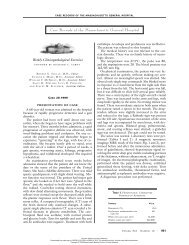
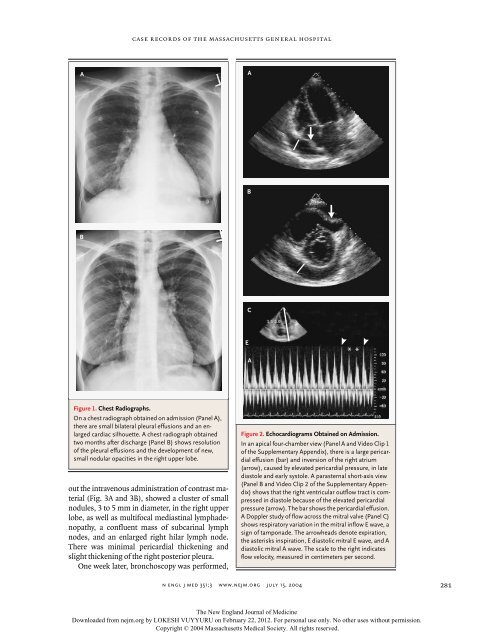
Figure 1. Chest Radiographs.<br />
case records of the massachusetts general hospital<br />
On a chest radiograph obtained on admission (Panel A),<br />
there are small bilateral pleural effusions and an enlarged<br />
cardiac silhouette. A chest radiograph obtained<br />
two months after discharge (Panel B) shows resolution<br />
of the pleural effusions and the development of new,<br />
small nodular opacities in the right upper lobe.<br />
out the intravenous administration of contrast material<br />
(Fig. 3A and 3B), showed a cluster of small<br />
nodules, 3 to 5 mm in diameter, in the right upper<br />
lobe, as well as multifocal mediastinal lymphadenopathy,<br />
a confluent mass of subcarinal lymph<br />
nodes, and an enlarged right hilar lymph node.<br />
There was minimal pericardial thickening and<br />
slight thickening of the right posterior pleura.<br />
One week later, bronchoscopy was performed,<br />
n engl j med 351;3<br />
E<br />
A<br />
B<br />
C<br />
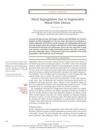
Figure 2. Echocardiograms Obtained on Admission.<br />
In an apical four-chamber view (Panel A and Video Clip 1<br />
of the Supplementary Appendix), there is a large pericardial<br />
effusion (bar) and inversion of the right atrium<br />
(arrow), caused by elevated pericardial pressure, in late<br />
diastole and early systole. A parasternal short-axis view<br />
(Panel B and Video Clip 2 of the Supplementary Appendix)<br />
shows that the right ventricular outflow tract is compressed<br />
in diastole because of the elevated pericardial<br />
pressure (arrow). The bar shows the pericardial effusion.<br />
A Doppler study of flow across the mitral valve (Panel C)<br />
shows respiratory variation in the mitral inflow E wave, a<br />
sign of tamponade. The arrowheads denote expiration,<br />
the asterisks inspiration, E diastolic mitral E wave, and A<br />
diastolic mitral A wave. The scale to the right indicates<br />
flow velocity, measured in centimeters per second.<br />
www.nejm.org july 15, <strong>2004</strong><br />
The New England Journal of Medicine<br />
Downloaded from nejm.org by LOKESH VUYYURU on February <strong>22</strong>, 2012. For personal use only. No other uses <strong>with</strong>out permission.<br />
Copyright © <strong>2004</strong> Massachusetts Medical Society. All rights reserved.<br />
A<br />
*<br />
*<br />
281