- Page 1 and 2: Color Atlas of Dental Medicine Edit
- Page 3: To my sons Philipp and Sebastian, a
- Page 6 and 7: VIII lar joint can come from condyl
- Page 8 and 9: Foreword Dr Bumann and Dr Lotzmann
- Page 10 and 11: XII Preface Medicine and dentistry
- Page 13 and 14: Table of Contents vii Forewords xii
- Page 15: Table of Contents xvii 231 Check-Bi
- Page 18 and 19: Introduction The Masticatory System
- Page 20 and 21: Introduction Functional Diagnostic
- Page 22 and 23: Introduction The Role of Dentistry
- Page 24 and 25: Primary Dental Evaluation The denta
- Page 26 and 27: darily by discomfort in the joints
- Page 28 and 29: Anatomy of the Masticatory System A
- Page 30 and 31: Embryology of the Temporomandibular
- Page 32 and 33: ounding bone. The fibrocartilaginou
- Page 34 and 35: However, the maturation process of
- Page 36 and 37: while the transforming growth facto
- Page 40 and 41: 22 Anatomy of the Masticatory Syste
- Page 42 and 43: 24 Anatomy of the Masticatory Syste
- Page 44 and 45: 26 Anatomy of the Masticatory Syste
- Page 46 and 47: 28 Anatomy of the Masticatory Syste
- Page 48 and 49: 30 Anatomy of the Masticatory Syste
- Page 50 and 51: 32 Anatomy of the Masticatory Syste
- Page 52 and 53: 34 Anatomy of the Masticatory Syste
- Page 54 and 55: 36 Anatomy of the Masticatory Syste
- Page 56 and 57: 38 Anatomy of the Masticatory Syste
- Page 58 and 59: 40 Anatomy of the Masticatory Syste
- Page 60 and 61: 42 Anatomy of the Masticatory Syste
- Page 62 and 63: 44 Anatomy of the Masticatory Syste
- Page 64 and 65: 46 Anatomy of the Masticatory Syste
- Page 66 and 67: 48 Anatomy of the Masticatory Syste
- Page 68 and 69: 50 Anatomy of the Masticatory Syste
- Page 70 and 71: 52 Anatomy of the Masticatory Syste
- Page 72 and 73: 54 Manual Functional Analysis The M
- Page 74 and 75: With identification of a specific l
- Page 76 and 77: The reverse side of the examination
- Page 78 and 79: Whenever a pain symptom is reported
- Page 80 and 81: Manual Fixation of the Head In addi
- Page 82 and 83: Active Movements and Passive Jaw Op
- Page 84 and 85: Active Movements and Passive Jaw Op
- Page 86 and 87: Differential Diagnosis of Restricte
- Page 88 and 89:
No crepitus no pain Adaptation of t
- Page 90 and 91:
In contrast with other definitions
- Page 92 and 93:
Crepitus during the protrusive move
- Page 94 and 95:
The three possible conditions of th
- Page 96 and 97:
Examination of the Joint Capsule an
- Page 98 and 99:
Joint-play Techniques 79 189 Hand g
- Page 100 and 101:
joint-play Techniques 81 197 Hand g
- Page 102 and 103:
Inferior Traction 83 205 Direction
- Page 104 and 105:
Regardless of these scientific find
- Page 106 and 107:
Examination of the Muscles of Masti
- Page 108 and 109:
Palpation of the Muscles of Mastica
- Page 110 and 111:
Palpation of the Muscles of Mastica
- Page 112 and 113:
Palpation of the Muscles of Mastica
- Page 114 and 115:
Referred Myofascial Pain 95 24A Myo
- Page 116 and 117:
Length of the Suprahyoid Structures
- Page 118 and 119:
In summary: Clicking sounds in the
- Page 120 and 121:
Investigation of Clicking Sounds 10
- Page 122 and 123:
Active Movements and Dynamic Compre
- Page 124 and 125:
Manual Translations 105 275 Examina
- Page 126 and 127:
Dynamic Compression during Retrusiv
- Page 128 and 129:
Group 2: Clicking Sounds due to Par
- Page 130 and 131:
Group 1 Lateral/medial portion of j
- Page 132 and 133:
No clicking sound during medial tra
- Page 134 and 135:
Partial or total disk displacement
- Page 136 and 137:
La ! Group 3 Cartilage hypertrophy
- Page 138 and 139:
Group 4 Disk displacement with term
- Page 140 and 141:
Clinical parameters for selection a
- Page 142 and 143:
These five techniques provide infor
- Page 144 and 145:
Neuromuscular Deprogramming Before
- Page 146 and 147:
cle tone. The centric condylar posi
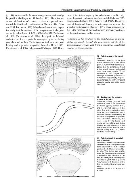
- Page 148 and 149:
from this centric occlusion the pat
- Page 150 and 151:
can already be excluded. Because po
- Page 152 and 153:
teeth to produce the present loadin
- Page 154 and 155:
Influence of Orthopedic Disorders o
- Page 156 and 157:
Panoramic Radiograph The panoramic
- Page 158 and 159:
Muscle tone can only be evaluated b
- Page 160 and 161:
Imaging Procedures Imaging procedur
- Page 162 and 163:
The panoramic radiograph of the tem
- Page 164 and 165:
Panoramic Radiographs of the Tempor
- Page 166 and 167:
Distortion Phenomena Distortions in
- Page 168 and 169:
Axial Cranial Radiograph Tomography
- Page 170 and 171:
Lateral Transcranial Radiograph The
- Page 172 and 173:
Computed Tomography of the Temporom
- Page 174 and 175:
_______________________ Three Dimen
- Page 176 and 177:
Three-Dimensional Models of Polyure
- Page 178 and 179:
T1-andT2-Weighting The magnetizing
- Page 180 and 181:
Practical Application of MRI Sectio
- Page 182 and 183:
Reproduction of Anatomical Detail i
- Page 184 and 185:
Classification of the Stages of Bon
- Page 186 and 187:
Disk Position in the Frontal Plane
- Page 188 and 189:
Morphology of the Pars Posterior Th
- Page 190 and 191:
Progressive Adaptation in T1- and T
- Page 192 and 193:
Disk Hypermobility Disk hypermobili
- Page 194 and 195:
Total Disk Displacement The inciden
- Page 196 and 197:
Disk Displacement without Repositio
- Page 198 and 199:
Partial Disk Displacement with Part
- Page 200 and 201:
Total Disk Displacement with Partia
- Page 202 and 203:
Posterior Disk Displacement As a ru
- Page 204 and 205:
Regressive Adaptation of Bony Joint
- Page 206 and 207:
The capacity for progressive adapta
- Page 208 and 209:
Avascular Necrosis Versus Osteoarth
- Page 210 and 211:
Metric (Quantitative) MRI Analysis
- Page 212 and 213:
Metric MRI Analysis 193 516 MRI of
- Page 214 and 215:
Three-Dimensional Imaging with MRI
- Page 216 and 217:
provide no significant new diagnost
- Page 218 and 219:
Indications for Imaging Procedures
- Page 220 and 221:
Mounting of Casts and Occlusal Anal
- Page 222 and 223:
Making of Impressions and Stone Cas
- Page 224 and 225:
Fabrication of Segmented Casts Segm
- Page 226 and 227:
Techniques for Recording the Centri
- Page 228 and 229:
tion of the intermaxillary position
- Page 230 and 231:
Centric Registration for Intact Den
- Page 232 and 233:
posterior teeth, then fine adjustme
- Page 234 and 235:
Jaw Relation Determination for Eden
- Page 236 and 237:
Attaching the Anatomical Transfer B
- Page 238 and 239:
Attaching the Anatomical Transfer B
- Page 240 and 241:
Mounting the Maxillary Cast using t
- Page 242 and 243:
Mounting the Maxillary Cast followi
- Page 244 and 245:
Mounting the Maxillary Cast followi
- Page 246 and 247:
Mounting the Mandibular Cast 227 61
- Page 248 and 249:
Axiosplit System 229 625 Fastening
- Page 250 and 251:
Check-Bite for Setting the Articula
- Page 252 and 253:
Occlusal Analysis on the Casts An o
- Page 254 and 255:
Occlusal Analysis on the Casts 235
- Page 256 and 257:
Occlusal Analysis Using Sectioned C
- Page 258 and 259:
Diagnostic Occlusal Reshaping of th
- Page 260 and 261:
Diagnostic Occlusal Reshaping of th
- Page 262 and 263:
Diagnostic Waxup In addition to dia
- Page 264 and 265:
Diagnostic Waxup 245 680 Completed
- Page 266 and 267:
One definite disadvantage is that i
- Page 268 and 269:
compare movement paths that were dr
- Page 270 and 271:
lateral movements serves to determi
- Page 272 and 273:
Axiography 253 703 Insertion of the
- Page 274 and 275:
Axiography 255 711 Attachment of th
- Page 276 and 277:
HIM-: Axiography 257 719 Tracing th
- Page 278 and 279:
Axiography 259 727 Recording the me
- Page 280 and 281:
Evaluating the Axiograms and Progra
- Page 282 and 283:
Effect of an Incorrectly Located Hi
- Page 284 and 285:
actual paths of movement within the
- Page 286 and 287:
Electronic Paraocclusal Axiograpy 2
- Page 288 and 289:
Diagnoses and Classifications The m
- Page 290 and 291:
Classification of Secondary Joint D
- Page 292 and 293:
Hyperplasia of the Coronoid Process
- Page 294 and 295:
Acute Arthritis Acute arthritis can
- Page 296 and 297:
Juvenile Chronic Arthritis The term
- Page 298 and 299:
Styloid or Eagle Syndrome Elongatio
- Page 300 and 301:
Disk Displacement with Condylar Nec
- Page 302 and 303:
Tumors in the Temporomandibular Joi
- Page 304 and 305:
Osteoarthritis Bony ankylosis ICD.9
- Page 306 and 307:
Joint Disorders—Bilaminar Zone an
- Page 308 and 309:
Total disk displacement withoyt rep
- Page 310 and 311:
Joint Disorders 291 Perforation of
- Page 312 and 313:
Sclerosing of lateral ligament Caps
- Page 314 and 315:
Joint Disorders—Ligaments Inserti
- Page 316 and 317:
Muscle Disorders Muscle Disorders 2
- Page 318 and 319:
Muscle Disorders 299 Muscle spasm F
- Page 320 and 321:
Principles of Treatment In this boo
- Page 322 and 323:
Nonspecific Treatment Nonspecific t
- Page 324 and 325:
Manipulative therapy is the essenti
- Page 326 and 327:
arch if the inclination of the ante
- Page 328 and 329:
Relationship between joint Surface
- Page 330 and 331:
Stabilization Splint The main purpo
- Page 332 and 333:
Repositioning Splint A repositionin
- Page 334 and 335:
-a*| Splint Therapy 315 834 Reduced
- Page 336 and 337:
changed by means of restorations fo
- Page 338 and 339:
If it has been necessary to perform
- Page 340 and 341:
Example 321 854 Occlusal splint the
- Page 342 and 343:
Illustration Credits Many of the pi
- Page 344 and 345:
Blumenfeld, I., Laufer, D., Livne,
- Page 346 and 347:
Hatcher, D. C, Faulkner, M. C, Hay,
- Page 348 and 349:
Moffet, B.: Histologic Aspects of T
- Page 350 and 351:
Voy, E.-D., Fuchs, M.: Anatomische
- Page 352 and 353:
Dworkin, S. F., LeResche, L, von Ko
- Page 354 and 355:
Lobbezoo-Scholte, A. M., De Leeuw,
- Page 356 and 357:
Shafagh, I., Yoder, J. L, Thayer, K
- Page 358 and 359:
Bumann, A., Schroder, C, Melchert,
- Page 360 and 361:
Harms S. E., Flamig D. P., Fisher C
- Page 362 and 363:
LiedbergJ., Panmekiate, S., Peterss
- Page 364 and 365:
Sahm, C, Witt, E.: Long-term result
- Page 366 and 367:
References 347 Mounting of Casts an
- Page 368 and 369:
Ferrario, V. F., Sforza, C, Gianni,
- Page 370 and 371:
Parkhouse, R. C: Medical complicati
- Page 372 and 373:
Karolyi, M.: Beobachtungen uber Pyo
- Page 374 and 375:
Index abrasion facets 234 adaptatio
- Page 376 and 377:
with disk displacement 281 dentin 8
- Page 378 and 379:
N nasion relator 251 neck pain 44 d


![SISTEM SENSORY [Compatibility Mode].pdf](https://img.yumpu.com/20667975/1/190x245/sistem-sensory-compatibility-modepdf.jpg?quality=85)


