

TMJ Disorders and Orofacial.pdf - E-Lib FK UWKS
TMJ Disorders and Orofacial.pdf - E-Lib FK UWKS
TMJ Disorders and Orofacial.pdf - E-Lib FK UWKS

Create successful ePaper yourself
Turn your PDF publications into a flip-book with our unique Google optimized e-Paper software.
Arterial Supply <strong>and</strong> Sensory Innervation of the Temporom<strong>and</strong>ibular Joint _____ 31^<br />
Arterial Supply <strong>and</strong> Sensory Innervation of the Temporom<strong>and</strong>ibular Joint<br />
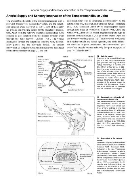
The arterial blood supply of the temporom<strong>and</strong>ibular joint is<br />
provided primarily by the maxillary artery <strong>and</strong> the superficial<br />
temporal artery (Boyer et al. 1964). Both of these arteries<br />
are also the principle supply for the muscles of mastication.<br />
Apart from the network of arteries surrounding it, the<br />
condyle is also supplied from the inferior alveolar artery<br />
through the bone marrow (Okeson 1998). The venous<br />
drainage is through the superficial temporal vein, the maxillary<br />
plexus, <strong>and</strong> the pterygoid plexus. The sensory<br />
innervation of the joint capsule <strong>and</strong> its receptors has already<br />
been addressed briefly on page 27. The tem-<br />
porom<strong>and</strong>ibular joint is innervated predominantly by the<br />
auriculotemporal, masseter, <strong>and</strong> temporal nerves (Klineberg<br />
et al. 1970, Harris <strong>and</strong> Griffin 1975). Proprioception occurs<br />
through four types of receptors (Thil<strong>and</strong>er 1961, Clark <strong>and</strong><br />
Wyke 1974, Zimny 1988): Ruffini mechanoreceptors (type I),<br />
pacinian corpuscles (type II), Golgi tendon organs (type III),<br />
<strong>and</strong> free nerve endings (type IV). These receptors are located<br />
in the joint capsule, the lateral ligament, <strong>and</strong> in the bilaminar<br />
zone <strong>and</strong> its genu vasculosum. The anteromedial portion<br />
of the capsule contains relatively few pain receptors, of<br />
type IV (Thil<strong>and</strong>er 1961).<br />
72 Arterial supply<br />
Diagram of the arterial blood supply<br />
of a left temporom<strong>and</strong>ibular<br />
joint (modified after Voy <strong>and</strong> Fuchs<br />
1980). The condyle is supplied with<br />
blood from all four sides. In addition,<br />
there are anastomoses with<br />
the inferior alveolar artery within<br />
the marrow spaces. Because of the<br />
abundant blood supply, avascular<br />
necrosis is rarely found in the<br />
condyle (Hatcher etal. 1997). Compression<br />
of the anterior vessels by<br />
anterior disk displacement (Schellhas<br />
et al. 1992) will not interfere<br />
with the condyle's blood supply.<br />
73 Sensory innervation of a left<br />
temporom<strong>and</strong>ibular joint<br />
The afferent nerve fibers arise from<br />
the m<strong>and</strong>ibular branch of the<br />
trigeminal nerve <strong>and</strong> exhibit four<br />
types of nerve endings. In rats, free<br />
nerve endings (type IV), which are<br />
potential pain receptors, have been<br />
found in the capsule, lateral ligament,<br />
bilaminar zone, <strong>and</strong> in the<br />
pars anterior <strong>and</strong> pars posterior of<br />
the disk (Ichikawa et al. 1990, Kido<br />
et al. 1991). This has not been verified<br />
for human disk structures,<br />
however.<br />
74 Innervation in the capsule<br />
region<br />
Schematic diagram of the different<br />
areas of innervation (modified from<br />
Ishibashi 1974, Schwarzer 1993).<br />
Activation of the type-IV receptors<br />
in the capsule increases the activity<br />
of sympathetic efferent fibers<br />
(Roberts <strong>and</strong> Elardo 1985). Because<br />
of the sympathetic innervation of<br />
the intrafusal muscle fibers (Grassi<br />
et al. 1993), a secondary rise in<br />
muscle tone is brought about by<br />
activation of the afferent fibers of<br />
the muscle spindles <strong>and</strong> the efferent<br />
α-motoneurons (Schwarzer<br />
1993).


![SISTEM SENSORY [Compatibility Mode].pdf](https://img.yumpu.com/20667975/1/190x245/sistem-sensory-compatibility-modepdf.jpg?quality=85)


