第48屆比較病理研討會 - 國立臺灣大學獸醫專業學院
第48屆比較病理研討會 - 國立臺灣大學獸醫專業學院
第48屆比較病理研討會 - 國立臺灣大學獸醫專業學院

You also want an ePaper? Increase the reach of your titles
YUMPU automatically turns print PDFs into web optimized ePapers that Google loves.
中華民國比較病理學會<br />
Chinese Society of Comparative Pathology<br />
第 48 次比較病理學研討會<br />
國立臺灣大學獸醫專業學院<br />
臺北市.臺灣<br />
中華民國 99 年 3 月 13 日<br />
48th Meeting of Comparative Pathology<br />
School of Veterinary Medicine, National Taiwan University<br />
Taipei, Taiwan<br />
March 13, 2010<br />
1
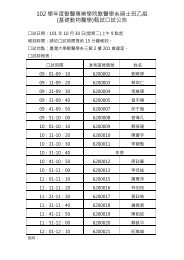
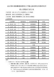
中華民國比較病理學會第 48 次比較病理學研討會議程表<br />
Schedule<br />
48th Meeting of the Chinese Society of Comparative Pathology<br />
時間:99 年 3 月 13 日(星期六) 09:00~17:00<br />
地點:國立臺灣大學獸醫學系 B01 演講廳<br />
地址:臺北市羅斯福路四段 1 號<br />
電話: 02-33663858<br />
2<br />
Date: March 13, 2010 (Sat) 09:00~17:00<br />
Location: B01, School of Vet Med, NTU<br />
Address: No. 1, Sec. 4, Roosevelt Road, Taipei<br />
Telephone: 02-33663858<br />
Time 時間 Schedule 議程 Moderator 主持<br />
08:30~09:00 Registratio 報到<br />
09:00~09:10 Opening Ceremony 致詞<br />
09:10~09:50<br />
09:50~10:30<br />
Keynote<br />
專題演講<br />
Keynote<br />
專題演講<br />
人類乳突病毒與肺癌<br />
李輝 研究員<br />
國家衛生研究院環境衛生與職業醫學研究組<br />
Individualized Cancer Therapy Has Come of Age<br />
劉興璟 助理教授<br />
臺北醫學大學臨床醫學研究所<br />
10:30~10:50 Coffee Break<br />
10:50~11:30<br />
11:30~12:00<br />
Keynote<br />
專題演講<br />
Case 334<br />
病例討論<br />
The Diagnosis and Study of Glioma<br />
李進成 主任<br />
新光醫院病理檢驗科<br />
Dr. T.T. Liu 劉婷婷 醫師<br />
Tzu Chi University & Tzu Chi General Hosipital<br />
佛教慈濟綜合醫院暨慈濟大學<br />
Dr. C.H. Liu<br />
劉振軒 理事長<br />
Dr. Y.H. Hsu<br />
許永祥 主任<br />
12:00~13:30 Lunch & Committee Meeting 午餐暨「中華民國比較病理學會理監事會議」<br />
13:30~14:00<br />
14:00~14:30<br />
14:30~15:00<br />
Case 335<br />
病例討論<br />
Case 336<br />
病例討論<br />
Case 337<br />
病例討論<br />
Dr. Y.L. Chen 陳燕麟 醫師<br />
Department of Pathology, Cardinal Tien Hospital<br />
天主教耕莘醫院病理科<br />
Dr. L.C. Chen 祝志平 醫師<br />
Department of Pathology, Lin Shin Hospital<br />
林新醫院病理科<br />
Dr. C.L. Hung 洪巧凌 獸醫師<br />
School of Veterinary Medicine, National Taiwan University<br />
國立臺灣大學獸醫專業學院<br />
15:00~15:20 Coffee Break<br />
15:20~15:50<br />
15:50~16:20<br />
Case 338<br />
病例討論<br />
Case 339<br />
病例討論<br />
Dr. C.H. Wu 吳介豪 獸醫師<br />
Dr. F.J. Leu<br />
呂福江 主任<br />
College of Vet. Med., National Chung-Hsing University<br />
國立中興大學獸醫學院 Dr. J.W. Liao<br />
Dr. S.W. Huang 黃思偉 獸醫師<br />
National Laboratory Animal Center<br />
國家實驗動物中心<br />
16:20~17:00 Town Hall Meeting 中華民國比較病理學會會員大會<br />
廖俊旺 教授
Table of Contents (目錄)<br />
A Schedule (議程表)…………………………………………………………………… 2<br />
B Table of Contents (目錄)…………………………………………………………… 3<br />
C Case Signalment…………………………………………………………………… 4<br />
D Case Diagnosis……………………………………………………………………… 5<br />
E Keynote Speakers…………………………………………………………………… 6<br />
F Comparative Pathology Case 334…………………………………………………… 7<br />
Comparative Pathology Case 335…………………………………………………… 12<br />
Comparative Pathology Case 336…………………………………………………… 16<br />
Comparative Pathology Case 337…………………………………………………… 20<br />
Comparative Pathology Case 338…………………………………………………… 23<br />
Comparative Pathology Case 339…………………………………………………… 28<br />
G 中華民國比較病理學會數位式組織切片影像資料庫…………………………… 32<br />
H 1~48 次研討會病例分類表………………………………………………………… 37<br />
I 中華民國比較病理學會章程……………………………………………………… 52<br />
J 會員資料更新服務………………………………………………………………… 56<br />
K 入會辦法…………………………………………………………………………… 58<br />
L Map (國立臺灣大學獸醫專業學院)………………………………………………… 59<br />
3
Case<br />
No.<br />
Case<br />
334<br />
Case<br />
335<br />
Case<br />
336<br />
Case<br />
337<br />
Case<br />
338<br />
Case<br />
339<br />
中華民國比較病理學會第 48 次比較病理學研討會<br />
Case Signalment<br />
48th Meeting of the Chinese Society of Comparative Pathology<br />
March 13, 2010<br />
Presenter Institution Slide No. Signalment<br />
Dr. T.T. Liu<br />
劉婷婷 醫師<br />
Dr. Y.L. Chen<br />
陳燕麟 醫師<br />
Dr. L.C. Chen<br />
祝志平 醫師<br />
Dr. C.L. Hung<br />
洪巧凌 獸醫師<br />
Dr. C.H. Wu<br />
吳介豪 獸醫師<br />
Dr. S.W. Huang<br />
黃思偉 獸醫師<br />
Tzu Chi University & Tzu Chi<br />
General Hosipital<br />
佛教慈濟綜合醫院暨慈濟大學<br />
Department of Pathology,<br />
Cardinal Tien Hospital<br />
天主教耕莘醫院病理科<br />
Department of Pathology, Lin<br />
Shin Hospital<br />
林新醫院病理科<br />
School of Veterinary<br />
Medicine, National Taiwan<br />
University<br />
國立臺灣大學獸醫專業學院<br />
College of Vet. Med., National<br />
Chung-Hsing University<br />
國立中興大學獸醫學院<br />
National Laboratory Animal<br />
Center<br />
國家實驗動物中心<br />
4<br />
A301-16 47- year-old woman<br />
CTH<br />
S10-233<br />
NTU09-788D<br />
C009-835<br />
S090982-b<br />
44-year-old female<br />
patient<br />
67-year-old male<br />
patient<br />
11-year-old, spayed<br />
female, mongrel<br />
canine<br />
4 year-old, male,<br />
hybrid rabbit<br />
5-month-old, male,<br />
Sprague Dawley rat
Case<br />
No.<br />
Case<br />
334<br />
Case<br />
335<br />
Case<br />
336<br />
Case<br />
337<br />
Case<br />
338<br />
Case<br />
339<br />
中華民國比較病理學會第 48 次比較病理學研討會<br />
Case Diagnosis<br />
48th Meeting of the Chinese Society of Comparative Pathology<br />
March 13, 2010<br />
Presenter Institution Slide No. Diagnosis<br />
Dr. T.T. Liu<br />
劉婷婷 醫師<br />
Dr. Y.L. Chen<br />
陳燕麟 醫師<br />
Dr. L.C. Chen<br />
祝志平 醫師<br />
Dr. C.L. Hung<br />
洪巧凌 獸醫師<br />
Dr. C.H. Wu<br />
吳介豪 獸醫師<br />
Dr. S.W. Huang<br />
黃思偉 獸醫師<br />
Tzu Chi University & Tzu Chi<br />
General Hosipital<br />
佛教慈濟綜合醫院暨慈濟大學<br />
Department of Pathology,<br />
Cardinal Tien Hospital<br />
天主教耕莘醫院病理科<br />
Department of Pathology,<br />
Lin Shin Hospital<br />
林新醫院病理科<br />
School of Veterinary<br />
Medicine, National Taiwan<br />
University<br />
國立臺灣大學獸醫專業學<br />
院<br />
College of Vet. Med.,<br />
National Chung-Hsing<br />
University<br />
國立中興大學獸醫學院<br />
National Laboratory Animal<br />
Center<br />
國家實驗動物中心<br />
5<br />
A301-16<br />
CTH<br />
S10-233<br />
NTU09-788D<br />
C009-835<br />
Metastatic<br />
infiltrating ductal<br />
carcinoma, liver<br />
Adenoid cystic<br />
carcinoma, grade II,<br />
Rt breast<br />
Malignant<br />
lymphoma, diffuse,<br />
large B-cell, right<br />
neck<br />
Pulmonary<br />
carcinoma,<br />
multicentric, lung<br />
Malignant<br />
melanoma, multiple<br />
organs metastasis<br />
S090982-b Mycoplasmosis
Keynote Speakers Chinese Society of Comparative Pathology, March 2010<br />
李輝 研究員<br />
現任:國家衛生研究院環境衛生與職業醫學研究組研究員<br />
中山醫學大學醫學院醫學分子毒理學教授<br />
學歷:中興大學食品科學研究所博士<br />
經歷:中山醫學大學醫學院生物化學系系主任<br />
中山醫學大學醫學院醫學分子毒理學研究所所長<br />
研究興趣:<br />
1. DDX3, LKB1 ,APE1, IL10 及 XPC 基因在 HPV 感染肺腫瘤化之角色。<br />
2. HPV16/18 E6 致癌蛋白在 EGFR 發生突變之角色。<br />
3. DNA repair (XPC, XRCC1, ERCC1, XRCC5, hOGG1, APE1)與抗氧化(MnSOD, GpX,<br />
Catalase)基因表現來評估抗癌藥之敏感性對個人化醫療之可能角色。<br />
4. 建立 E6 轉殖鼠探討 HPV E6 致癌蛋白在肺腫瘤化之角色。<br />
5. Micro RNA 在 HPV 感染肺腫瘤化之角色。<br />
劉興璟 助理教授<br />
現任:臺北醫學大學臨床醫學研究所助理教授<br />
台北市立萬芳醫院血液腫瘤科主治醫師<br />
學歷:臺北醫學大學醫學系<br />
明尼蘇達大學博士<br />
經歷:臺北市立仁愛醫院內科醫師<br />
美國明尼蘇達大學醫院血液腫瘤科臨床研究員<br />
臺北醫學大學細胞及分子生物學研究所助理教授<br />
內科、血液科及腫瘤內科專科醫師<br />
研究興趣:<br />
癌症生物學<br />
幹細胞生物學<br />
李進成 主任<br />
現任:新光醫院病理檢驗科主治醫師<br />
輔仁大學醫學系兼任副教授<br />
學歷:英國倫敦大學神經學研究所博士<br />
經歷:<br />
1. 林口長庚醫院內科住院醫師<br />
2. 台大醫院病理科住院醫師<br />
3. 英國倫敦大學神經研究所神經病理科住院醫師<br />
4. 馬偕醫院病理科住院醫師<br />
5. 台北市立中興醫院解剖病理總醫師<br />
6. 新光醫院病理檢驗科主任<br />
6
Case Number: 334 Chinese Society of Comparative Pathology, March 2010<br />
Liu, T.T. (劉婷婷), M.D.; Hsu, Yung-Hsiang (許永祥), M.D.<br />
Buddhist Tzu-Chi General Hospital and Tzu-Chi University (佛教慈濟綜合醫院暨慈濟大學)<br />
CASE HISTORY:<br />
Signalment: 47- year-old woman<br />
Clinical History :<br />
The 47-year-old woman was a victim of infiltrating ductal carcinoma of right breast, stage IV<br />
since 2000. She started to have regular OPD follow-up at HuaLien Buddhist Tzu Chi General<br />
Hospital since May 2007. Xeloda was discontinued at that time. Right shoulder pain was<br />
complained with a tender point. However, all image study including bone scan showed no<br />
relevant lesion, except one suspected metastatic lesion in lower S1. Tamoxifen was<br />
discontinued and shifted to Arimidex on May 31, 2007. In the beginning of June, a 3.5 x 2.5 x<br />
2 cm non-tender bony prominence was noted over the right sternal angle. Lymphadenopathy<br />
with differential diagnosis of granulomatous disease was showed by CT scan. Therefore a<br />
biopsy for right chest wall was performed and revealed only necrotic tissue on July 2007.<br />
Shoulder pain and right anterior lateral chest pain with local tenderness persisted, but the<br />
following image study only showed T7 and T8 metastatic lesion. In Dec. 2007, marked<br />
hepatomegaly was noted in an OPD visit. Abdominal sonar revealed multiple metastatic lesions.<br />
She was admitted to this hospital since January 2008 for some cancer metastatic bone pain and<br />
abdominal fullness due to liver metastasis, and readmission to the ward for 3 times for<br />
continuous anti-cancer therapy in 2008. On the CT scan of Aug. 2008, progressive diffuse<br />
metastatic hepatic masses as compared with previous study on Jul. 2008. A CT guided biopsy<br />
of liver also confirmed metastatic ductal carcinoma from breast, and immunohistochemical<br />
study of the tumor cells showing ER (+), PR (+++), P53 (++) and HER-2/neu (-) on Aug. 2008.<br />
She received chemotherapy with Vinorelbine + CDDP since 2008-8-21. The abdominal CT<br />
scan of 2008-11-29 revealed slight shrinkage of liver size and the liver metastases from breast<br />
cancer but still prominent viable tumors with decompensated hepatic function and portal<br />
hypertension. She was admitted because of progressive abdominal fullness, lower leg edema<br />
for one week and black stool for 2-3 days on Dec. 29 2008. Two weeks later she was<br />
discharged and transferred to hospice ward for palliative care.<br />
Due to progressive jaundice, generalized weakness, lymphedema of lower extremities and<br />
abdominal fullness, she was admitted for symptomatic treatment on Feb. 5, 2009.<br />
Transfusion of albumin was done for two days due to hypoalbuminemia. Lymph massage per<br />
day was given during this hospitalization. The patient's condition went downhill day by day<br />
with cachexia formation. On Feb. 22, drowsy consciousness, breathing with ruffle sounds and<br />
air hunger were noted. She expired on Feb. 23, 2009.<br />
7
Gross Finding:<br />
At autopsy, she was 64.5 kg in weight and 155 cm in length. Old surgical scar of modified<br />
radical mastectomy on right chest wall, generalized jaundice and bilateral leg pitting edema<br />
were noted. A port-A catheter over left subclavian area is found. Opening the chest and<br />
abdomen, bilateral pleural adhesion with marked adhesion in the right side, mild (about 100<br />
C.C) serous effusion of bilateral pleural cavity respectively and over 3600 C.C serous ascites<br />
were obtained. The position of the dome of bilateral diaphragm was over 5th ICS respectively.<br />
The heart weighed 400 gm. The wall thickness of right ventricle was 0.3 cm and the one of<br />
left was 1.1 cm. The circumferences of tricuspid, pulmonary, mitral and aortic valves were 9.5<br />
cm, 6.0 cm, 9.5 cm and 5.5 cm respectively. Pericardial adhesion with scanty pericardial<br />
effusion, fibrinous pericarditis with bread and butter appearance over pericardial space are<br />
noticed. The lumen of the aorta showed no atherosclerotic change. The right lung weighed 500<br />
gm and the left one weighed 490 gm. There were three nodules was seen in the pleural<br />
surface of RLL. On cut, focal red consolidation involved both lungs. The liver showed<br />
hepatomegaly (1700 gm, 28 x 17 x 9.5 cm in size) with multiple disseminated tumorous lesions<br />
with marked fibrosis mimicking cirrhotic nodules. The unremarkable gallbladder measured 5<br />
x 2 x 1.8 cm in size. The pancreas showed grossly unremarkable change. The spleen weighed<br />
430 gm (14.7 x 9 x 5.5 cm in size) with smooth capsule and no tumor nodule. The right<br />
kidney weighed 100 gm (10.5 x 5.5 x 3.2 cm in size) and the left one weighed 130 gm (11 x 5.9<br />
x 3.1 cm in size). In the G-I tract, hemorrhagic gastritis with petechiae on mucosa was found.<br />
The brain weighed 1300 gm and pituitary gland 0.85 gm (1.5 x 0.9 x 0.8 cm in size). There<br />
are small tumor metastatic foci over the surface of cerebellum and posterior portion of right<br />
frontal lobe. On serial sections, there are scattered foci of yellowish hued tumor metastasis<br />
within the cerebellum (the largest locus measuring up to 2 cm in dimension), left posterior pons,<br />
right occipital lobe and left basal ganglia.<br />
8
Case Number: 334 Chinese Society of Comparative Pathology, March 2010<br />
Liu, T.T. (劉婷婷), M.D.; Hsu, Yung-Hsiang (許永祥), M.D.<br />
Buddhist Tzu-Chi General Hospital and Tzu-Chi University (佛教慈濟綜合醫院暨慈濟大學)<br />
CASE RESULT:<br />
Microscopic Findings:<br />
We reviewed S2007-08454 and S2008-10461. Multiple glandular cancer cells infiltrated the<br />
chest wall diagnostic of metastatic infiltrating ductal carcinoma proved by<br />
immunohistochemisty stain with CK. The liver showed metastatic infiltrating ductal<br />
carcinoma with marked desmoplasia and focal tumor necrosis with ER (+), PR (+++), P53 (++)<br />
& HER-2/neu (-). We detail studies all samples slides. This poorly differentiated infiltrating<br />
ductal carcinoma penetrated into pituitary gland, left pleura, pericardium, left hilar lymph<br />
nodes, bilateral ovaries, adrenal gland ,T8 vertebra proved by CK immunohistochemisty stain,<br />
leptomenix and brain parenchyma of right frontal lobe, right occipital lobe, left pons, left basal<br />
ganglion and cerebellum. The most intrinsting finding was in the liver. It showed massive<br />
multiple metastatic cancer nests with marked desmoplasia mimicking cirrhosis. In addition,<br />
multiple ischemic hepatocyte necrosis with intra-hepatic and sinusoid cholestasis was<br />
consistent with hepatic failure clinically. The cirrhosis complication included varicose vein of<br />
esophagus & congestive splenomegaly. The brain stem showed Alzeimer’s type II astrocytes<br />
formation indicated hepatic encephalopathy clinically. The kidney showed hypokalemia<br />
associated vacuolar degeneration of proximal tubes, bile nephrosis and nephrocalcinosis.<br />
Metastatic calcification involved capillary wall of kidney, renal tubules and mucosa of stomach<br />
due to multiple bony destruction. The bone marrow showed erythroid hyperplasia with<br />
megaloblastic change. The spleen also showed extra-medullary hemopoiesis. The other<br />
findings including alveolar hemorrhage,edema and focal terminal bronchopneumonia of RLL &<br />
LLL & chronic cholecystitis and cholesterolosis of gall bladder.<br />
Morphologic Diagnosis: Metastatic infiltrating ductal carcinoma of liver<br />
Immunohistochemisty Stain: ER (+), PR (+++), P53 (++) & HER-2/neu (-).<br />
Comments:<br />
Although involvement of the liver is observed in 50% of metastatic malignancies, few patients<br />
survive long enough to develop cirrhosis from biliary obstruction perviously, 24 cases of<br />
carcinoma mimicking cirrhosis have been reported in the literature. Primary neoplasms<br />
included breast, colon, pancreas, lung, stomach, kidney, lymphoma, and adrenal gland.<br />
Including this case report, the breast was the primary site. Final admission to the hospital was<br />
usually associated with gastrointestinal bleeding, jaundice, ascites, or other symptoms of<br />
9
hepatic failure,such as our case. The course of all patients was rapidly downhill, with<br />
uncontrolled esophageal bleeding or hepatic failure as the terminal event. These signs of<br />
hepatic parenchymal disease are frequently seen in patients with hepatic metastases, but<br />
manifestations of portal hypertension are rare. The liver involved with metastases is usually<br />
larger than normal. Characteristically, the surface of the liver showed many lobulations of<br />
various sizes. The characteristic histologic feature was the filtration, and intravascular and<br />
lymphatic intense, diffuse fibrosis of the liver parenchyma- embolization resulting in portal<br />
hypertension and rapidly fatal course, an additional of carcinomatous involvement of the liver<br />
resulting in a clinical picture of cirrhosis and autopsy findings of portal hypertension secondary<br />
to metastatic carcinoma of the breast is reported, such as our case. Perusal of the literature and<br />
our own institutional autopsy series indicates the relative infrequency of this complication of<br />
metastatic malignancy. Aggressive therapy directed against the malignant process might alter<br />
the course of this invariably fatal complication if tumor infiltration rather than fibrosis is a<br />
major cause of vascular obstruction. At least in patients with breast carcinoma, where usually<br />
widespread dissemination of metastatic disease is observed, there seems little indication for<br />
aggressive surgical management of the resulting portal hypertension.<br />
Conclusion:<br />
This 47-year-old woman suffered from right breast infiltrating ductal carcinoma with multiple<br />
metastases. This cancer also metastatic into liver mimicking cirrhosis resulted in portal<br />
hypertension and severe ascites formation. Although chemotherapy was performed, finally the<br />
breast cancer still disseminated to multiple organs. The cause of death was due to hepatic<br />
failure.<br />
Virtual Slide:<br />
http://140.112.96.83:82/CSCP/48CSCP/case334/2731.svs/view.apml?ahide=1&alayer=0&aregion=0<br />
10
References:<br />
1. Borja ER, Hori JM, Pugh RP. Metastatic carcinomatosis of the liver mimicking cirrhosis: case report and review of<br />
the literature. Cancer. 1975; 35(2):445-9.<br />
2. Viguier J, De Muret A, Bacq Y. Ascites due to portal hypertension from breast cancer- related metastatic liver<br />
infiltration] Gastroenterol Clin Biol. 2006;30(6-7):903-5.<br />
3. Martelli O, Coppola L, De Quarto AL, Palma M, Sarmiento R, Foggi CM. Fulminant hepatic failure caused by<br />
diffuse intrasinusoidal metastatic liver disease: a case report. Tumori. 2000; 86(5):424-7.<br />
11
Case Number: 335 Chinese Society of Comparative Pathology, March 2010<br />
Chen, Yen-Lin (陳燕麟), M.D.; 江蓉華, M.D.; Leu, Fur-Jiang (呂福江), M.D., Ph.D.; Suen, J.H.<br />
(孫政宏), M.D.;林進耀, M.D..<br />
Department of Pathology, Cardinal Tien Hospital (天主教耕莘醫院病理科)<br />
CASE HISTORY:<br />
Signalment : 44-year-old female patient<br />
Clinical History:<br />
This is a 44 years old woman with a chief complaint of right breast mass at 6 o’clock position.<br />
The mass has been years and she felt getting bigger than before. There was no pain about mass<br />
but discomfort when menstruation. The echo showed a hypoechoic lesion with 1.7 x 1.8 cm in<br />
size and fibroadenoma was suggested. Mammography was not done. No other underlying<br />
disease was noted. The patient received excision surgery and post OP follow up for 4 months<br />
was good.<br />
Gross Finding:<br />
The specimen consisted of a piece of tissue measuring 4 x 3.5 x 3 cm in size and 25 gm in<br />
weight without lymph nodes, fixed in formalin. Grossly, nodule lesion with microcysts displayed<br />
soft in consistency and the cut surface was gray-white in color. Representative parts were taken<br />
for sections.<br />
Laboratory Results:<br />
CBC/DC: WNL<br />
Biochemistry (sugar, Ca, BUN, Cr, Na, K, Cl, AST, ALT) : WNL<br />
12
Case Number: 335 Chinese Society of Comparative Pathology, March 2010<br />
Chen, Yen-Lin (陳燕麟), M.D.; 江蓉華, M.D.; Leu, Fur-Jiang (呂福江), M.D., Ph.D.; Suen, J.H.<br />
(孫政宏), M.D.;林進耀, M.D..<br />
Department of Pathology, Cardinal Tien Hospital (天主教耕莘醫院病理科)<br />
CASE RESULT:<br />
Histopathologic Finding:<br />
The sections show picture of adenoid cystic carcinoma with well differentiated tumor cells. It<br />
consists of a mixture of proliferating glands (adenoid component) with eosinophilic granular<br />
secretion and basement membrane components (“pseudoglandular” or cylindromatous<br />
component). There are cribriform, tubular and focal solid (
The majority of lesions measure between 1 and 3 cm. Most ACCs are circumscribed or nodular<br />
grossly. Small cystic areas are not unusual, especially in lesions smaller than 5 cm. The lesions<br />
have been variously described as gray, pale yellow, tan, and pink. ACC arises from<br />
myoepithelium-like cells and ducts. It consists of a mixture of proliferating glands (adenoid<br />
component) and basement membrane components (“pseudoglandular” or cylindromatous<br />
component). A variety of microscopic growth patterns have been described as cribriform, solid,<br />
tubular, reticular (trabecular), and basaloid. Shrinkage artifacts occur relatively often in adenoid<br />
cystic carcinoma and may be mistaken for lymphatic tumor emboli. ACC can be stratified into<br />
three grades on the basis of the proportion of solid growth within the lesion (grade I, no solid<br />
elements; grade II, less than 30% solid; and grade III, more than 30% solid).<br />
More than 90% of ACC cells expressed c-kit and it is not expressed in invasive cribriform<br />
carcinoma and collagenous spherulosis. P63 is expressed in ACC with cribriform pattern and<br />
collagenous spherulosis with ring-like pattern but not in invasive cribriform carcinoma.<br />
P63 c-kit<br />
Adenoid cystic carcinoma + (cribriform) +<br />
Invasive cribriform carcinoma - -<br />
Collagenous spherulosis + (ring-like) -<br />
Axillary and distant metastases are rare with only 4 of 182 (1.7%) lymph nodes are positive. It<br />
may be considered in patients with high-grade lesions or if the tumor is larger than 3 cm.<br />
15%–31% recurrence within 2.3–11.9 years. Tumors with a solid component (grades II and III)<br />
tended to be larger than those without a solid element (grade I), and that tumors with a solid<br />
element were more likely to have recurrences. Lung is the most common distant metastases<br />
site.<br />
Virtual Slide:<br />
http://140.112.96.83:82/CSCP/48CSCP/case335/2726.svs/view.apml?ahide=1&alayer=0&aregion=0<br />
14
References:<br />
1. Halil Alis, Hakan Yigitbas, et al. Multifocal adenoid cystic carcinoma of the breast: an unusual presentation, J<br />
can chir, Vol. 51, No 2, avril 2008<br />
2. David L. Page, et al. Adenoid cystic carcinoma of breast, a special histopathologic type with excellent prognosis,<br />
Breast Cancer Research and Treatment (2005) 93: 189–190<br />
3. Joseph T Rabban, Rebecca S Swain, et al. Immunophenotypic overlap between adenoid cystic carcinoma and<br />
collagenous spherulosis of the breast: potential diagnostic pitfalls using myoepithelial markers, Modern<br />
Pathology (2006) 19, 1351–1357<br />
4. Sandy Azoulay, Marick Lae, et al. KIT is highly expressed in adenoid cystic carcinoma of the breast, a basal-like<br />
carcinoma associated with a favorable outcome , Modern Pathology (2005) 18, 1623–1631<br />
5. M Pia-Foschini, et al. Salivary gland-like tumors of the breast: surgical and molecular pathology, Journal of<br />
Clinical Pathology; Jul 2003; 56, 7<br />
6. Grazia Arpino, et al. Adenoid Cystic Carcinoma of the Breast Molecular Markers, Treatment, and Clinical<br />
Outcome, CANCER April 15, 2002 / Volume 94 / Number 8<br />
7. World Health Organization Classification of Tumors, Pathology & Genetics, Tumors of the Breast and Female<br />
Genital Organs, 2003<br />
8. Rosen's Breast Pathology, 3rd Edition<br />
15
Case Number: 336 Chinese Society of Comparative Pathology, March 2010<br />
祝志平 1 , M.D.; Hsu, Yung-Hsiang (許永祥) 2 , M.D.<br />
1<br />
Department of Pathology, Lin Shin Hospital (林新醫院病理科)<br />
2<br />
Buddhist Tzu-Chi General Hospital and Tzu-Chi University (佛教慈濟綜合醫院暨慈濟大學)<br />
CASE HISTORY:<br />
Signalment: 67-year-old male patient<br />
Clinical History:<br />
A 67 year old male suffered from right neck mass for a month. The tumor increased size rapidly.<br />
Then he went to the Dalin Branch, Tzu Chi General Hospital (大林慈濟醫院) for help. The<br />
tumor was biopsied and Ct performed. Then he was brought to our hospital for second opion.<br />
The past history included rheumatous arthritis with drug control at LMD and subtotal<br />
thyroidectomy at Tao-Liao (Uhn-Lin) 30 years ago. Physical examination showedd supple neck<br />
The 5 biopsy tissues, up to 0.8 x 0.6 x 0.3 cm were all for section.<br />
Clinical Courses:<br />
1. Jan 18- 23, 2010: Lymphoma, Stage IIIa, CHOP x 1. (WBC= 7880, Hb= 11.4) (endoxan,<br />
adriblastina, oncovia, prenisolone)<br />
2. Feb. 4- 9: Neutropenic fever (WBC= 690, eosinophil= 0, Hb= 11.9) (LN= 2-3, 0.5 x 0.5 cm.)<br />
3. Feb. 25-27: change CHOP to R-CEOP x 1. (Mabthera) (Endoxan, Epirubicin, Oncovia,<br />
Prednisolone)<br />
Bone marrow Biopsy (Jan 18, 2010):<br />
Cellularity: moderate hypocellular marrow with increased fat cells.<br />
Cell elements: intact without excess of blast, est, < 5 %.<br />
Erythroids: normoblastic maturation with normal hemoglobinization.<br />
Granulopoiesis: normal with normal differentiation.<br />
Lymphocytes: est. 16 % mature lymphocytes.<br />
Foreign cell: not found.<br />
Impression: lymphoma, B-cell type, without marrow infiltration.<br />
LN Smear (Jan 18, 2010):<br />
It showed presence of a lot of young lymphoblast est. > 70 % of total nucleated cells. Some of<br />
them showed presence of cytoplasmic vacuolization and nuclear indentation. Histiocyte<br />
increased in count and morphologically normal.<br />
Impression: lymphoma, poorly differentiated, B-cell type. Not favor Hodgkin's disease.<br />
16
Case Number: 336 Chinese Society of Comparative Pathology, March 2010<br />
祝志平 1 , M.D.; Hsu, Yung-Hsiang (許永祥) 2 , M.D.<br />
1<br />
Department of Pathology, Lin Shin Hospital (林新醫院病理科)<br />
2<br />
Buddhist Tzu-Chi General Hospital and Tzu-Chi University (佛教慈濟綜合醫院暨慈濟大學)<br />
CASE RESULT:<br />
Histopathologic Finding:<br />
Solid nests of neoplastic lymphoid cells with large, vesicular nuclei and prominent nucleoli.<br />
Immunohistochemistry Surveys:<br />
1. SW10700233: CK13, CK17; CD5, p53, CD31: (-)<br />
2. CD10, CD20: (+)<br />
In Situ Hybridization: EBER: (-)<br />
Diagnosis: Soft tissue, neck, right, biopsy - malignant lymphoma, diffuse, Large B-cell.<br />
Differential Diagnosis<br />
1. Nasopharyngeal Carcinoma.<br />
2. Methotrexate- associated lymphoproliferative disorders.<br />
3. EB virus-associated anaplastic large cell variant of diffuse large B-Cell type non-Hodgkin's<br />
lymphoma. (ALCL)(DLBCL)<br />
Diagnostic Criteria:<br />
Low power<br />
1. Complete sinus and interfollicular involvement.<br />
2. Infiltration of perinodal tissues<br />
High power<br />
1. Variable cytology<br />
2. large cell with vesicular nuclei, prominent nucleoli, cytoplasm variable.<br />
3. Bizarre nuclei may be present.<br />
4. Mitoses common.<br />
Immunophenotype:<br />
1. CD19, CD20, CD22 , CD79a (+).<br />
2. CD45 usually +<br />
3. May express CD10 (25-50 %) or CD5 ((10 %)<br />
4. Bcl-2 express in 30-50 %-associated with adverse disease free survival.<br />
17
Discussion:<br />
Lymphomas associated with EB virus arising in acquired immunodeficiency syndrome in<br />
post-organ transplatation states and during MTX therapy for RA has been describes and the<br />
associated of EBV with Hodgkin's disease and ALCL also has been described. 1. RA is at an<br />
increased risk of developing malignancy lymphoma, including EBV-positive lymphoma,<br />
independent of drug treatment. The number of LPDs in RA treated with methotrexate (MTX)<br />
is increasing. The etiology remains unclear, but the major role of MTX in the development of<br />
these LPDs has been demonstrated by the spontaneous remission in some RA patients<br />
following MTX withdrawal. 2. Latent viral infection of B-lymphocytes causes cell cycle<br />
dysregulation through the physical interaction of latent viral protein with normal p53 protein<br />
and through transcription activation of the p53 gene. This may be important in virus-induced<br />
lymphomagenesis.<br />
Virtual Slide:<br />
http://140.112.96.83:82/CSCP/48CSCP/case336/2739.svs/view.apml?ahide=1&alayer=0&aregion=0<br />
References:<br />
1. E Thomas, DH Brewster:Risk of malignancy among patients with rheumatic conditions. Int J Cancer88 497-502,<br />
2000.<br />
2. E Salloum, DL Cooper: Spontaneous regression of lymphoproliferative disorders in patients treated with<br />
methotrexate for rheumatoid arthritis and other rheumatic diseases. J Clin Oncol 14: 1943-1949, 1996.<br />
3. Y Hirose, Y Masaki: Epstein-Barr Virus-associated anaplatic large cell variant of diffuse large B-cell type<br />
non-Hodgkin's lymphoma with concurrent p53 protein expression. Int J Hematol 77: 499-502, 2003..<br />
4. OW Kamel, LM Weiss: Brief report: reversible lymphoma associated EBV occcuring during MTX therapy for RA<br />
and dermatomyositis. N Engl J Med 328: 1317-1321, 1993.<br />
18
Case Number: 337 Chinese Society of Comparative Pathology, March 2010<br />
Hung, C.L. (洪巧凌), D.V.M.; Chang, P.H. (張本恆), D.V.M., Ph.D.<br />
School of Veterinary Medicine, National Taiwan University (國立臺灣大學獸醫專業學院)<br />
CASE HISTORY:<br />
Signalment: 11-year-old, spayed female, mongrel canine<br />
Clinical History:<br />
The patient presented with decreased spirit and appetite, fever, abdominal enlargement, pale<br />
mucous membrane, panting and weakness. Diarrhea 2-3 times per day had been noted since<br />
one week ago while anorexia had been noted for 4 days.<br />
Gross finding:<br />
There were three 0.3×0.3 cm grey-white foci randomly dispersed on the left anterior<br />
pulmonary lobe with irregular shape and locally extensive black patches on the right lobes.<br />
After sectioned, the white focus was well-circumscribed, and presented as a white patch of<br />
0.2×0.3 cm in diameter, without changing the outline of lung.<br />
19
Case Number: 337 Chinese Society of Comparative Pathology, March 2010<br />
Hung, C.L. (洪巧凌), D.V.M.; Chang, P.H. (張本恆), D.V.M., Ph.D.<br />
School of Veterinary Medicine, National Taiwan University (國立臺灣大學獸醫專業學院)<br />
CASE RESULT:<br />
Histopathologic Finding:<br />
There is an area showing hypercellularity in the alveolar spaces. The cells are cuboidal, low<br />
columnar or irregular shaped, and arranged in single cell or papillary-like clusters. Variable<br />
amount of eosinophilic, fibrillar cytoplasm and a lower profile of intercellular junction are noted.<br />
The nuclei are located eccentrically or in the center, appearing round, ovoid or bizarre with<br />
normochromic or vesicular chromatin pattern. Binucleated cells are occasionally seen.<br />
Anisocytosis and anisokaryosis are prominent with no mitotic figures.<br />
Special Stains:<br />
The tumor cells show CK positive with some individual cells showing vimentin positive.<br />
Morphologic Diagnosis: Pulmonary carcinoma, multicentric, anterior left lobe, lung, canine.<br />
Comment:<br />
In this case, we described an 11-year-old mongrel dog with pulmonary carcinoma in the left<br />
anterior lung lobe. Pulmonary neoplasia is an infrequent finding in animals. Frequencies<br />
reported in the overall canine population range from 0.1 to 0.9 percent; however, in aging dogs,<br />
the occurrence of pulmonary tumors may reach 25 percent. According to the reports, dogs with<br />
primary lung tumors were 10.8 years old. However, anaplastic carcinomas tend to occur in<br />
younger dogs. No sex predilection has been reported in dogs. Breeds at an increased risk<br />
included of boxer, Doberman pinscher, Australian shepherd, Irish setter and Bernese mountain<br />
dog.<br />
Because of the rarity in veterinary studies, the division of the primary pulmonary tumors is not<br />
yet fully elucidated with clear-cut definition. In general, primary pulmonary carcinomas are<br />
divided into adenocarcinoma, squamous cell carcinoma, adenosquamous carcinoma, small cell<br />
carcinoma, large cell carcinoma and combined carcinoma. Adenocarcinoma is the most<br />
common histological type in dogs and cats. There are reports suggesting that adenocarcinoma<br />
made up 74 percent to 77 percent of all primary lung tumors. Other reports suggest<br />
adenocarcinoma is more common in people and cats, and bronchioloalveolar carcinoma is more<br />
common in dogs. Squamous cell carcinoma is the most common pulmonary neoplasm in people,<br />
and is less common in companion animals, representing 6 percent and 4 percent of all of canine<br />
and feline lung tumors, respectively.<br />
20
Whether a single primary growth presents in the dog, they are more frequent in the right lobes<br />
with an additional predilection in the caudal lung. Tumors of large airway origin tend to grow<br />
near the hilus and are often solitary, aggressive and large, while bronchoalveolar tumors are<br />
peripheral and can appear multicentric. The case reported here was submitted from one of the<br />
multicentric lesions at the peripheral of the anterior left lung. As a result, the tumor was more<br />
likely originated from bronchoalveola.<br />
Microscopically, the neoplastic cells are cuboidal, low columnar or irregular shaped and<br />
arranged in an infiltrative pattern as single cell or papillary-like clusters. Variable amounts of<br />
eosinophilic, fibrillar cytoplasm and a lower profile of intercellular junction are noted. Under<br />
immunohistochemical staining, the tumor cells revealed generally CK positive, indicating its<br />
epithelial origin. There were several round individual cells with abundant cytoplasm showing<br />
vimentin positive, suggesting the involvement of alveolar macrophage. According to the growth<br />
pattern and cellular morphology, the possibilities of small cell carcinoma and combined<br />
carcinoma were ruled out. However, the mixed pattern and bizarre cellular components of the<br />
tumor made the final diagnosis challenging and difficult to group it into a specific subtype of<br />
primary pulmonary tumors. The tumor was temporarily diagnosed as pulmonary carcinoma in a<br />
dog.<br />
Virtual Slide:<br />
http://140.112.96.83:82/CSCP/48CSCP/case337/3060.svs/view.apml?ahide=1&alayer=0&aregion=0<br />
Reference:<br />
1. Colby TV, Koss MN, and Travis WD. Tumors of the lower respiratory tract. In: Atlas of tumor pathology. 3rd<br />
series. Armed Forces Institute of Pathology, Washinton D.C. 1995.<br />
2. Dungworth DL, Hauser B, Hahn FF, Wilson DW, Haenichen T, and Harkema JR. Histological classification of<br />
tumors of the respiratory system of domestic animals. In: The WHO International Histological Classificatioin of<br />
Tumors of Domestic Animals. 2nd series. Vol.VI, Armed Forces Institute of Pathology, Washinton D.C. 1999.<br />
21
3. Ramos-Vara JA, Miller MA, and Johnson GC. Usefulness of thyroid Transcription factor-1<br />
immunohistochemical staining in the differential diagnosis of primary pulmonary tumors of dogs. Vet Pathol.<br />
42(3): 315-320, 2005<br />
4. Wilson DW, Dungworth DL. Tumors of the respiratory tract. In: Meuten DJ, ed. Tumors of domestic animals.<br />
4th edi. Ames: Iowa State Press, 380-392, 2002.<br />
22
Case Number: 338 Chinese Society of Comparative Pathology, March 2010<br />
Wu, C.H. (吳介豪) 1 , D.V.M.; Kao, J.P. (高如栢) 2 D.V.M., M.S.; Chang, S.C. (張仕杰) 2 , D.V.M.,<br />
Ph.D.; Yang, N.Y. (楊甯雅) 2 , D.V.M.; Yang, C.C. (楊崇君) 2 , D.V.M.; Chang, W.F. (張文發) 3 ,<br />
D.V.M.; Liao, J.W. (廖俊旺) 1, 3 , D.V.M., Ph.D.<br />
1 2 3<br />
Graduate Institute of Veterinary Pathology, Veterinary Medical Teaching Hospital & Animal<br />
Disease Diagnostic Center, National Chung Hsing University ( 1 中興大學獸醫病理生物學研究<br />
所、 2 獸醫教學醫院暨 3 動物疾病診斷中心)<br />
CASE HISTORY:<br />
Signalment: 4 year-old, male, hybrid rabbit<br />
Case History:<br />
A black-pigmented, ulcerated mass on the base of the left ear was found a hybrid pet rabbit.<br />
Clinical findings were normal. The mass was surgically excised from the rabbit for<br />
histopathologic evaluation. However, the rabbit appeared gradually depression, anorexia,<br />
dyspnea, and rapid clinical deterioration, and then died after three month of the surgery.<br />
Gross Finings:<br />
The biopsy mass was taken from the rabbit and was measured approximately 0.6 cm in<br />
diameter, involving the skin and subcutaneous tissues at the base of the left ear from surgery.<br />
The mass on the cut surface showed a diffusely black with ulceration and hemorrhage. At<br />
necropsy, rabbit revealed multifocal and coalescing soft, black nodules, up to 0.5 cm in<br />
diameter, and nodules were found throughout the ear, heart, liver, lung, kidney, diaphragm and<br />
lymph nodes and deeply infiltrated into the cartilage of the left ear. Unfortunately, brain was<br />
not taken due to owner request. The tissues were fixed by 10% formalin and for<br />
histopathological diagnosis.<br />
23
Case Number: 338 Chinese Society of Comparative Pathology, March 2010<br />
Wu, C.H. (吳介豪) 1 , D.V.M.; Kao, J.P. (高如栢) 2 D.V.M., M.S.; Chang, S.C. (張仕杰) 2 , D.V.M.,<br />
Ph.D.; Yang, N.Y. (楊甯雅) 2 , D.V.M.; Yang, C.C. (楊崇君) 2 , D.V.M.; Chang, W.F. (張文發) 3 ,<br />
D.V.M.; Liao, J.W. (廖俊旺) 1, 3 , D.V.M., Ph.D.<br />
1 2 3<br />
Graduate Institute of Veterinary Pathology, Veterinary Medical Teaching Hospital & Animal<br />
Disease Diagnostic Center, National Chung Hsing University ( 1 中興大學獸醫病理生物學研究<br />
所、 2 獸醫教學醫院暨 3 動物疾病診斷中心)<br />
CASE RESULT:<br />
Histopathologic Finding:<br />
In the biopsy tissue, neoplastic cells on the subcutis of ear were arranged as round to spindle<br />
shapes. Tumor cells had eosinophilic, poorly delineated cytoplasm with brown to black pigment<br />
deposition and basophilic, oval to fusiform, nuclei. Some nuclei had multiple nucleoli. Mitotic<br />
figures were not obvious observed.<br />
In the tissues obtained from necropsy, tumor cells in the subcutis and multiple organs<br />
displayed an interwoven or whorled pattern of fusiform cells. Furthermore, neoplastic cells<br />
were also pleomorphic, polyhedral to fusiform shapes that infiltrated and surrounded normal<br />
tissues. They had foamy, eosinophilic, poorly delineated cytoplasm and basophilic, oval to<br />
fusiform, nuclei. They contained variable amounts of brown to black intracytoplasmic pigment<br />
granules. Some nuclei had multiple nucleoli. Mitotic figures were numerous. Metastatic<br />
malignant melanomas were found in the multiple organs included heart, liver, lung, kidney,<br />
diaphragm and lymph nodes. No brain was submitted to the pathology.<br />
Immunohistochemistry Staining:<br />
Slides of various organs with tumor lesions were treated with immunohistochemical kits of<br />
Tyrosine hydroxylase (1:200x, CHEMICO, AB152) and Dopamine transporter (1: 1000x,<br />
CHEMICO, MAB369), for one hour and then 30 min with Detection System (rabbit/mouse,<br />
peroxidase/DAB) (Dako REAL EnVision, Glostrup, Denmark) and counterstained with<br />
hematoxylin and mounted. Two additional monoclonal primary antibodies against S-100 (DAKO,<br />
Z0311), and vimentin (Biogenex, San Ramon, California, USA) are preparing for the IHC<br />
staining.<br />
Positive reactions of Tyrosine hydroxylase and Dopamine transporter were noted in tumor cells,<br />
included subcutis and metastatic organs. Two additional monoclonal primary antibodies against<br />
S-100 and vimentin are preparing for the IHC staining.<br />
Morphologic Diagnosis: Malignant Melanoma with Multiple Organs Metastasis<br />
24
Differential Diagnosis:<br />
1. Fibrosarcoma<br />
2. Histiocytoma<br />
Discussion:<br />
Melanotic tumors were from genetically altered epidermal melanocytes or melanoblasts derived<br />
from embryonal neuroectoderm. In humans and dogs, genetic factors, chromosome mutation,<br />
and exposure to ultraviolet light are thought to be the development of malignant melanoma<br />
(Benjamin et al., 2007). They are characterized as rapidly aggressive metastasis from primary<br />
tumor site to regional lymph nodes, liver and lung, and through hematogenously or via<br />
lymphatic (Kim et al., 2009). In pet rabbits, risk factors for melanomas have not been described.<br />
Dermal melanomas have been reported in a variety of domestic and wild animals. However,<br />
malignant melanoma is rare in rabbits (Hotchkiss et al., 1994). More male rabbits were found<br />
malignant melanoma than female, and the lesions were localized in head, pinna, extremities<br />
and perineum. Age is from 2 to 8 years old. The recurrence had not been described in rabbits<br />
(Von Bomhard et al., 2007). In this case, a 4-year-old male hybrid pet rabbit had consistency.<br />
The rabbit developed recurrence and metastasis, after the mass from the base of the ear were<br />
surgically excised.<br />
Multiple black foci of metastasis were observed in the ear, heart, liver, lung, kidney, diaphragm<br />
and lymph nodes with infiltration of the cartilage of the ear. All the masses of the rabbit<br />
appeared black grossly, and pigment was prominent histologically.<br />
Tyrosinase and DOPA, a key enzyme in melanin biosynthesis, is a specific and sensitive marker<br />
for the detection of melanocytic lesions in formalin-fixed paraffin wax-embedded human<br />
tissues (Hofbauer et al., 1998). In this case, immunohistochmical stained positive of Tyrosinase<br />
and DOPA indicated the melanocytic cells. Histochemical silver stain for melanin and<br />
immunohistochemical stain of vimentin and S-100 are used for the diagnosis of melanoma,<br />
especially amelanotic melanoma (Sandusky et al., 1985). These stains are nonspecific.<br />
Antibodies to tyrosinase, melanosomal proteins (HMB-45), and specific melanocyte antigens<br />
(Melan A) are routinely used for diagnosis of human melanocytic tumors (Ramos-Vara et al.,<br />
2000). Criteria for the diagnostic immunohistochemistry of the melanocytic tumors are<br />
suggested as Vimentin (+), HMB45 (+), S-100 (+), Melan-A (+), Tyrosinase (+), CD63 (+),<br />
PNL2 (+), Nestin (+/-), CD117 (+/-), Pan-cytokeratin (-), PCNA or Ki-67 (high in melanoma, but<br />
very low in nervous cells) (Tuffaha, 2008).<br />
Treatment of melanoma in rabbits used to surgically excise. In the treatment of metastatic<br />
melanoma, it had not been described in pet rabbits. Nevertheless, some therapies have been<br />
used in the treatment of melanoma in dogs. Chemotherapy and radiotherapy often kill tumor<br />
25
cells by initiating a genetic suicide mechanism (apoptosis). And immunotherapy enhances a<br />
response by the body's immune cells to identify and destroy cancer cells by mechanisms that<br />
rely on direct cytotoxicity or apoptotic cell death (Modiano et al., 1999). For example,<br />
development of vaccine for canine malignant melanoma, such as allogeneic whole-cell tumor<br />
vaccine (Alexander et al., 2006), xenogeneic DNA vaccine (Bergman et al., 2006), human<br />
tyrosinase DNA vaccine (Liao et al., 2007) and melanoma cell surface antigen GD3 vaccine<br />
(Milner et al., 2006). These were proven with clinical response in the treatment of canine<br />
malignant melanoma. Recently, alternating electric tumor treating fields (TTFields) were shown<br />
to disrupt cancer cell replication that effectively inhibiting metastatic spread of tumors to the<br />
lung in the study of mice and New Zealand White rabbits (Kirson et al., 2009).<br />
Virtual Slide:<br />
http://140.112.96.83:82/CSCP/48CSCP/case338/2730.svs/view.apml?ahide=1&alayer=0&aregion=0<br />
References:<br />
1. Alexander AN, Huelsmeyer MK, Mitzey A, Dubielzig RR, Kurzman ID, Macewen EG, Vail DM. Development of<br />
an allogeneic whole-cell tumor vaccine expressing xenogeneic gp100 and its implementation in a phase II<br />
clinical trial in canine patients with malignant melanoma. Cancer Immunol Immunother. 55(4):433-42, 2006.<br />
2. Benjamin CL, Melnikova VO, and Ananthaswamy HN. Models and Mechanisms in Malignant Melanoma.<br />
Molecular Carcinogenesis. 46:671-678, 2007.<br />
3. Bergman PJ, Camps-Palau MA, McKnight JA, Leibman NF, Craft DM, Leung C, Liao J, Riviere I, Sadelain M,<br />
Hohenhaus AE, Gregor P, Houghton AN, Perales MA, Wolchok JD. Development of a xenogeneic DNA vaccine<br />
program for canine malignant melanoma at the Animal Medical Center. Vaccine. 24: 4582-4585, 2006.<br />
4. Hofbauer GF, Kamarashev J, Geertsen R, Böni R, Dummer R. Tyrosinase immunoreactivity in formalin-fixed,<br />
paraffin-embedded primary and metastatic melanoma: frequency and distribution. J Cutan Pathol. 25: 204–209,<br />
1998.<br />
5. Hotchkiss CE, Norden H, Colins BR, and Ginn PE. Malignant melanoma in two rabbits. Laboratory Animal<br />
Science. 44(4): 377-379, 1994.<br />
6. Kim DY, Royal AB, and Villamil JA. Disseminated melanoma in a dog with involvement of leptomeninges and<br />
bone marrow. Vet Pathol. 46: 80-83, 2009.<br />
7. Kirson ED, Giladi M, Gurvich Z, Itzhaki A, Mordechovich D, Schneiderman RS, Wasserman Y, Ryffel B, Goldsher<br />
26
D, Palti Y. Alternating electric fields (TTFields) inhibit metastatic spread of solid tumors to the lungs. Clin Exp<br />
Metastasis. 26:633-640, 2009.<br />
8. Liao JCF, Gregor P, Wolchok JD, Orlandi F, Craft D, Leung C, Houghton AN, and Bergman PJ. Vaccination with<br />
human tyrosinase DNA induces antibody responses in dogs with advanced melanoma. Cancer Immun. 6(8):<br />
1-17, 2007.<br />
9. Milner RJ, Salute M, Crawford C, Abbot JR, Farese J. The immune response to disialoganglioside GD3<br />
vaccination in normal dogs: a melanoma surface antigen vaccine. Vet Immunol Immunopathol. 15;114(3-4):<br />
84-273, 2006.<br />
10. Modiano JF, Ritt MG, Wojcieszyn J. The molecular basis of canine melanoma: pathogenesis and trends in<br />
diagnosis and therapy. J Vet Intern Med. 13(3):74-163, 1999.<br />
11. Ramos-Vara JA, Beissenherz ME, Miller MA, Johnson GC, Pace LW, Fard A, Kottler SJ. Retrospective study of<br />
338 canine oral melanomas with clinical, histologic, and immunohistochemical review of 129 cases. Vet Pathol.<br />
37(6):597-608, 2000.<br />
12. Sandusky GE Jr, Carlton WW, Wightman KA. Immunohistochemical staining for S100 protein in the diagnosis<br />
of canine amelanotic melanoma. Vet Pathol. 22(6):577-81, 1985.<br />
13. Tuffaha MSA. 2008. Diagnostic immunohistochemistry. In: Phenotypic and Genotypic Diagnosis of<br />
Malignancies: An Immunohistochemical and Molecular Approach. Tuffaha MSA. Eds, Chapter 2, Wiley-VCH<br />
press, p. 22.<br />
14. Von Bomhard W, Goldschmidt MH, Shofer FS, Perl L, Rosenthal KL, and Mauldin EA. Cutaneous neoplasms in<br />
pet rabbits: a retrospective study. Vet Pathol. 44: 579-588, 2007.<br />
27
Case Number: 339 Chinese Society of Comparative Pathology, March 2010<br />
Huang, Szu-Wei (黃思偉), D.V.M.; Ho, Pei-Yin (何蓓音), D.V.M.; Chen, Yo-Lin (陳幼岭), D.V.M.;<br />
Lee, Kan-Hung (李泔泓), D.V.M.; Liang, Chung-Tiang (梁鍾鼎), D.V.M.<br />
National Laboratory Animal Center (國家實驗動物中心)<br />
CASE HISTORY:<br />
Signalment: 5–month-old, male, Sprague Dawley rat.<br />
Clinical History:<br />
The rat came from one medical center in southern Taiwan. Labored breathing, rattling,<br />
snuffling were noted, especially after hand-scratching on the back.<br />
Gross Findings:<br />
Affected pulmonary cranioventral areas showed dark plum-colored, and parenchyma<br />
consolidation multifocally. Increased catarrhal exudates in trachea and bronchial lumen were<br />
noted. No other lesion was noted.<br />
Laboratory Results:<br />
1. ELISA - positive (1/3)<br />
2. PCR - negative (0/1)<br />
28
Case Number: 339 Chinese Society of Comparative Pathology, March 2010<br />
Huang, Szu-Wei (黃思偉), D.V.M.; Ho, Pei-Yin (何蓓音), D.V.M.; Chen, Yo-Lin (陳幼岭), D.V.M.;<br />
Lee, Kan-Hung (李泔泓), D.V.M.; Liang, Chung-Tiang (梁鍾鼎), D.V.M.<br />
National Laboratory Animal Center (國家實驗動物中心)<br />
CASE RESULT:<br />
Histopathological Findings:<br />
Histopathologically, peribronchiolar mononuclear cells cuffing was noted. These cells were<br />
large, ovoid to polyhedral, bizarre, quite a few of them showed starry sky and mitotic<br />
appearance. In addition, variable degree of neutrophils, lymphocytes and macrophages were<br />
plugging in the underlying lamina propria of nasoturbinates respiratory epithelium and<br />
epiglottis multifocally.<br />
Diagnosis:<br />
1. Lung and larynx: Bronchiolitis and laryngitis, mucopurulent, moderate, subacute.<br />
2. Lung, nasoturbinate and epiglottis: lymphoid tissues hyperplasia, peribronchiolar and<br />
nasoturbinates, Sprague Dawley rat, etiology - consistent with Mycoplasma pulmonis<br />
infection.<br />
Discussion:<br />
Mycoplasmosis is primarily caused by Mycoplasma pulmonis which is more sentisitive to rats.<br />
Mycoplasmosis has been called: murine pneumonitis, infectious catarrh, enzootic<br />
bronchiectasis, chronic respiratory disease (CRD), and chronic murine pneumonia.<br />
Transmission is by the transplacental and aerosol between cagemates. Thus, mycoplasma is<br />
one of the diseases that are hard to be cleaned from rat colonies. Concurrent infections with<br />
Sendai virus, Sialodacryoadenitis virus (SDAV), Cilia-associated respiratory (CAR) bacillus,<br />
opportunistic secondary bacteria including Pasteurella pneumotropica, ammonia concentrations<br />
at the cage level of greater than 25 ppm may also enhance the progression of the disease<br />
(National Research Council, 1991).<br />
Serology and culture are widely used in the diagnosis of M. pulmonis infection, but<br />
discrepancies sometimes occur (Cassell et al., 1981). An advantage of ELISA is the low<br />
incidence of non-specific or false positive reactions as compared with haemagglutination<br />
inhibition (HI) (Kraft and Meyer, 1986). Discrepant results for M. pulmonis infection obtained by<br />
different serological tests may be due to reactive substances in the serum, such as lysozyme,<br />
antinuclear antibodies, protease and bacterial products (LaRegina et al., 1987).<br />
Culture of M. pulmonis from tracheobronchial lavage fluid showed 89.6% positivity in rats and<br />
29
36.5% positivity in mice in non-barrier-maintained facilities (Timenetsky and DeLuca, 1998).<br />
For routine monitoring of M. pulmonis, the preferred use of time-consuming culture procedures<br />
as opposed to serological testing is applicable only in acute or early infection. One-third of<br />
infected animals do not yield M. pulmonis in culture (Kraft et al., 1982). Culture and<br />
histopathology may be misleading in evaluating a colony of rodents for mycoplasma infection,<br />
particularly when the prevalence is low (Cox et al., 1988). M. pulmonis infection in the chronic<br />
stage is readily detected histopathologically (Kraft et al., 1982; Goto et al., 1994). In addition,<br />
immunohistochemical techniques using murine sera containing specific antibody have been<br />
developed for detecting Mycoplasma pulmonis infections in immunodeficient mice (Liang et al.,<br />
2004).<br />
The laboratory rodents in Taiwan are contaminated with numerous infectious agents.<br />
Specifically, mouse colonies are affected by mouse parvovirus, MHV, TMEV, Mycoplasma<br />
pulmonis, and PVM, and rat colonies carry sialodacryoadenitis virus, PVM, Kilham rat virus, rat<br />
parvovirus, Mycoplasma pulmonis, or Syphacia spp. (Liang et al., 2009). Mycoplasma pulmonis<br />
infection used to be very common in mouse (35% to 91%) and rat (8% to 78%) colonies in North<br />
America in the 1990s (Casebolt et al., 1988; Kraft and Meyer, 1990; Won et al.,2003), but its<br />
prevalence and incidence has declined since then (Pritchett-Corning et al.,2009) and Taiwan<br />
alike(Liang et al.,2009).<br />
Virtual Slide:<br />
http://140.112.96.83:82/CSCP/48CSCP/case339/2728.svs/view.apml?ahide=1&alayer=0&aregion=0<br />
References:<br />
1. Casebolt DB, Lindsey JR, Cassell GH. 1988. Prevalence rates of infectious agents among commercial breeding<br />
populations of rats and mice. Lab Anim Sci, 38:327–329.<br />
2. Cassell GH, Lindsey JR, Davis JK, Davidson MK, Brown MB, Mayo JG. 1981. Detection of natural Mycoplasma<br />
pulmonis infection in rats and mice by an enzyme linked immunosorbent assay (ELISA).Lab Anim Sci, 31,<br />
676–682.<br />
30
3. Cox NR, Davidson MK, Davis JK, Lindsey JR, Cassell GH. 1988. Natural mycoplasmal infections in<br />
isolator-maintained LEW/Tru rats. Lab Anim Sci, 38, 381–388.<br />
4. Goto K, Kunita S, Terada E, Itoh T. 1994. Comparison of polymerase chain reaction and culture methods for<br />
detection of Mycoplasma pulmonis from nasal, tracheal and oral swab samples of rats. Exp Anim, 43, 413–415.<br />
5. Kraft V, Meyer B, Thunert A, Deerberg F, Rehm S. 1982. Diagnosis of Mycoplasma pulmonis infection of rats by<br />
an indirect immunofluorescence test compared with 4 other diagnostic methods. Lab Anim Sci, 16, 369–373.<br />
6. Kraft V, Meyer B. 1986. Diagnosis of murine infections in relation to test methods employed. Lab Anim Sci, 36,<br />
271–276.<br />
7. Kraft V, Meyer B. 1990. Seromonitoring in small laboratory animal colonies, a 5-year survey: 1984–1988. Z<br />
Versuchstierkd 33:29–35.<br />
8. LaRegina M, Lonigro J, Steffen E. 1987. A comparison of three ELISA systems for the detection of Mycoplasma<br />
pulmonis antibody in rats. Lab Anim Sci, 37, 331–334.<br />
9. Liang CT, Shih A, Chang YH, Liu CW, Huang YL, Huang WT, Kuang CH, Lee KH, Zhuo YX, Ho SY, Liao SL,<br />
Liang SC, Yu CK. 2009. Microbiological Contamination of Laboratory Mice and Rats in Taiwan from 2004 to<br />
2007. J Amer Assoc Lab Anim Sci 48: 381-386.<br />
10.Liang CT, Wu SC, Huang Y T, Lin YC, Chang WJ, Chou JY, Liang SC, Liu CH. 2004. Immunohistochemical<br />
diagnosis of mouse hepatitis virus and Mycoplasma pulmonis infection with murine antiserum. J Comp Pathol<br />
131:214-220.<br />
11. National Research Council.1991. Infectious Disease of Mice and Rats, a Report of the Institute of Laboratory<br />
Animal Resources, Committee on Infectious Diseases of Mice and Rats, National Academy Press,<br />
WashingtonDC, pp. 33–163.<br />
12. Pritchett-Corning KR, Cosentino J, Clifford CB. 2009. Contemporaary prevalence of infectious agents in<br />
laboratory mice and rats. Lab Anim 43:165–173.<br />
13.Timenetsky J, DeLuca RR. 1998. Detection of Mycoplasma pulmonis from rats and mice of Sa˜o Paulo/SP, Brazil.<br />
Lab Anim Sci, 48, 210–213.<br />
14.Won YS, Jeong ES, Park HJ, Lee CH, Nam KH, Kim HC, Hyun BH, Lee SK, Choi YK. 2006. Microbiological<br />
contamination of laboratory mice and rats in Korea from 1999 to 2003. Exp Anim 55:11–16.<br />
31
中華民國比較病理學會數位式組織切片影像資料庫<br />
How-To Access Chinese Society of Comparative Pathology Virtual Slides<br />
at the Web Library in NTU Vet Med Digital Pathology Lab<br />
Chinese Society of Comparative Pathology slides are now digitalized and accessible to all<br />
participants through the internet and a web browser (see below for detail instruction).<br />
1. Please make sure that your web browser (e.g. Internet Explorer, Firefox or Safari) is equipped<br />
with "flash player." If not, it can be added from http://www.adobe.com/products/flashplayer/ for<br />
free.<br />
2. Please go to the NTU Vet Med Digital Pathology Lab web site at<br />
http://140.112.96.83:82/CSCP/ with your web browser.<br />
3. A pop-up window appears to ask for "User name" and "Password." Enter “guest" for both<br />
boxes.<br />
32
4. Choose a Comparative Pathology meeting (e.g. 48CSCP)<br />
5. Pick any case you'd like to read (e.g. case335).<br />
33
6. Click on "WebVeiwer" right lower to the slide thumbnail.<br />
7. You now can control the "internet microscope" to view the slide with your mouse and the<br />
control-icons at the lower center corner of the window. The signalment of the case is shown in<br />
the “Annotation” column on the right.<br />
34
8. To maximize your viewing window, you may choice to hide the “Annotation” column by click<br />
on the square box at the right upper corner of the window.<br />
9. The highest resolution is at 40X objective at any corner of the slide showing on the<br />
thumbnail. There is a red square in the thumbnail to tell you where you are on the slide.<br />
35
9. You may also choice to read the slides with a free "ImageScope" software by clicking on "<br />
ImageScope " left lower to the slide thumbnail. Follow the instruction appears in a pop-up<br />
window to download and install the software if you have not done so.<br />
10. Some of us find that our viewing experience on the slides is better with the "ImageScope"<br />
software than with a web browser.<br />
36
分 類 病例<br />
編號<br />
中華民國比較病理學會<br />
第一次至第四十八次比較病理學研討會病例分類一覽表<br />
診 斷 動物別 提 供 單 位<br />
腫 瘤 1. Myxoma Dog 美國紐約動物醫學中<br />
心<br />
2. Chordoma Ferret 美國紐約動物醫學中<br />
心<br />
3. Ependymoblastoma Human 長庚紀念醫院<br />
8. Synovial sarcoma Pigeon 美國紐約動物醫學中<br />
心<br />
18. Malignant lymphoma Human 長庚紀念醫院<br />
19. Malignant lymphoma Wistar rat 國家實驗動物繁殖及<br />
研究中心<br />
24. Metastatic thyroid carcinoma Human 省立新竹醫院<br />
25. Chordoma Human 新光吳火獅紀念醫院<br />
34. Interstitial cell tumor Dog 中興大學獸醫學系<br />
35. Carcinoid tumor Human 長庚紀念醫院<br />
36. Hepatic carcinoid Siamese cat 美國紐約動物醫學中<br />
心<br />
38. Pheochromocytoma Ferret 美國紐約動物醫學中<br />
心<br />
39. Extra adrenal pheochromocytoma Human 新光吳火獅紀念醫院<br />
40. Mammary gland fibroadenoma Rat 國家實驗動物繁殖及<br />
研究中心<br />
41. Fibroadenoma Human 省立豐原醫院<br />
42. Canine benign mixed type mammary<br />
gland tumor<br />
Pointer bitch 中興大學獸醫學系<br />
43. Phyllodes tumor Human 台中榮民總醫院<br />
44. Canine oral papilloma Dog 國立臺灣大學獸醫專<br />
業學院<br />
45. Squamous cell papilloma Human 中國醫藥學院<br />
47. Lung: metastatic carcinoma associated<br />
with cryptococcal infection.<br />
Liver: metastatic carcinoma.<br />
Adrenal gland, right: carcinoma<br />
(primary)<br />
Human 三軍總醫院<br />
56. Gastrointestinal stromal tumor Human 台中榮民總醫院<br />
37
59. Colonic adenocarcinoma Dog 美國紐約動物醫學中<br />
心<br />
62. Submucosal leiomyoma of stomach Human 頭份為恭紀念醫院<br />
64. 1.Adenocarcinoma of sigmoid colon<br />
2.Old schistosomiasis of rectum<br />
Human 省立新竹醫院<br />
71. Myelolipoma Human 天主教耕莘醫院<br />
72. Reticulum cell sarcoma Mouse 國家實驗動物繁殖及<br />
研究中心<br />
73. Hepatocellular carcinoma Human 新光吳火獅紀念醫院<br />
74. Hepatocellular carcinoma induced by Wistar strain 台灣省農業藥物毒物<br />
aflatoxin B1<br />
rats<br />
試驗所<br />
81. Angiomyolipoma Human 羅東博愛醫院病理科<br />
82. Inverted papilloma of prostatic urethra Human 省立新竹醫院<br />
84. Nephrogenic adenoma Human 國泰醫院<br />
86. Multiple myeloma with systemic<br />
amyloidosis<br />
Human 佛教慈濟綜合醫院<br />
87. Squamous cell carcinoma of renal pelvis<br />
and calyces with extension to the ureter<br />
38<br />
Human 台北病理中心<br />
88. Fibroepithelial polyp of the ureter Human 天主教耕莘醫院<br />
90. Clear cell sarcoma of kidney Human 台北醫學院<br />
93. Mammary gland adenocarcinoma,<br />
complex type , with chondromucinous<br />
differentiation<br />
94. 1.Breast, left, modified radical<br />
mastectomy, showing papillary<br />
carcinoma, invasive<br />
2.Nipple, left, modified radical<br />
mastectomy, papillary carcinoma,<br />
invasive<br />
3.Lymph node, axillary, left,<br />
lymphadenectomy, palillary carcinoma,<br />
metaststic<br />
Dog 國立臺灣大學獸醫專業<br />
Human 羅東聖母醫院<br />
95. Transmissible venereal tumor Dog 中興大學獸醫學系<br />
96. Malignant lymphoma, large cell type,<br />
diffuse, B-cell phenotype<br />
Human 彰化基督教醫院<br />
97. Carcinosarcomas Tiger 台灣養猪科學研究所<br />
98. Mucinous carcinoma with intraductal<br />
carcinoma<br />
Human 省立豐原醫院<br />
99. Mammary gland adenocarcinoma, type Mouse 國家實驗動物繁殖及研
B, with pulmonary metastasis,<br />
BALB/cBYJ mouse<br />
100. Malignant fibrous histiocytoma and<br />
paraffinoma<br />
102. Pleomorphic adenoma (benign mixed<br />
tumor)<br />
39<br />
中心<br />
Human 中國醫藥學院<br />
Human 佛教慈濟綜合醫院<br />
103. Atypical central neurocytoma Human 新光吳火獅紀念醫院<br />
104. Cardiac schwannoma SD rat 國家實驗動物繁殖及<br />
研究中心<br />
109. Desmoplastic infantile ganglioglioma Human 高雄醫學院<br />
107. 1.Primary cerebral malignant<br />
lymphoma<br />
2.Acquired immune deficiency<br />
syndrome<br />
Human 台北市立仁愛醫院<br />
111. Schwannoma Human 三軍總醫院<br />
114. Osteosarcoma Dog 美國紐約動物醫學中<br />
心<br />
115. Mixed germ-cell stromal tumor, mixed Dog 美國紐約動物醫學中<br />
sertoli cell and seminoma-like cell<br />
tumor<br />
心<br />
116. Krukenberg’s Tumor Human 台北病理中心<br />
117. Primary insular carcinoid tumor arising<br />
from cystic teratoma of ovary.<br />
Human 佛教慈濟綜合醫院<br />
119. Polypoid adenomyoma Human 大甲李綜合醫院<br />
120. Gonadal stromal tumor Human 天主教耕莘醫院<br />
122. Gestational choriocarcinoma Human 彰化基督教醫院<br />
123. Ovarian granulosa cell tumor Horse 中興大學獸醫學系<br />
129. Kaposi's sarcoma Human 華濟醫院<br />
131. Basal cell carcinoma (BCC) Human 羅東聖母醫院<br />
132. Transmissible venereal tumor Dog 國立臺灣大學獸醫專<br />
業學院<br />
137 Canine Glioblastoma Multiforme in Dog 中興大學獸醫病理研<br />
Cerebellopontine Angle<br />
究所<br />
143 Osteosarcoma associated with metallic<br />
implants<br />
Dog 紐約動物醫學中心<br />
144 Radiation-induced osteogenic sarcoma Human 佛教慈濟綜合醫院<br />
145 Osteosarcoma, osteogenic Dog 國立臺灣大學獸醫專<br />
業學院<br />
146 Pleomorphic rhabdomyosarcoma Human 行政院衛生署新竹醫<br />
院
147 Papillary Mesothelioma of pericardium Leopard 屏東科大學獸醫學系<br />
148 Cystic ameloblastoma Human 台北醫學院<br />
149 Giant cell tumor of bone Canine 中興大學獸醫學院<br />
150 Desmoplastic small round cell tumor<br />
(DSRCT)<br />
40<br />
Human 華濟醫院<br />
152 Hepatocellular carcinoma Human 羅東聖母醫院<br />
158 Hemangiopericytoma Human 羅東聖母醫院<br />
160 Cardiac fibroma Human 高雄醫學大學病理學<br />
科<br />
166 Nephroblastoma Rabbit 紐約動物醫學中心<br />
168 Nephroblastoma Pig 台灣動物科技研究所<br />
169 Nephroblastoma with rhabdomyoblastic<br />
differentiation<br />
Human 高雄醫學大學病理科<br />
172 Spindle cell sarcoma Human 羅東聖母醫院<br />
174 Juxtaglomerular cell tumor Human 新光醫院病理檢驗科<br />
190 Angiosarcoma Human 高雄醫學大學病理學<br />
科<br />
192 Cardiac myxoma Human 彰化基督教醫院病理<br />
科<br />
194 Kasabach-Merrit syndrome Human 佛教慈濟綜合醫院<br />
195 Metastatic hepatocellular carcinoma,<br />
right atrium<br />
Human 新光醫院病理科<br />
197 Papillary fibroelastoma of aortic valve Human 新光醫院病理科<br />
198 Extraplacental chorioangioma Human 天主教耕莘醫院<br />
208 Granulocytic sarcoma (Chloroma) of<br />
uterine cervix<br />
210 Primary non-Hodgkin's lymphoma of<br />
bone, diffuse large B cell, right humerus<br />
Human 高雄醫學大學病理學<br />
科<br />
Lymphoma 彰化基督教醫院病理<br />
科<br />
213 Lymphoma, multi-centric type Dog 中興大學獸醫系<br />
214 CD30 (Ki-1)-postitive anaplastic large<br />
cell lymphoma (ALCL)<br />
Human 新光醫院病理科<br />
215 Lymphoma, mixed type Koala 國立臺灣大學獸醫專<br />
業學院<br />
217 Mucosal associated lymphoid tissue Cat 國立臺灣大學獸醫專<br />
(MALT) lymphoma, small intestine<br />
業學院<br />
218 Nasal type NK/T cell lymphoma Human 高雄醫學大學病理科<br />
222 Acquired immunodeficiency syndrome<br />
(AIDS)with disseminated Kaposi's<br />
sarcoma<br />
Human 佛教慈濟綜合醫院<br />
224 Epithelioid sarcoma Human 彰化基督教醫院病理
226 Cutaneous B cell lymphoma , eyelid ,<br />
bilateral<br />
227 Extramammary Paget's disease<br />
(EMPD)<br />
of the scrotum<br />
228 Skin, back, excision, CD30+diffuse<br />
large B cell lymphoma, Soft tissue, leg ,<br />
side not stated, excision, vascular<br />
leiomyoma<br />
231 Malignant melanoma, metastasis to<br />
intra-abdominal cavity<br />
41<br />
科<br />
Human 羅東聖母醫院病理科<br />
Human 萬芳北醫皮膚科,病理<br />
科<br />
Human 高雄醫學大學附設醫<br />
院病理科<br />
Human 天主教耕莘醫院<br />
232 Vaccine-associated rhabdomyosarcoma Cat 國立臺灣大學獸醫專<br />
業學院<br />
233 1. Pleura: fibrous plaque, 2. Lung: Human 高雄醫學大學附設中<br />
adenocarcinoma, 3. Brain: metastatic<br />
adenocarcinoma<br />
和醫院病理科<br />
235 1. Neurofibromatosis, type I<br />
2. Malignant peripheral nerve sheath<br />
tumor (MPNST)<br />
Human 佛教慈濟綜合醫院<br />
239 Glioblastoma multiforme Human 羅東聖母醫院<br />
240 Pineoblastoma Wistar rat 綠色四季<br />
241 Chordoid meningioma Human 高醫病理科<br />
243 Infiltrating lobular carcinoma of left<br />
breast with meningeal carcinomatosis<br />
and brain metastasis<br />
Human 佛教慈濟綜合醫院<br />
245 Microcystic Meningioma. Human 天主教耕莘醫院<br />
247 Well-differentiated fetal<br />
adenocarcinoma without lymph node<br />
metastasis<br />
Human 新光吳火獅紀念醫院<br />
249 Adenocarcinoma of lung. Human 羅東聖母醫院<br />
252 Renal cell carcinoma Canine 國立臺灣大學獸醫專<br />
業學院<br />
253 Clear cell variant of squamous cell Human 高雄醫學大學附設中<br />
carcinoma, lung<br />
和醫院病理科<br />
256 Metastatic adrenal cortical carcinoma Human 天主教耕莘醫院<br />
258 Hashimoto's thyroiditis with diffuse Human 高雄醫學大學附設中<br />
large B cell lymphoma and papillary<br />
carcinoma<br />
和醫院病理科<br />
262 Medullar thyroid carcinoma Canine 國立臺灣大學獸醫專
42<br />
業學院<br />
264 Merkel cell carcinoma Human 羅東博愛醫院<br />
266 Cholangiocarcinoma Human 天主教耕莘醫院<br />
268 Sarcomatoid carcinoma of renal pelvis Human 佛教慈濟綜合醫院<br />
269 Mammary Carcinoma Canine 中興大學獸醫學系<br />
270 Metastatic prostatic adenocarcinoma Human 天主教耕莘醫院<br />
271 Malignant canine peripheral nerve Canine 國立臺灣大學獸醫專<br />
sheath tumors<br />
業學院<br />
272 Sarcomatoid carcinoma, lung Human 羅東聖母醫院<br />
273 Vertebra,T12,laminectomy, metastatic<br />
adenoid cystic carcinoma<br />
Human 彰化基督教醫院<br />
274 rhabdomyosarcoma Canine 國立臺灣大學獸醫專<br />
業學院<br />
275 Fetal rhabdomyosarcoma SD Rat 中興大學獸醫學系<br />
276 Adenocarcinoma, metastatic, iris, eye Human 高雄醫學大學<br />
277 Axillary lymph node metastasis from an<br />
occult breast cancer<br />
Human 羅東博愛醫院病理科<br />
278 Hepatocellular carcinoma Human 國軍桃園總醫院<br />
279 Feline diffuse iris melanoma Faline 中興大學獸醫學系<br />
280 Metastatic malignant melanoma in the<br />
brain and inguinal lymph node<br />
Human 佛教慈濟綜合醫院<br />
281 Tonsil Angiosarcoma Human 羅東博愛醫院病理科<br />
282 Malignant mixed mullerian tumor Human 天主教耕莘醫院<br />
283 Renal cell tumor Rat 中興大學獸醫學系<br />
284 Multiple Myeloma Human 佛教慈濟綜合醫院<br />
285 Myopericytoma Human 新光吳火獅紀念醫院<br />
287 Extramedullary plasmacytoma with Canine 國立臺灣大學獸醫專<br />
amyloidosis<br />
業學院<br />
288 Metastatic follicular carcinoma Human 羅東聖母醫院病理科<br />
289 Primitive neuroectodermal tumor<br />
(PNET), T-spine.<br />
Human 羅東博愛醫院病理科<br />
292 Hemangioendothelioma of bone Human 佛教慈濟綜合醫院<br />
293 Malignant tumor with perivascular<br />
epithelioid differentiation, favored<br />
malignant PEComa<br />
Human 彰化基督教醫院<br />
297 Mucin-producing cholangiocarcinoma Human 基隆長庚醫院<br />
300 Cutaneous epitheliotropic lymphoma Canine 國立臺灣大學獸醫專<br />
業學院<br />
301 Cholangiocarcinoma Felis Lynx 國立臺灣大學獸醫專<br />
業學院
302 Lymphoma Canine 國立臺灣大學獸醫專<br />
業學院<br />
303 Solitary fibrous tumor Human 彰化基督教醫院<br />
304 Multiple sarcoma Canine 國立臺灣大學獸醫專<br />
業學院<br />
306 Malignant solitary firous tumor of<br />
pleura<br />
Human 佛教慈濟綜合醫院<br />
307 Carcinoma with thymus-like element Human 彰濱秀傳紀念醫院<br />
308 Medullary carcinoma of right lobe of<br />
thyroid<br />
Human 彰化基督教醫院<br />
309 Thyroid carcinosarcoma with cartilage Canine 國立臺灣大學獸醫專<br />
and osteoid formation<br />
業學院<br />
312 Systemic T- lymphocytic<br />
Koala 國立臺灣大學獸醫專<br />
leukemia/lymphoma<br />
業學院<br />
313 Neuroendocrine carcinoma of liver Human 佛教慈濟綜合醫院<br />
314 Parachordoma Human 羅東博愛醫院病理科<br />
315 Carcinoma ex pleomorphic adenoma,<br />
submandibular gland<br />
Human 天主教耕莘醫院<br />
316 Melanoma, tongue Canine 國立臺灣大學獸醫專<br />
業學院<br />
317 Renal cell carcinoma, papillary type Canine 國立臺灣大學獸醫專<br />
業學院<br />
323 Metastatic papillary serous<br />
cystadenocarcinoma, abdomen<br />
Human 國軍桃園總醫院<br />
324 Malignant gastrointestinal stromal tumor Human 天主教耕莘醫院<br />
329 Sclerosing stromal tumor Human 彰化基督教醫院<br />
330 Pheochromocytoma Human 天主教耕莘醫院<br />
334 Metastatic infiltrating ductal carcinoma,<br />
liver<br />
Human 佛教慈濟綜合醫院<br />
335 Adenoid cystic carcinoma, grade II, Rt<br />
breast<br />
Human 天主教耕莘醫院<br />
336 Malignant lymphoma, diffuse, large<br />
B-cell, right neck<br />
Human 林新醫院<br />
337<br />
Pulmonary carcinoma, multicentric<br />
Dog 國立臺灣大學獸醫專<br />
業學院<br />
338 Malignant melanoma, multiple organs Rabbit 國立中興大學獸醫學<br />
metastasis<br />
院<br />
細菌 6. Tuberculosis Monkey 國立臺灣大學獸醫專<br />
業學院<br />
43
7. Tuberculosis Human 省立新竹醫院<br />
12. H. pylori-induced gastritis Human 台北病理中心<br />
13. Pseudomembranous colitis Human 省立新竹醫院<br />
26. Swine salmonellosis Pig 中興大學獸醫學系<br />
27. Vegetative valvular endocarditis Pig 台灣養猪科學研究所<br />
28. Nocardiosis Human 台灣省立新竹醫院<br />
29. Nocardiosis Largemouth 屏東縣家畜疾病防治<br />
bass 所<br />
32. Actinomycosis Human 台灣省立豐原醫院<br />
33. Tuberculosis Human 苗栗頭份為恭紀念醫<br />
院<br />
53. Intracavitary aspergilloma and cavitary<br />
tuberculosis, lung.<br />
Human 羅東聖母醫院<br />
54. Fibrocalcified pulmonary TB, left Apex.<br />
Mixed actinomycosis and aspergillosis<br />
lung infection with abscess DM,<br />
NIDDM.<br />
44<br />
Human 林口長庚紀念醫院<br />
58. Tuberculous enteritis with perforation Human 佛教慈濟綜合醫院<br />
61. Spirochetosis Goose 國立嘉義農專獸醫科<br />
63. Proliferative enteritis (Lawsonia Porcine 屏東縣家畜疾病防治<br />
intracellularis infection)<br />
所<br />
68. Liver abscess (Klebsillae pneumoniae) Human 台北醫學院<br />
77. 1. Xanthogranulomatous<br />
inflammation with nephrolithiasis,<br />
kidney, right.<br />
2. Ureteral stone, right.<br />
Human 羅東聖母醫院<br />
79. Emphysematous pyelonephritis Human 彰化基督教醫院<br />
89. 1. Severe visceral gout due to kidney<br />
damaged<br />
2. Infectious serositis<br />
Goose 中興大學獸醫學系<br />
108. Listeric encephalitis Lamb 屏東縣家畜疾病防治<br />
所<br />
113. Tuberculous meningitis Human 羅東聖母醫院<br />
134. Swine salmonellosis with meningitis Swine 中興大學獸醫學系<br />
135. Meningoencephalitis, fibrinopurulent Swine 國家實驗動物繁殖及<br />
and lymphocytic, diffuse, subacute,<br />
moderate, cerebrum, cerebellum and<br />
brain stem, caused by Streptococcus<br />
spp. infection<br />
研究中心<br />
140 Coliform septicemia of newborn calf Calf 屏東縣家畜疾病防治
161 Porcine polyserositis and arthritis<br />
(Glasser's disease)<br />
162 Mycotic aneurysm of jejunal artery<br />
secondary to infective endocarditis<br />
170 Chronic nephritis caused by Leptospira<br />
spp<br />
45<br />
所<br />
Pig 中興大學獸醫學院<br />
Human 佛教慈濟綜合醫院<br />
Pig 中興大學獸醫學院<br />
173 Ureteropyelitis and cystitis Pig 中國化學製藥公司<br />
254 Pulmonary actinomycosis. Human 天主教耕莘醫院<br />
259 Tuberculous peritonitis Human 彰化基督教醫院病理<br />
科<br />
260 Septicemic salmonellosis Piglet 屏東科技大學獸醫系<br />
261 Leptospirosis Human 佛教慈濟綜合醫院<br />
267 Mycobacteriosis Soft turtles 屏東科技大學獸醫系<br />
290 Staphylococcus spp. infection Formosa 中興大學獸醫病理學<br />
Macaque 研究所<br />
291 Leptospirosis Dog 國立臺灣大學獸醫專<br />
業學院<br />
296 Leptospirosis Human 佛教慈濟綜合醫院<br />
305 Cryptococcus and Tuberculosis Human 彰濱秀傳紀念醫院<br />
319 Placentitis, Coxiella burnetii Goat 台灣動物科技研究所<br />
321 Pneumonia, Buirkholderia pseudomallei Goat 屏東縣家畜疾病防治<br />
所<br />
339 Mycoplasmosis Rat 國家實驗動物中心<br />
病毒 21. Newcastle disease Chickens 國立臺灣大學獸醫專<br />
業學院<br />
22. Herpesvirus infection Goldfish 國立臺灣大學獸醫專<br />
業學院<br />
30. Demyelinating canine distemper<br />
encephalitis<br />
Dog 台灣養猪科學研究所<br />
31. Adenovirus infection Malayan sun 國立臺灣大學獸醫專<br />
bears 業學院<br />
50. Porcine cytomegalovirus infection Piglet 台灣省家畜衛生試驗<br />
所<br />
55. Infectious laryngo-tracheitis<br />
Broilers 國立屏東技術學院獸<br />
(Herpesvirus infection)<br />
醫學系<br />
69. Pseudorabies (Herpesvirus infection) Pig 台灣養猪科學研究所<br />
78. Marek's disease in native chicken Chicken 屏東縣家畜疾病防治<br />
所<br />
92. Foot- and- mouth disease (FMD) Pig 屏東縣家畜疾病防治
101. Swine pox Pig 屏東科技大學獸醫學<br />
系<br />
110. Pseduorabies Piglet 國立屏東科技大學<br />
112. Avian encephalomyelitis Chicken 國立中興大學<br />
128. Contagious pustular dermatitis Goat 屏東縣&台東縣家畜<br />
疾病防治所<br />
130. Fowl pox and Marek's disease Chicken 中興大學獸醫學系<br />
133. Japanese encephalitis Human 佛教慈濟綜合醫院<br />
136 Viral encephalitis, polymavirus infection Lory 美國紐約動物醫學中<br />
心<br />
138 1.Aspergillus spp. encephalitis and Dog 國立臺灣大學獸醫專<br />
myocarditis<br />
2.Demyelinating canine distemper<br />
encephalitis<br />
業學院<br />
153 Enterovirus 71 infection Human 彰化基督教醫院<br />
154 Ebola virus infection African Green 行政院國家科學委員<br />
monkey 會實驗動物中心<br />
155 Rabies Longhorn 國立臺灣大學獸醫專<br />
Steer 業學院<br />
163 Parvoviral myocarditis Goose 屏東科技大學獸醫學<br />
系<br />
199 SARS Human 台大醫院病理科<br />
200 TGE virus swine 臺灣動物科技研究所<br />
201 Feline infectious peritonitis(FIP) Feline 國立臺灣大學獸醫專<br />
業學院<br />
209 Chicken Infectious Anemia (CIA) Layer 屏東防治所<br />
219 1.Lymph node:Lymphdenitis, with<br />
lymphocytic depletion and<br />
intrahistiocytic basophilic cytoplasmic<br />
inclusion bodies. Etiology consistent<br />
with Porcine Circovirus(PCV)infection.<br />
2.Lung: Bronchointerstitial<br />
pneumonia,moderate,<br />
lymphoplasmacytic, subacute.<br />
Pig 臺灣動物科技研究所<br />
220 Cytomegalovirus colitis Human 彰化基督教醫院病理<br />
科<br />
221 Canine distemper virus<br />
Canine 國家實驗動物繁殖及<br />
Canine adenovirus type ll co-infection<br />
研究中心<br />
223 1. Skin, mucocutaneous junction (lip): Goat 台灣動物科技研究所<br />
46<br />
所
Cheilitis, subacute, diffuse, sever, with<br />
epidermal pustules, ballooning<br />
degeneration, proliferation, and<br />
eosinophilic intracytoplasmic inclusion<br />
bodies, Saanen goat.<br />
2. Haired skin: Dermatitis, proliferative,<br />
lymphoplasmacytic, subacute, diffuse,<br />
sever, with marked epidermal pustules,<br />
ballooning degeneration, acanthosis,<br />
hyperkeratosis, and eosinophilic<br />
intracytoplasmic inclusion bodies.<br />
238 Hydranencephaly Cattle 國立屏東科技大學獸<br />
醫學系<br />
248 Porcine Cytomegalovirus (PCMV) Swine 國立屏東科技大學獸<br />
infection<br />
醫學系<br />
250 Porcine respiratory disease complex<br />
(PRDC) and polyserositis, caused by<br />
co-infection with pseudorabies (PR)<br />
virus, porcine circovirus type 2 (PCV 2),<br />
porcine reproductive and respiratory<br />
syndrome (PRRS) virus and Salmonella<br />
typhimurium.<br />
Swine 屏東縣家畜疾病防所<br />
255 Vaccine-induced canine distemper gray foxes 國立臺灣大學獸醫專<br />
業學院<br />
265 Bronchointerstitial pneumonia Swine 國立臺灣大學獸醫專<br />
(PCV II infection)<br />
業學院<br />
295 Feline infectious peritonitis (FIP) Cat 中興大學獸醫病理所<br />
黴菌 23. Chromomycosis Human 台北病理中心<br />
47. Lung: metastatic carcinoma associated<br />
with cryptococcal infection.<br />
Liver: metastatic carcinoma.<br />
Adrenal gland, right: carcinoma<br />
(primary)<br />
Human 三軍總醫院<br />
48. Adiaspiromycosis Wild rodents 國立臺灣大學獸醫專<br />
業學院<br />
52. Aspergillosis Goslings 屏東縣家畜疾病防治<br />
所<br />
53. Intracavitary aspergilloma and cavitary<br />
tuberculosis, lung.<br />
Human 羅東聖母醫院<br />
54. Fibrocalcified pulmonary TB, left Apex. Human 林口長庚紀念醫院<br />
47
寄生<br />
蟲<br />
Mixed actinomycosis and aspergillosis<br />
lung infection with abscess DM,<br />
NIDDM.<br />
105. Mucormycosis<br />
Diabetes mellitus<br />
48<br />
Human 佛教慈濟綜合醫院<br />
127. Eumycotic mycetoma Human 佛教慈濟綜合醫院<br />
138 1.Aspergillus spp. encephalitis and Dog 國立臺灣大學獸醫專<br />
myocarditis<br />
2.Demyelinating canine distemper<br />
encephalitis<br />
業學院<br />
298 Systemic Candidiasis Tortoise 中興大學獸醫學院<br />
322 Allergic fungal sinusitis Human 羅東博愛醫院<br />
326 Meningoencephalitis, Aspergillus flavus Cat 國立臺灣大學獸醫專<br />
業學院<br />
331 Histoplasmosis Human 花蓮慈濟醫院病理科<br />
332 Pulmonary Blastomycosis Rat 中興大學獸醫學院<br />
14. Dirofilariasis Dog 台灣省家畜衛生試驗<br />
所<br />
15. Pulmonary dirofilariasis Human 台北榮民總醫院<br />
20. Sparganosis Human 台北榮民總醫院<br />
46. Feline dirofilariasis Cat 美國紐約動物醫學中<br />
心<br />
49. Echinococcosis Human 台北榮民總醫院<br />
60. Intestinal capillariasis Human 台北馬偕醫院<br />
64. 1.Adenocarcinoma of sigmoid colon<br />
2.Old schistosomiasis of rectum<br />
Human 省立新竹醫院<br />
66. Echinococcosis<br />
Chapman , s<br />
zebra<br />
國立臺灣大學獸醫專<br />
業學院<br />
67. Hepatic ascariasis and cholelithiasis Human 彰化基督教醫院<br />
106. Parasitic meningoencephalitis, caused<br />
by Toxocara canis larvae migration<br />
Dog 臺灣養猪科學研究所<br />
139 Disseminated strongyloidiasis Human 佛教慈濟綜合醫院<br />
141 Eosinophilic meningitis caused by Human 台北榮民總醫院病理<br />
Angiostrongylus cantonensis<br />
檢驗部<br />
156 Parastrongylus cantonensis infection Formosan<br />
gem-faced<br />
civet<br />
中興大學獸醫學院<br />
157 Capillaria hepatica,<br />
Angiostongylus cantonensis<br />
Norway Rat 行政院農業委員會農<br />
業藥物毒物試驗所
202 Colnorchiasis Human 高雄醫學院附設醫院<br />
203 Trichuriasis Human 彰化基督教醫院<br />
204 Psoroptes cuniculi infection (Ear mite) Rabbit 農業藥物毒物試驗所<br />
205 Pulmonary dirofilariasis Human 和信治癌中心醫院<br />
206 Capillaries philippinesis Human 和信治癌中心醫院<br />
207 Adenocarcinoma with schistosomiasis Human 佛教慈濟綜合醫院<br />
286 Etiology- consistent with Spironucleus<br />
(Hexamita) muris<br />
49<br />
Rat 國家實驗動物中心<br />
327 Dermatitis, mange infestation Serow 中興大學獸醫學院<br />
328 Trichosomoides crassicauda, urinary<br />
bladder<br />
Rat 國家實驗動物中心<br />
原蟲 4. Cryptosporidiosis Goat 台灣養猪科學研究所<br />
立克<br />
次體<br />
15. Amoebiasis Lemur fulvus 台灣養猪科學研究所<br />
16. Toxoplasmosis Squirrel 台灣養猪科學研究所<br />
17. Toxoplasmosis Pig 屏東技術學院獸醫學<br />
系<br />
51. Pneumocystis carinii pneumonia Human 台北病理中心<br />
57. Cecal coccidiosis Chicken 中興大學獸醫學系<br />
65. Cryptosporidiosis Carprine 台灣養猪科學研究所<br />
211 Avian malaria, African black-footed<br />
penguin<br />
Avian 臺灣動物科技研究所<br />
242 Neosporosis Cow 國立屏東科技大學獸<br />
醫學系<br />
263 Intestinal amebiasis Human 彰化基督教醫院病理<br />
科<br />
320 Cutaneous leishmaniasis Human 佛教慈濟綜合醫院<br />
325 Myocarditis/encephalitis, Toxoplasma Wallaby 國立臺灣大學獸醫專<br />
gondii<br />
業學院<br />
229 Necrotizing inflammation due to scrub<br />
typhus<br />
251 Scrub typhus with diffuse alveolar<br />
damage in bilateral lungs.<br />
皮膚 216 Cytophagic histiocytic panniculitis with<br />
terminal hemophagocytic syndrome<br />
Human 佛教慈濟綜合醫院<br />
Human 佛教慈濟綜合醫院<br />
Human 佛教慈濟綜合醫院<br />
其它 9. Perinephric pseudocyst Cat 國立臺灣大學獸醫專<br />
業學院<br />
10. Choledochocyst Human 長庚紀念醫院<br />
11. Bile duct ligation Rat 中興大學獸醫學系<br />
37. Myositis ossificans Human 台北醫學院
75. Acute yellow phosphorus intoxication Rabbits 中興大學獸醫學系<br />
76. Polycystic kidney bilateral and renal Cat 美國紐約動物醫學中<br />
failure<br />
心<br />
151 Osteodystrophia fibrosa Goat 台灣養猪科學研究所<br />
&台東縣家畜疾病防<br />
治所<br />
80. 1.Glomerular sclerosis and hyalinosis, SHR rat 國防醫學院 & 國家<br />
segmental, focal, chronic, moderate<br />
實驗動物繁殖及研究<br />
2.Benign hypertension<br />
中心<br />
83. Phagolysosome-overload nephropathy SD rats 實驗動物繁殖及研究<br />
中心<br />
85. Renal amyloidosis Dog 台灣養猪科學研究所<br />
89. 1.Severe visceral gout due to kidney<br />
damaged<br />
2.Infectious serositis<br />
Goose 中興大學獸醫學系<br />
91. Hypervitaminosis D Orange-rumpe 國立臺灣大學獸醫專<br />
d agoutis 業學院<br />
118. Cystic endometrical hyperplasia Dog 臺灣養猪科學研究所<br />
121. Cystic subsurface epithelial structure<br />
(SES)<br />
Dog 國科會實驗動物中心<br />
124. Superficial necrolytic dermatitis Dog 美國紐約動物醫學中<br />
心<br />
125. Solitary congenital self-healing<br />
histiocytosis<br />
Human 羅東博愛醫院病理科<br />
126. Alopecia areata Mouse 實驗動物繁殖及研究<br />
中心<br />
142 Avian encephalomalacia (Vitamin E Chicken 國立屏東科技大學獸<br />
deficiency)<br />
醫學系<br />
159 Hypertrophic cardiomyopathy Pig 國立臺灣大學獸醫專<br />
業學院<br />
165 Chinese herb nephropathy Human 三軍總醫院病理部及<br />
腎臟科<br />
167 Acute pancreatitis with rhabdomyolysis Human 佛教慈濟綜合醫院<br />
171 Malakoplakia Human 彰化基督教醫院<br />
183 Darier's disease Human 高雄醫學大學病理科<br />
191 1. Polyarteritis nodosa<br />
Feline 國立臺灣大學獸醫專<br />
2. Hypertrophic Cardiomyopathy<br />
業學院<br />
193 Norepinephrin cardiotoxicity Cat 台中榮總<br />
196 Cardiomyopathy (Experimental) Mice 綠色四季<br />
212 Kikuchi disease (histiocytic necrotizing Lymphandeniti 天主教耕莘醫院<br />
50
lymphandenitis) s<br />
225 Calcinosis circumscripta, soft tissue of Dog 國立臺灣大學獸醫專<br />
the right thigh, dog<br />
業學院<br />
230 Hemochromatosis, liver, bird Bird 國立臺灣大學獸醫專<br />
業學院<br />
234 Congenital hyperplastic goiter Holstein calves 屏東縣家畜疾病防治<br />
所<br />
236 Hepatic lipidosis (fatty liver) Rats 中興大學獸醫學病理<br />
學研究所<br />
237 Arteriovenous malformation (AVM) of<br />
cerebrum<br />
Human 天主教耕莘醫院<br />
244 Organophosphate induced delayed<br />
neurotoxicity<br />
257 Severe lung fibrosis after chemotherapy<br />
in a child with Ataxia- Telangiectasia<br />
51<br />
Hens 中興大學獸醫學病理<br />
學研究所<br />
Human 佛教慈濟綜合醫院<br />
294 Arteriovenous malformation of the left Dog 國立臺灣大學獸醫專<br />
hindlimb<br />
業學院<br />
299 Polioencephalomalacia Caprine 屏東家畜疾病防治所<br />
310 Thyroid Follicular Hyperplasia<br />
Porcine 屏東縣家畜疾病防治<br />
(hyperplastic goiter)<br />
所<br />
311 Melamine and cyanuric acid<br />
Rat 國立中興大學獸醫學<br />
contaminated pet food induced<br />
nephrotoxicity<br />
院<br />
318 Alfatoxicosis Canine 國立臺灣大學獸醫專<br />
業學院<br />
333 Lordosis, C6 to C11 Penguin 國立臺灣大學獸醫專<br />
業學院
中華民國比較病理學會章程<br />
第一章 總則<br />
第一條 本會定名為中華民國比較病理學會,英文名稱為 Chinese Society of<br />
Comparative Pathology (CSCP) (以下簡稱本會)<br />
第二條 本會依內政部人民團體法設立,為非營利目的之社會團體,以結合人類醫學與<br />
動物醫學資源,提倡比較病理學之研究與發展,交換研究教學心得,聯絡會員<br />
友誼及促進國際間比較醫學之交流為宗旨。<br />
第三條 本會以全國行政區域為組織區域,會址設於主管機關所在地區,並得報經主管<br />
機關核准設主分支機構。前項分支機構組織簡則由理事會擬訂,報請主管機關<br />
核准後行之。會址及分支機構之地址於設置及變更時應報請主管機關核備。<br />
第四條 本會之任務如左:<br />
一、 提倡比較病理學之研究與發展。<br />
二、 舉辦學術演講會、研討會及相關訓練課程。<br />
三、 建立國內比較醫學相關資料庫。<br />
四、 發行比較病理學相關刊物。<br />
五、 促進國內、外比較醫學之交流。<br />
六、 其他有關比較病埋學術發展之事項。<br />
第五條 本會之主管機關為內政部。目的事業主管機關依章程所訂之宗旨與任務,主要<br />
為行政院衛生署及農業委員會,其目的事業應受各該事業主管機關之指導與監<br />
督。<br />
第二章 會員<br />
第六條 本會會員申請資格如下:<br />
一、 一般會員:贊同本會宗旨,年滿二十歲,具有國內外大專院校( 或<br />
同等學歷)生命科學及其它相關科系畢業資格或高職畢業從<br />
事生命科學相關工作滿兩年者。<br />
二、 學生會員:贊同本會宗旨,在國內、外大專院校生命科學或其它<br />
相關科系肄業者 (檢附學生身份證明)。<br />
三、 贊助會員:贊助本會工作之團體或個人。<br />
四、 榮譽會員:凡對比較病理學術或會務之推展有特殊貢獻,經理事<br />
會提名並經會員大會通過者。<br />
前項一、二、三項會員申請時應填具入會申請書,經一般會員二人之推薦,經<br />
理事會通過,並繳納會費。學生會員身份改變成一般會員時,得再補繳一般會<br />
員入會費之差額後,即成為一般會員,榮譽會員免繳入會費與常年會費。<br />
第七條 一般會員有表決權、選舉權、被選舉與罷免權,每一會員為一權。贊助會員、<br />
52
學生會員與榮譽會員無前項權利。<br />
第八條 會員有遵守本會章程、決議及繳納會費之義務。<br />
第九條 會員有違反法令、章程或不遵守會員大會決議時,得經理事會決議,予以警告<br />
或停權處分,其危害團體情節重大者,得經會員大會決議予以除名。<br />
第十條 會員喪失會員資格或經會員大會決議除名者,即為出會。<br />
第十一條 會員得以書面敘明理由向本會聲明退會。但入會費與當年所應繳納的常年會費<br />
不得申請退費。<br />
第三章 組織及職員<br />
第十二條 本會以會員大會為最高權力機構。<br />
第十三條 會員大會之職權如下:<br />
一、 訂定與變更章程。<br />
二、 選舉及罷免理事、監事。<br />
三、 議決入會費、常年會費、事業費及會員捐款之方式。<br />
四、 議決年度工作計畫、報告、預算及決算。<br />
五、 議決會員之除名處置。<br />
六、 議決財產之處分。<br />
七、 議決本會之解散。<br />
八、 議決與會員權利義務有關之其他重大事項。<br />
第十四條<br />
前項第八款重大事項之範圍由理事會訂定之。<br />
本會置理事十五人,監事五人,由會員選舉之,分別成立理事會、監事會。<br />
選舉前項理事、監事時,依計票情形得同時選出候補理事五人,候補監<br />
事一人,遇理事或監事出缺時,分別依序遞補之。<br />
本屆理事會得提出下屆理事及監事候選人參考名單。<br />
第十五條 理事會之職權如下:<br />
一、 審定會員之資格。<br />
二、 選舉及罷免常務理事及理事長。<br />
三、 議決理事、常務理事及理事長之辭職。<br />
四、 聘免工作人員。<br />
五、 擬訂年度工作計畫、報告、預算及決算。<br />
六、 其他應執行事項。<br />
第十六條 理監事置常務理事五人,由理事互選之,並由理事就常務理<br />
事中選舉一人為理事長。<br />
理事長對內綜理監督會議,對外代表本會,並擔任會員大會、<br />
理事會主席。<br />
理事長因事不能執行職務時,應指定常務理事一人代理之,<br />
未指定或不能指定時,由常務理事互推一人代理之。<br />
理事長或常務理事出缺時,應於一個月內補選之。<br />
第十七條 監事會之職權如左:<br />
53
一、監察理事會工作之執行。<br />
二、審核年度決算。<br />
三、選舉及罷免常務監事。<br />
四、議決監事及常務監事之辭職。<br />
五、其他應監察事項。<br />
第十八條 監事會置常務監事一人,由監事互選之,監察日常會務,並<br />
擔任監事會主席。<br />
常務監事因事不能執行職務時,應指定監事一人代理之,未<br />
指定或不能指定時,由監事互推一人代理之。監事會主席(常<br />
務監事)出缺時,應於一個月內補選之。<br />
第十九條 理事、監事均為無給職,任期三年,連選得連任。理事長之<br />
連任以一次為限。<br />
第二十條 理事、監事有下列情事之一者,應即解任:<br />
一、喪失會員資格。<br />
二、因故辭職經理事會或監事會決議通過者。<br />
三、被罷免或撤免者。<br />
四、受停權處分期間逾任期二分之一者。<br />
第二十一條 本會置祕書長一人,承理事長之命處理本會事務,令置其他<br />
工作人員若干人,由理事長提名經理事會通過後聘免之,並<br />
報主管機關備查。但祕書長之解聘應先報主管機關核備。<br />
前項工作人員不得由選任之職員(理監事)擔任。<br />
工作人員權責及分層負責事項由理事會令另定之。<br />
第二十二條 本會得設各種委員會、小組或其它內部作業組織,其組織簡<br />
則由理事會擬定,報經主機關核備後施行,變更時亦同。<br />
第二十三條 本會得由理事會聘請無給顧問若干人,其聘期與理事、監事<br />
之任期同。<br />
第四章 會議<br />
第二十四條 會員大會分定期會議與臨時會議兩種,由理事長召集,召集時<br />
除緊急事故之臨時會議外應於十五日前以書面通知之。定期會<br />
議每年召開一次,臨時會議於理事會過半數認為必要,或經會<br />
員五分之一以上之請,或監事會半數函請召集時召開之。<br />
第二十五條 會員不能親自出席會員大會時,得以書面委託其他會員代理,<br />
每一會員以代理一人為限。<br />
第二十六條 會員大會之決議,以出席人數過半之同意行之。但章程之訂定與變更、會員之<br />
除名、理事及監事之罷免、財產之處置、本會之解散及其他與會權利義務有關<br />
之重大事項應有出席人數三分之二以上同意。但本會如果辦理法人登記後,章<br />
54
程之變更應以出席人數四分之三以上之同或全體會員三分之二以上書面之同意<br />
行之。<br />
第二十七條 理事會及監事會至少每六個月各舉行會議一次,必要時得召開聯席會議或臨時<br />
會議。<br />
前項會議召集時除臨時會議外。應於七日以以書面通知,會議之決議各以理事、監事過<br />
半數之出席,出席人較多數之同意行之。<br />
第二十八條 理事應出席理事會議,監事應出席監事會議,不得委託出席;理<br />
事、監事連續二次無故缺席理事會、監事會者,視同辭職。<br />
第五章 經費及會計<br />
第二十九條 本會經費來源如下:<br />
一、入會費:一般會員新台幣壹仟元,學生會員壹佰元,贊助會員伍仟元,於<br />
入會時繳納.<br />
二、常年會費:一般會員新台幣五佰元,學生會員壹佰元.<br />
三、事業費.<br />
四、會員捐款.<br />
五、委託收益.<br />
六、基金及其孳息.<br />
七、其他收入.<br />
第三十條 本會會計年度以國曆年為準,自每年一月一日起至十二月三十<br />
一日止。<br />
第三十一條 本會每年於會計年度開始前二個月由理事會編造年度工作計劃、收支預算表、<br />
員工待遇表,提會員大會通過(會員大會因故未能如期召開者,先提理監事聯<br />
席會議通過),於會計年度開始前報主管機關核備.並於會計年度終了後二個月<br />
內由理事會編造年度工作報告、收支決算表、現金出納表、資產負債表、財產<br />
目錄及基金收支表,送監事會審核後,造具審核意見書送還理事會,提會員大<br />
會通過,於三月底前報主管機關核備(會員大會未能如期召開者,需先報主管<br />
機關備查).<br />
第三十二條 本會解散後,剩餘財產歸屬所在地之地方自治團體或主管機關指定之<br />
機關團體所有.<br />
第三十三條 本章程未規定事項,悉依有關法令規定辦理。<br />
第三十四條 本章程經大會通過,報經主管機關核備後施行,變更時亦同。<br />
第三十五條 本章程經本會民國八十五年二月四日第一屆第一次會員大會通過,並報經內政<br />
部 85 年 3 月 14 日台(85)內社字第 8507009 號函准予備查。<br />
55
各位會員:<br />
會 員 資 料 更 新 服 務<br />
您好!如果您的會員資料有更新或誤刊情形,麻煩您填妥表格後寄回學會秘書處或<br />
電話連絡:<br />
中華民國比較病理學會秘書處<br />
10617 臺北市大安區羅斯福路四段 1 號<br />
國立臺灣大學獸醫系三館 106 室 蕭世烜秘書長 收<br />
Tel: (02) 33663858<br />
Fax: (02) 23682423<br />
e-mail address: shsiao1@ntu.edu.tw<br />
------------------------------中華民國比較病理學會----------------------------<br />
會員資料更改卡<br />
姓 名:<br />
最高學歷:<br />
會員類別:一般會員<br />
學生會員<br />
贊助會員<br />
服務單位:<br />
永久地址:<br />
通訊地址:<br />
職 稱:<br />
電 話: 傳 真:<br />
E-Mail Address:<br />
56
中華民國比較病理學會<br />
誠摯邀請您加入<br />
入 會 辦 法<br />
一、本會會員申請資格為:<br />
(一)一般會員:贊同本會宗旨,年滿二十歲,具有國內外大專院校(或同等學歷)生<br />
命科學及其它相關科系畢業資格或高職畢業從事生命科學相關工作滿兩年者。<br />
(二)學生會員:贊同本會宗旨,在國內、外大專院校生命科學或其他相關科系肄業者<br />
(請檢附學生身份證明)。<br />
(三)贊助會員:贊助本會工作之團體或個人。<br />
(四)榮譽會員:凡對比較病理學術或會務之推廣有特殊貢獻,經理事會提名並經會員<br />
大會通過者。<br />
二、會員:<br />
(一)入 會 費:一般會員新台幣一仟元,學生會員一佰元,贊助會員伍仟元,於入會<br />
時繳納。<br />
(二)常年會費:一般會員新台幣伍佰元,學生會員一佰元。<br />
【註:學生會員身份變更為一般會員時,只需繳交一般會員之常年會費】<br />
三、請填妥入會申請表郵寄或傳真方式寄回中華民國比較病理學會秘書處收。<br />
地址:10617 臺北市大安區羅斯福路四段 1 號 國立臺灣大學獸醫系三館 106 室<br />
蕭世烜秘書長 收<br />
電話:02-33663858、傳真 02-23682423。<br />
57
姓<br />
名<br />
學<br />
歷<br />
主<br />
要<br />
經<br />
歷<br />
現<br />
職<br />
地<br />
址<br />
中<br />
文<br />
英<br />
文<br />
1.<br />
2.<br />
3.<br />
4.<br />
通訊:<br />
戶籍:<br />
Email:<br />
茲贊同<br />
中華民國比較病理學會入會申請及會員卡<br />
性<br />
別<br />
□男 出生日期<br />
□女 身份字號<br />
會員身份:□一般;□學生;□贊助<br />
58<br />
民國 年 月 日<br />
會籍電腦編號:<br />
出<br />
生<br />
地<br />
省<br />
縣/市<br />
稱謂:□醫師;□獸醫師;□先生;□小姐;□教授;<br />
□主任;□研究員;□<br />
研<br />
究<br />
興<br />
趣<br />
1.<br />
2.<br />
3.<br />
機關名稱 職稱 起 止<br />
貴會宗旨妳加入為會員嗣後並願遵守一切規章共圖發展<br />
此致<br />
中華民國比較病理學會<br />
電話:<br />
申請人: 簽章<br />
介紹人: 簽章<br />
介紹人: 簽章<br />
中華民國 年 月 日<br />
年 月 年 月<br />
年 月 年 月<br />
年 月 年 月<br />
年 月 年 月<br />
審核結果
獸醫三館<br />
校園出入口<br />
59