The trisomy 18 syndrome - Orphanet Journal of Rare Diseases
The trisomy 18 syndrome - Orphanet Journal of Rare Diseases
The trisomy 18 syndrome - Orphanet Journal of Rare Diseases

Create successful ePaper yourself
Turn your PDF publications into a flip-book with our unique Google optimized e-Paper software.
Cereda and Carey <strong>Orphanet</strong> <strong>Journal</strong> <strong>of</strong> <strong>Rare</strong> <strong>Diseases</strong> 2012, 7:81<br />
http://www.ojrd.com/content/7/1/81<br />
REVIEW Open Access<br />
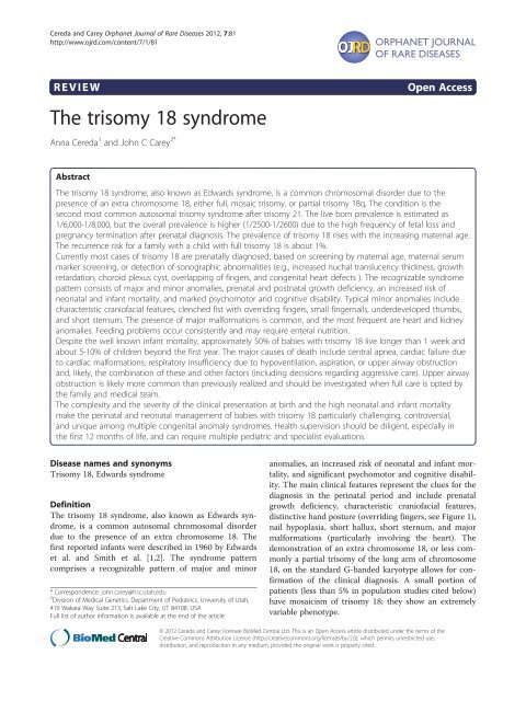
<strong>The</strong> <strong>trisomy</strong> <strong>18</strong> <strong>syndrome</strong><br />
Anna Cereda 1 and John C Carey 2*<br />
Abstract<br />
<strong>The</strong> <strong>trisomy</strong> <strong>18</strong> <strong>syndrome</strong>, also known as Edwards <strong>syndrome</strong>, is a common chromosomal disorder due to the<br />
presence <strong>of</strong> an extra chromosome <strong>18</strong>, either full, mosaic <strong>trisomy</strong>, or partial <strong>trisomy</strong> <strong>18</strong>q. <strong>The</strong> condition is the<br />
second most common autosomal <strong>trisomy</strong> <strong>syndrome</strong> after <strong>trisomy</strong> 21. <strong>The</strong> live born prevalence is estimated as<br />
1/6,000-1/8,000, but the overall prevalence is higher (1/2500-1/2600) due to the high frequency <strong>of</strong> fetal loss and<br />
pregnancy termination after prenatal diagnosis. <strong>The</strong> prevalence <strong>of</strong> <strong>trisomy</strong> <strong>18</strong> rises with the increasing maternal age.<br />
<strong>The</strong> recurrence risk for a family with a child with full <strong>trisomy</strong> <strong>18</strong> is about 1%.<br />
Currently most cases <strong>of</strong> <strong>trisomy</strong> <strong>18</strong> are prenatally diagnosed, based on screening by maternal age, maternal serum<br />
marker screening, or detection <strong>of</strong> sonographic abnormalities (e.g., increased nuchal translucency thickness, growth<br />
retardation, choroid plexus cyst, overlapping <strong>of</strong> fingers, and congenital heart defects ). <strong>The</strong> recognizable <strong>syndrome</strong><br />
pattern consists <strong>of</strong> major and minor anomalies, prenatal and postnatal growth deficiency, an increased risk <strong>of</strong><br />
neonatal and infant mortality, and marked psychomotor and cognitive disability. Typical minor anomalies include<br />
characteristic crani<strong>of</strong>acial features, clenched fist with overriding fingers, small fingernails, underdeveloped thumbs,<br />
and short sternum. <strong>The</strong> presence <strong>of</strong> major malformations is common, and the most frequent are heart and kidney<br />
anomalies. Feeding problems occur consistently and may require enteral nutrition.<br />
Despite the well known infant mortality, approximately 50% <strong>of</strong> babies with <strong>trisomy</strong> <strong>18</strong> live longer than 1 week and<br />
about 5-10% <strong>of</strong> children beyond the first year. <strong>The</strong> major causes <strong>of</strong> death include central apnea, cardiac failure due<br />
to cardiac malformations, respiratory insufficiency due to hypoventilation, aspiration, or upper airway obstruction<br />
and, likely, the combination <strong>of</strong> these and other factors (including decisions regarding aggressive care). Upper airway<br />
obstruction is likely more common than previously realized and should be investigated when full care is opted by<br />
the family and medical team.<br />
<strong>The</strong> complexity and the severity <strong>of</strong> the clinical presentation at birth and the high neonatal and infant mortality<br />
make the perinatal and neonatal management <strong>of</strong> babies with <strong>trisomy</strong> <strong>18</strong> particularly challenging, controversial,<br />
and unique among multiple congenital anomaly <strong>syndrome</strong>s. Health supervision should be diligent, especially in<br />
the first 12 months <strong>of</strong> life, and can require multiple pediatric and specialist evaluations.<br />
Disease names and synonyms<br />
Trisomy <strong>18</strong>, Edwards <strong>syndrome</strong><br />
Definition<br />
<strong>The</strong> <strong>trisomy</strong> <strong>18</strong> <strong>syndrome</strong>, also known as Edwards <strong>syndrome</strong>,<br />
is a common autosomal chromosomal disorder<br />
due to the presence <strong>of</strong> an extra chromosome <strong>18</strong>. <strong>The</strong><br />
first reported infants were described in 1960 by Edwards<br />
et al. and Smith et al. [1,2]. <strong>The</strong> <strong>syndrome</strong> pattern<br />
comprises a recognizable pattern <strong>of</strong> major and minor<br />
* Correspondence: john.carey@hsc.utah.edu<br />
2 Division <strong>of</strong> Medical Genetics, Department <strong>of</strong> Pediatrics, University <strong>of</strong> Utah,<br />
419 Wakara Way Suite 213, Salt Lake City, UT 84108, USA<br />
Full list <strong>of</strong> author information is available at the end <strong>of</strong> the article<br />
anomalies, an increased risk <strong>of</strong> neonatal and infant mortality,<br />
and significant psychomotor and cognitive disability.<br />
<strong>The</strong> main clinical features represent the clues for the<br />
diagnosis in the perinatal period and include prenatal<br />
growth deficiency, characteristic crani<strong>of</strong>acial features,<br />
distinctive hand posture (overriding fingers, see Figure 1),<br />
nail hypoplasia, short hallux, short sternum, and major<br />
malformations (particularly involving the heart). <strong>The</strong><br />
demonstration <strong>of</strong> an extra chromosome <strong>18</strong>, or less commonly<br />
a partial <strong>trisomy</strong> <strong>of</strong> the long arm <strong>of</strong> chromosome<br />
<strong>18</strong>, on the standard G-banded karyotype allows for confirmation<br />
<strong>of</strong> the clinical diagnosis. A small portion <strong>of</strong><br />
patients (less than 5% in population studies cited below)<br />
have mosaicism <strong>of</strong> <strong>trisomy</strong> <strong>18</strong>; they show an extremely<br />
variable phenotype.<br />
© 2012 Cereda and Carey; licensee BioMed Central Ltd. This is an Open Access article distributed under the terms <strong>of</strong> the<br />
Creative Commons Attribution License (http://creativecommons.org/licenses/by/2.0), which permits unrestricted use,<br />
distribution, and reproduction in any medium, provided the original work is properly cited.
Cereda and Carey <strong>Orphanet</strong> <strong>Journal</strong> <strong>of</strong> <strong>Rare</strong> <strong>Diseases</strong> 2012, 7:81 Page 2 <strong>of</strong> 14<br />
http://www.ojrd.com/content/7/1/81<br />
Figure 1 A boy with full <strong>trisomy</strong> <strong>18</strong> in early infancy and at<br />
one year. Note the characteristic hand feature with the over-riding<br />
fingers, the tracheostomy, and his engaging smile. He is now over 2<br />
years <strong>of</strong> age and is quite stable medically, gaining weight, sitting up,<br />
and participating in the many activities <strong>of</strong> his family.<br />
Epidemiology<br />
Trisomy <strong>18</strong> is the second most common autosomal <strong>trisomy</strong><br />
<strong>syndrome</strong> after <strong>trisomy</strong> 21.<br />
Several population studies have been performed in<br />
different countries including Australia, Europe and<br />
North America that estimate the prevalence <strong>of</strong> <strong>trisomy</strong><br />
<strong>18</strong> [3-9]. On the basis <strong>of</strong> these investigations the live<br />
birth prevalence <strong>of</strong> <strong>trisomy</strong> <strong>18</strong> ranges from 1/3600 to<br />
1/10,000 with the best overall estimate in liveborns as<br />
1 in 6,000 [3,6].<br />
It is well known that <strong>trisomy</strong> <strong>18</strong> pregnancies have a<br />
high risk <strong>of</strong> fetal loss and stillbirth [10,11]; furthermore,<br />
currently most diagnoses are made in the prenatal<br />
period based on screening by maternal age or maternal<br />
serum marker screening and amniocentesis, followed by<br />
pregnancy termination in a significant percentage <strong>of</strong><br />
cases [9]. Because <strong>of</strong> this, the overall prevalence (considering<br />
stillborn infants, terminated pregnancies, and liveborn<br />
infants) <strong>of</strong> <strong>trisomy</strong> <strong>18</strong> would be expected to be<br />
higher than live birth prevalence. A seminal population<br />
study in the United Kingdom in 1996 reported an overall<br />
prevalence <strong>of</strong> 1/4272 and a liveborn prevalence <strong>of</strong><br />
1/8333 [4]; the overall frequency detected in Hawaii<br />
from a similar study was 1/2123 with a liveborn frequency<br />
<strong>of</strong> 1/7900 [5]. Recent investigations showed an<br />
increase <strong>of</strong> the overall prevalence <strong>of</strong> <strong>trisomy</strong> <strong>18</strong> over<br />
the last 20 years due to increased maternal age [9]; however,<br />
a decrease <strong>of</strong> liveborn frequency was observed<br />
because <strong>of</strong> the increased use <strong>of</strong> prenatal diagnosis and<br />
the high rate <strong>of</strong> pregnancy termination after the prenatal<br />
diagnosis [7,9]. In these more recent studies overall<br />
prevalence was estimated as 1/2500 in United States [7]<br />
and as 1/2600 in United Kingdom [9]; liveborn prevalence<br />
was estimated as 1/8600 in United States [7] and<br />
as 1/10,000 in United Kingdom [9].<br />
<strong>The</strong> prevalence at birth is higher in females compared<br />
to males (F:M %, 60.4), but this discordance is not<br />
present if the sex ratio is calculated among fetuses electively<br />
terminated (F:M % 48:51.) [7]. Moreover the frequency<br />
<strong>of</strong> fetal loss is higher for males compared to<br />
females [10,11]. Furthermore, liveborn females showed<br />
better survival compared to males [4,6].<br />
Etiology and pathogenesis<br />
<strong>The</strong> <strong>trisomy</strong> <strong>18</strong> (or Edwards <strong>syndrome</strong>) phenotype results<br />
from full, mosaic, or partial <strong>trisomy</strong> <strong>18</strong>q [4,12-15].<br />
Complete or full <strong>trisomy</strong> <strong>18</strong> is the most common form<br />
(about 94% <strong>of</strong> cases); in this situation every cell contains<br />
three entire copies <strong>of</strong> chromosome <strong>18</strong>.<br />
Most authorities have suggested that the extra<br />
chromosome is present because <strong>of</strong> nondisjunction. In<br />
parent-<strong>of</strong>-origin analyses the extra chromosome is most<br />
<strong>of</strong>ten <strong>of</strong> maternal origin, the result <strong>of</strong> an error during<br />
the segregation <strong>of</strong> chromosomes in meiosis or postzygotic<br />
mitosis. About 50% <strong>of</strong> the nondisjunctional errors<br />
in oogenesis occur in meiosis II, unlike other human<br />
trisomies where the malsegregation is more frequent in<br />
meiosis I [16-19]. In the minority <strong>of</strong> cases in which the<br />
extra chromosome has a paternal origin, the error is<br />
the result <strong>of</strong> a postzygotic error. <strong>The</strong> cause <strong>of</strong> nondisjunction<br />
is unknown. Recently a higher prevalence<br />
<strong>of</strong> methylene tetrahydr<strong>of</strong>olate reductase gene (MTHFR)<br />
polymorphisms in mothers <strong>of</strong> <strong>trisomy</strong> <strong>18</strong> fetuses compared<br />
with other groups was reported [20] but this result<br />
has not been replicated.<br />
As in the other common autosomal trisomies, the frequency<br />
<strong>of</strong> nondisjunctional errors increases with advancing<br />
maternal age. Savva et al., studied the maternal age<br />
specific live birth prevalence <strong>of</strong> <strong>trisomy</strong> <strong>18</strong>: the frequency<br />
is constant until age 30, then increases exponentially<br />
before beginning to become constant again at age<br />
45 [21]. <strong>The</strong> observed increased overall prevalence <strong>of</strong> <strong>trisomy</strong><br />
<strong>18</strong> in the last years is likely due to changes in the<br />
maternal age distribution during this time period [9,21].<br />
A small positive association <strong>of</strong> paternal age with <strong>trisomy</strong><br />
<strong>18</strong>, similar to that observed in Down <strong>syndrome</strong>, has also<br />
been observed [22].<br />
In individuals carrying mosaic <strong>trisomy</strong> <strong>18</strong> (less than<br />
5% <strong>of</strong> cases), both a complete <strong>trisomy</strong> <strong>18</strong> and a normal<br />
cell line exist. <strong>The</strong> phenotype is extremely variable, ranging<br />
from complete <strong>trisomy</strong> <strong>18</strong> phenotype with early<br />
mortality to apparently phenotypically normal adults, in<br />
which the mosaicism is detected after the diagnosis<br />
<strong>of</strong> complete <strong>trisomy</strong> <strong>18</strong> in a child [23-27]. <strong>The</strong>re is no<br />
correlation between the percentage <strong>of</strong> <strong>trisomy</strong> <strong>18</strong> cells in<br />
either blood cells or skin fibroblasts and the severity <strong>of</strong><br />
clinical manifestations and intellectual disabilities [24].<br />
Tucker et al., [24] provided a comprehensive review <strong>of</strong>
Cereda and Carey <strong>Orphanet</strong> <strong>Journal</strong> <strong>of</strong> <strong>Rare</strong> <strong>Diseases</strong> 2012, 7:81 Page 3 <strong>of</strong> 14<br />
http://www.ojrd.com/content/7/1/81<br />
all published cases <strong>of</strong> <strong>trisomy</strong> <strong>18</strong> mosaicism in their recent<br />
paper and reported on 3 new cases.<br />
In the partial <strong>trisomy</strong> form only a segment <strong>of</strong> the<br />
chromosome <strong>18</strong> long arm is present in triplicate, <strong>of</strong>ten<br />
resulting from a balanced translocation or inversion carried<br />
by one parent. This type <strong>of</strong> <strong>trisomy</strong> accounts for approximately<br />
2% <strong>of</strong> cases presenting with the Edwards<br />
phenotype. <strong>The</strong> location and the extent <strong>of</strong> the triplicated<br />
segment and the possible associated deletion <strong>of</strong> genomic<br />
material due to unbalanced translocation can explain the<br />
variable phenotype associated with partial <strong>trisomy</strong> [12].<br />
<strong>The</strong> region <strong>of</strong> long arm <strong>of</strong> chromosome <strong>18</strong> extending<br />
from q11.2 has been proposed as the critical region for<br />
<strong>trisomy</strong> <strong>18</strong> phenotype, but some controversial data have<br />
been reported [28,29]. Boghosian-Sell et al. hypothesized<br />
the presence <strong>of</strong> two critical regions along the long arm<br />
<strong>of</strong> chromosome <strong>18</strong>: one proximal region lying within<br />
<strong>18</strong>q12.1-<strong>18</strong>q21.2 and another one more distal lying<br />
within <strong>18</strong>q22.33-<strong>18</strong>qter [29]. <strong>The</strong> same authors reported<br />
two patients with <strong>trisomy</strong> <strong>of</strong> <strong>18</strong>q11.2 to terminus not<br />
showing the complete pattern <strong>of</strong> <strong>trisomy</strong> <strong>18</strong>; the patients<br />
had better survival and growth. <strong>The</strong>refore, some role for<br />
genes on the short arm or <strong>18</strong>q11.1 region in the expression<br />
<strong>of</strong> full phenotype cannot be excluded.<br />
Antenatal diagnosis<br />
Currently in the North America and Europe most cases<br />
<strong>of</strong> <strong>trisomy</strong> <strong>18</strong> are prenatally diagnosed, based on screening<br />
by maternal age, maternal serum marker screening,<br />
or detection <strong>of</strong> sonographic abnormalities during the<br />
second and third trimester [9,30]. <strong>The</strong> prenatal diagnosis<br />
<strong>of</strong> <strong>trisomy</strong> <strong>18</strong> leads to the decision <strong>of</strong> pregnancy termination<br />
in 86% <strong>of</strong> cases [9]. Knowledge <strong>of</strong> the survival<br />
where termination is not chosen is important as well,<br />
because the parents will seek this information and this<br />
knowledge can influence the management at the time <strong>of</strong><br />
delivery and in the neonatal period [12].<br />
First trimester non invasive screening based on maternal<br />
age, serum markers and sonographic “s<strong>of</strong>t markers”<br />
demonstrated high sensitivity for diagnosis <strong>of</strong> <strong>trisomy</strong> <strong>18</strong><br />
[31-33], and it is now being applied routinely. <strong>The</strong> levels<br />
<strong>of</strong> human chorionic gonadotropin, unconjugated estriol,<br />
and alpha-fetoprotein are significantly lower in pregnancies<br />
with <strong>trisomy</strong> <strong>18</strong> compared to normal pregnancy<br />
[31]. <strong>The</strong> most common s<strong>of</strong>t sonographic markers<br />
detected in the late first/early second trimester are the<br />
increased nuchal translucency thickness and the absence<br />
or hypoplasia <strong>of</strong> the nasal bone [34-36]; the screening by<br />
assessment <strong>of</strong> nuchal fold and nasal bone identifies<br />
66.7% <strong>of</strong> cases with <strong>trisomy</strong> <strong>18</strong> (and 13) [36]. By including<br />
the evaluation <strong>of</strong> reversed flow in the ductus venosus<br />
and the tricuspid valve regurgitation, the detection rate<br />
increases to 83.3% [36]. Furthermore, some structural<br />
anomalies can be detected by ultrasound screening<br />
during the first trimester; the most common are omphalocele<br />
(21%), abnormal posturing <strong>of</strong> the hands (6%),<br />
megacystis (4%) and abnormal four-chamber view <strong>of</strong> the<br />
heart (4%) [35]. Early-onset fetal growth retardation can<br />
be detected in 26% <strong>of</strong> cases [36], but becomes more evident<br />
in the second trimester [30,37]. <strong>The</strong> detection<br />
rate <strong>of</strong> combined late first trimester screening (nuchal<br />
translucency, pregnancy-associated plasma protein and<br />
free beta-hCG) and second trimester quadruple screening<br />
(serum alpha-fetoprotein, total hCG, unconjugated<br />
estriol and inhibin A) is at least 78% sensitive [32,33].<br />
Many studies have been published in the last 15 years<br />
regarding the prenatal pattern <strong>of</strong> ultrasound findings in<br />
<strong>trisomy</strong> <strong>18</strong> fetuses in the second and third trimester<br />
[30,35-39]. One or more sonographic anomalies are<br />
detected in over 90% <strong>of</strong> fetuses; two or more abnormalities<br />
are present in 55% <strong>of</strong> cases [38]. <strong>The</strong> prenatal sonographic<br />
pattern <strong>of</strong> <strong>trisomy</strong> <strong>18</strong> is characterized by growth<br />
retardation, polyhydramnios, “strawberry-shaped” cranium<br />
(brachycephaly and narrow frontal cranium), choroid<br />
plexus cyst, overlapping <strong>of</strong> hands fingers (second<br />
and fifth on third and fourth respectively), congenital<br />
heart defects, omphalocele, and single umbilical artery<br />
[30,35-39]. <strong>The</strong> prevalence <strong>of</strong> growth retardation and<br />
polyhydramnios increases with gestational age: 28% and<br />
29% in the second trimester and 87% and 62% in the<br />
third trimester, respectively [37]. More than 30% <strong>of</strong><br />
fetuses show hands abnormalities [39], and one third <strong>of</strong><br />
cases have a single umbilical artery [37]. Furthermore,<br />
the mothers <strong>of</strong>ten noted a decrease in fetal movement<br />
compared to their normal pregnancies [37]. Choroid<br />
plexus cyst (CPC) is detected in about 50% <strong>of</strong> <strong>trisomy</strong> <strong>18</strong><br />
fetuses [39]; in the most <strong>of</strong> cases (80-90%) it is associated<br />
with other sonographic anomalies [37,39], but in<br />
a small percentage <strong>of</strong> pregnancies (11% according to<br />
Cho et al. 2010) carrying <strong>trisomy</strong> <strong>18</strong> fetus, CPC can be<br />
the only abnormality detected at ultrasound screening.<br />
Choroid plexus cyst can be also a transitory finding in<br />
normal fetuses; it has been reported that, among fetuses<br />
that show CPC at second trimester sonographic screening,<br />
only about 5% have <strong>trisomy</strong> <strong>18</strong> [37,40,41]. Because<br />
<strong>of</strong> these reasons, there is not a clear consensus in the<br />
medical literature on whether to <strong>of</strong>fer amniocentesis<br />
after the discovery <strong>of</strong> choroid cyst, particularly when it is<br />
an isolated finding [37,42-46].<br />
Trisomy <strong>18</strong> pregnancies have a high risk <strong>of</strong> fetal loss<br />
and stillbirth [10,11,37]. <strong>The</strong> probability <strong>of</strong> survival to<br />
term increases with the increase <strong>of</strong> gestational age: 28%<br />
at 12 weeks, 35% at <strong>18</strong> weeks and 41% at 20 weeks [10].<br />
Fetal losses are uniformly distributed throughout gestation<br />
after 24 weeks without a clustering <strong>of</strong> fetal demises<br />
at a particular gestational age [11,37]. Cases detected by<br />
abnormal sonographic findings are more likely to result<br />
in a miscarriage or stillbirth [37]. Furthermore, the
Cereda and Carey <strong>Orphanet</strong> <strong>Journal</strong> <strong>of</strong> <strong>Rare</strong> <strong>Diseases</strong> 2012, 7:81 Page 4 <strong>of</strong> 14<br />
http://www.ojrd.com/content/7/1/81<br />
frequency <strong>of</strong> miscarriage or stillbirth is higher (up to<br />
tw<strong>of</strong>old according to Niedrist et al., [47]) for males compared<br />
to females [10,47].<br />
Genetic counseling<br />
When prenatal or neonatal diagnosis <strong>of</strong> <strong>trisomy</strong> <strong>18</strong> is<br />
made, the counseling <strong>of</strong> the family should be realistic,<br />
but not desolate. <strong>The</strong> parents can find it difficult to<br />
accept the lack <strong>of</strong> certainty <strong>of</strong> the newborn situation,<br />
but they have to be prepared for both the probability<br />
<strong>of</strong> death and the possibility <strong>of</strong> living [48]. Because the<br />
parents have to make practical decisions concerning<br />
resuscitation, surgery and life support, all options for<br />
newborn management should be explained. <strong>The</strong> complex<br />
issues regarding perinatal management are covered<br />
in more detail below.<br />
Facilitating the family getting in touch with family support<br />
groups can be helpful: they can share experiences,<br />
thoughts and concerns regarding health problems <strong>of</strong><br />
their children, and daily situations that they are coping<br />
with. Table 1 shows the known support groups in North<br />
America, Europe, Japan, and Australia.<br />
<strong>The</strong> recurrence risk, for a family with a child with<br />
complete <strong>trisomy</strong> <strong>18</strong> is usually stated as 1% [12]. Parental<br />
mosaicism has been reported in a few cases [24-27].<br />
Furthermore, recurrence <strong>of</strong> different trisomies in the<br />
same family has been reported [49]. Empirically calculated<br />
risks suggest that the recurrence risk seems to<br />
be less than 1%, but higher than the age-specific background<br />
risk [50,51]. <strong>The</strong> recurrence risk in families<br />
with partial <strong>trisomy</strong> <strong>18</strong> could be higher compared with<br />
full <strong>trisomy</strong> <strong>18</strong>, depending on the presence <strong>of</strong> a genomic<br />
rearrangement (translocation or inversion) in one <strong>of</strong><br />
the parents.<br />
Clinical description<br />
<strong>The</strong> clinical pattern <strong>of</strong> <strong>trisomy</strong> <strong>18</strong> is characterized by<br />
prenatal growth deficiency, specific crani<strong>of</strong>acial features<br />
and other minor anomalies, major malformations, and<br />
marked psychomotor and cognitive developmental delay.<br />
<strong>The</strong> growth delay starts in prenatal period and continues<br />
after the birth, and most <strong>of</strong> the time is associated<br />
with feeding problems that may require enteral<br />
nutrition. Specific growth charts for <strong>trisomy</strong> <strong>18</strong> are<br />
available [49] and are published on the SOFT US and<br />
UK web pages (see Table 1) for printing and placement<br />
in the child’s chart. Postnatal onset microcephaly is usually<br />
present.<br />
Typical crani<strong>of</strong>acial features include dolichocephaly,<br />
short palpebral fissures, micrognathia, (see Figure 1)<br />
external anomalies <strong>of</strong> the ears, and redundant skin at<br />
the back <strong>of</strong> the neck.<br />
Other characteristic clinical findings are the clenched<br />
fist with overriding fingers (index finger overlapping the<br />
third and 5th finger overlapping the 4 th –see Figure 1),<br />
which is particularly distinctive, small fingernails, underdeveloped<br />
thumbs, short sternum, and club feet. Presence<br />
<strong>of</strong> major malformations is common, and any organ<br />
and system can be affected. Structural heart defects<br />
occur in over 90% <strong>of</strong> infants. Table 2 summarizes the<br />
most common major (medically significant) malformations<br />
detected in <strong>trisomy</strong><strong>18</strong> from various sources.<br />
Differential diagnosis<br />
<strong>The</strong> clinical pattern <strong>of</strong> <strong>trisomy</strong> <strong>18</strong> is quite well-defined,<br />
and it is rarely misdiagnosed [12]. <strong>The</strong>re are some overlapping<br />
features with Pena-Shokeir <strong>syndrome</strong> type 1 or<br />
<strong>syndrome</strong>s with fetal akinesia sequence (because <strong>of</strong> polyhydramnios<br />
and joint contractures including overriding<br />
fingers), with distal arthrogryposis type 1 (because <strong>of</strong><br />
the similar finger positioning) and with CHARGE <strong>syndrome</strong><br />
(because <strong>of</strong> the overlapping <strong>of</strong> major malformations).<br />
<strong>The</strong> not well characterized and co-called<br />
condition known as pseudo<strong>trisomy</strong> <strong>18</strong> <strong>syndrome</strong> [53]<br />
probably belongs to the group <strong>of</strong> disorders with fetal<br />
akinesia sequence.<br />
Table 1 International parent support groups for <strong>trisomy</strong> <strong>18</strong><br />
Country Support Group Web site<br />
Australia SOFT <strong>of</strong> Australia http://members.optushome.com.au/s<strong>of</strong>taus<br />
Europe Chromosome <strong>18</strong> Registry and Research Society (Europe) http://www.chromosome<strong>18</strong>eur.org<br />
France Valentin APAC Association de Porteurs d'Anomalies Chromosomiques http://www.valentin-apac.org<br />
Germany LEONA e.V. - Verein für Eltern chromosomal geschädigter Kinder http://www.leona-ev.de<br />
Ireland SOFT <strong>of</strong> Ireland http://s<strong>of</strong>tireland.com<br />
Italy SOFT Italia http://www.trisomia.org/index.html<br />
Japan <strong>The</strong> Trisomy <strong>18</strong> Support Group http://<strong>18</strong><strong>trisomy</strong>.com<br />
United Kingdom SOFT <strong>of</strong> United Kingdom http://www.s<strong>of</strong>t.org.uk<br />
United States USA Support Group SOFT http://www.<strong>trisomy</strong>.org<br />
United States Trisomy <strong>18</strong> Foundation http://www.<strong>trisomy</strong><strong>18</strong>.org<br />
United States <strong>The</strong> Chromosome <strong>18</strong> Registry and Research Society http://www.chromosome<strong>18</strong>.org
Cereda and Carey <strong>Orphanet</strong> <strong>Journal</strong> <strong>of</strong> <strong>Rare</strong> <strong>Diseases</strong> 2012, 7:81 Page 5 <strong>of</strong> 14<br />
http://www.ojrd.com/content/7/1/81<br />
Table 2 Common major structural malformations in the <strong>trisomy</strong> <strong>18</strong> <strong>syndrome</strong><br />
Frequency Organ/System Prevalent type <strong>of</strong> malformation<br />
Common (>75%) heart septal defects, patent ductus arteriosus, and polyvalvular disease<br />
Frequent (25-75%) genitourinary horseshoe kidney<br />
Less frequent (5-25%) gastrointestinal omphalocele, esophageal atresia with tracheo-esophageal fistula,<br />
pyloric stenosis, Meckel diverticulum<br />
central nervous system cerebellar hypoplasia, agenesis <strong>of</strong> corpus callosum, polymicrogyria,<br />
spina bifida<br />
crani<strong>of</strong>acial or<strong>of</strong>acial clefts<br />
eye microphthalmia, coloboma, cataract, corneal opacities<br />
limb radial aplasia/hypoplasia<br />
From Jones [52], Baty et al. [49].<br />
Natural history/prognosis<br />
Survival after birth and neonatal management<br />
Perinatal and neonatal management <strong>of</strong> fetuses and newborn<br />
diagnosed with <strong>trisomy</strong> <strong>18</strong> is multifaceted issue for<br />
a variety <strong>of</strong> reasons: the complexity and, most <strong>of</strong> the<br />
time, the severity <strong>of</strong> the clinical presentation at birth; the<br />
need <strong>of</strong> parents and care providers to urgently make<br />
decisions in care <strong>of</strong> the baby; the inevitable ethical implications<br />
due to the well known high neonatal and infant<br />
mortality, and the significant developmental disability<br />
in the surviving children that characterize this unique<br />
(together with <strong>trisomy</strong> 13) condition.<br />
<strong>The</strong>re is a high percentage <strong>of</strong> fetuses dying during<br />
labor (38.5%), and the preterm frequency (35%) is higher<br />
compared to general population [30]. An increased incidence<br />
<strong>of</strong> cesareans has been reported [4,54], even if in<br />
the previous obstetric literature avoidance <strong>of</strong> delivery by<br />
cesarean was recommended [55,56].<br />
<strong>The</strong> first study about postnatal survival <strong>of</strong> children<br />
with <strong>trisomy</strong> <strong>18</strong> was published in 1967: Weber reported<br />
a mean survival <strong>of</strong> 70 days [57]. Most <strong>of</strong> the ensuing<br />
population studies showed a shorter survival, likely because,<br />
with prenatal and neonatal diagnosis, it is now<br />
possible to diagnose many cases, which would have died<br />
prior to detection in the past [3].<br />
Most recent studies report a median survival <strong>of</strong> 3-14.5<br />
days, a percentage <strong>of</strong> survival at 24 hours <strong>of</strong> 60%-75%,<br />
at 1 week <strong>of</strong> 40%-60%, at 1 month <strong>of</strong> 22%-44%, at<br />
6 months <strong>of</strong> 9%-<strong>18</strong>%, and after 1 year <strong>of</strong> 5%-10%<br />
[3,4,6,12,13,15,49,54,58-62]. To summarize, approximately<br />
50% <strong>of</strong> babies with <strong>trisomy</strong> <strong>18</strong> live longer than 1 week,<br />
and 5-10% <strong>of</strong> children survive beyond the first year.<br />
Because these figures document that 1 in 10 to 1 in 20<br />
babies live to their first birthday, the commonly used<br />
term, “lethal abnormality”, is inaccurate, misleading, and<br />
inappropriate [12].<br />
<strong>The</strong> major causes <strong>of</strong> death are sudden death due<br />
to central apnea, cardiac failure due to cardiac malforxmations<br />
and respiratory insufficiency due to<br />
hypoventilation, aspiration, upper airway obstruction or,<br />
likely, the combination <strong>of</strong> these and other factors<br />
[4,12,13,15,49,54,58,59,63-65]. A recent study reported<br />
a >100 times higher risk <strong>of</strong> mortality in neonatal period<br />
and in the first years <strong>of</strong> life for children with <strong>trisomy</strong> <strong>18</strong><br />
compared to infants born without birth defects [8].<br />
Upper airway obstruction is likely more common than<br />
previously realized and should be investigated when<br />
full care is opted by the family and medical team. <strong>The</strong><br />
factors underlying the potential <strong>of</strong> survival are not<br />
known; the presence <strong>of</strong> heart defects does not seem<br />
to affect long-term survival [6]. However a recent trend<br />
toward consideration <strong>of</strong> performing cardiac surgery<br />
may alter that premise as surgery may play a role in preventing<br />
pulmonary hypertension, a point not investigated<br />
in determining the notion that heart defects do<br />
not affect survival [6]. A longer survival for females<br />
compared to males has been reported, as in the prenatal<br />
period [4,6].<br />
Because <strong>of</strong> the elevated risk <strong>of</strong> mortality in the first<br />
month <strong>of</strong> life and the presence <strong>of</strong> significant developmental<br />
disability in the surviving children, historically<br />
there has been a consensus among care providers that<br />
<strong>trisomy</strong> <strong>18</strong> be considered a condition for which non<br />
intervention in the newborn was indicated [65,66].<br />
Nevertheless, the most recent American Academy <strong>of</strong><br />
Pediatrics neonatal resuscitation guidelines omit <strong>trisomy</strong><br />
<strong>18</strong> from the list <strong>of</strong> examples <strong>of</strong> conditions for which<br />
resuscitation is not indicated [67]. A recent survey <strong>of</strong><br />
the opinion <strong>of</strong> American neonatologists on newborn<br />
care <strong>of</strong> <strong>trisomy</strong> <strong>18</strong> infants reported that 44% would<br />
intervene mostly because <strong>of</strong> parental wishes to support<br />
the baby [68].<br />
A recent Japanese study documented the survival rate<br />
in a group <strong>of</strong> <strong>trisomy</strong> <strong>18</strong> newborn to which intensive<br />
care were <strong>of</strong>fered: the median survival time (152.5 days)<br />
and survival rate at 12 months [25%] were higher compared<br />
to those reported in the previous studies, but the<br />
survival over 2 years (4%) was similar to the 5-10% usually<br />
reported as 1-year survival rate [54]. To our knowledge<br />
this is the only study that addresses the question<br />
<strong>of</strong> infant survival if full intervention (short <strong>of</strong> cardiac<br />
surgery) is <strong>of</strong>fered.
Cereda and Carey <strong>Orphanet</strong> <strong>Journal</strong> <strong>of</strong> <strong>Rare</strong> <strong>Diseases</strong> 2012, 7:81 Page 6 <strong>of</strong> 14<br />
http://www.ojrd.com/content/7/1/81<br />
In this study the authors also investigated the pathophysiology<br />
to death in patients who had intensive treatment;<br />
they distinguish between underlying factors<br />
associated with death and final modes <strong>of</strong> death. <strong>The</strong><br />
common underlying factors associated with death were<br />
congenital heart defects and heart failure, and pulmonary<br />
hypertension. On the other hand, the final<br />
modes <strong>of</strong> death were sudden cardiac or cardiopulmonary<br />
arrest and events related to progressive pulmonary<br />
hypertension [54]. From these observations, it becomes<br />
clear that apnea and withdrawal <strong>of</strong> treatment could be<br />
considered the major cause <strong>of</strong> death when a patient with<br />
<strong>trisomy</strong> <strong>18</strong> was managed with purely comfort care.<br />
When a patient with <strong>trisomy</strong> <strong>18</strong> has intensive treatment,<br />
the common causes <strong>of</strong> death are altered, and survival<br />
does increase.<br />
<strong>The</strong> senior author had pointed out in an Editorial [69]<br />
in 2006 that there existed a dire need to have a dialogue<br />
regarding the ethical issues surrounding the management<br />
and care <strong>of</strong> infants and children with <strong>trisomy</strong> <strong>18</strong>.<br />
Such a dialogue seems to be occurring in recent years:<br />
the publication <strong>of</strong> the McGraw and Perlman paper [68]<br />
mentioned above and the Ethics Rounds, a Special Article<br />
in Pediatrics in 2011 [70], both discuss the key<br />
themes and controversies that needed current discussion.<br />
<strong>The</strong> former paper indicated that the majority <strong>of</strong><br />
neonatologists polled in the study would not resuscitate<br />
a newborn in the delivery room who had <strong>trisomy</strong> <strong>18</strong> and<br />
a heart defect. <strong>The</strong> authors stated a concern about a<br />
trend away from the “best interest <strong>of</strong> the child” standard<br />
and towards parental opinion. In the more recent<br />
Special Article two neonatologists and a parent discuss<br />
their views on the management <strong>of</strong> a baby with <strong>trisomy</strong><br />
<strong>18</strong> and a heart defect surrounding the decision to have<br />
cardiac surgery [70]. While the doctors and the parent<br />
disagreed on many points, one <strong>of</strong> the doctors and the<br />
Editor state that “deference to the parents” is generally<br />
the best course (unless the child is “suffering” from the<br />
ongoing treatment) in situations <strong>of</strong> unclear outcome.<br />
<strong>The</strong>se papers and the published responses to them in<br />
Pediatrics suggest that a dialogue is in fact now occurring.<br />
Another recently published paper by Wilfond and Carey<br />
[71], a case-based discussion <strong>of</strong> the issues and themes<br />
involved in the management <strong>of</strong> <strong>trisomy</strong> <strong>18</strong> (and related<br />
conditions), also illustrates this point <strong>of</strong> an emerging dialogue.<br />
<strong>The</strong> reader is referred to these papers for further<br />
discussion <strong>of</strong> the relevant issues.<br />
One <strong>of</strong> the key themes at the center <strong>of</strong> the controversy<br />
is the question <strong>of</strong> so-called “quality <strong>of</strong> life” <strong>of</strong> children<br />
and their families when a child has <strong>trisomy</strong> <strong>18</strong>. We will<br />
discuss this issue in the Unresolved Questions section<br />
below as little data exist in the scientific literature on<br />
this topic.<br />
Growth and feeding<br />
Prenatal growth retardation is one <strong>of</strong> the most frequent<br />
prenatal finding in <strong>trisomy</strong> <strong>18</strong> [30,35-39]; the mean birth<br />
weight is 1700-<strong>18</strong>00 g at a mean gestational age <strong>of</strong> 37<br />
weeks [4,54]. Weight and height continue to be below<br />
the third centile in the postnatal period; growth charts<br />
specific for the condition has been published [49] and<br />
are available on the SOFT web pages for the both the<br />
US and UK support groups (see Table 1). Head circumference<br />
also tends to be below the third centile [12].<br />
Most <strong>of</strong> the children have feeding difficulties that <strong>of</strong>ten<br />
require tube feeding in the neonatal period or placement<br />
<strong>of</strong> gastrostomy in the older children (at average age <strong>of</strong> 8<br />
months) [49]. Both sucking and swallowing problems<br />
can be present. Usually the skill <strong>of</strong> oral feeding if<br />
achieved is achieved in infancy, and not later [12,49].<br />
If it is unclear if an infant can or cannot protect her<br />
airway, a swallow study can be performed to determine<br />
the safety <strong>of</strong> oral feedings.<br />
Gastroesophageal reflux is a significant medical problem<br />
because <strong>of</strong> both its high prevalence and its<br />
potential consequences, like irritability, recurrent pneumonia<br />
and aspiration [12]. Aspiration due to gastroesophageal<br />
reflux or during feeding is included among the<br />
causes <strong>of</strong> early death [4,12,13,49,54,58,59,61-63].<br />
Gastrointestinal malformations, such as esophageal<br />
atresia with tracheo-esophageal fistula, occur with<br />
increased frequency but are not a common feature in <strong>trisomy</strong><br />
<strong>18</strong>; pyloric stenosis has been reported and should<br />
be considered in the older infant with vomiting [12]. Occasionally<br />
the newborn with <strong>trisomy</strong> <strong>18</strong> can have or<strong>of</strong>acial<br />
clefts that may contribute to feeding problems [12].<br />
Cardiovascular<br />
Larger series <strong>of</strong> infants with the <strong>syndrome</strong> show that<br />
80%-100% <strong>of</strong> patients with <strong>trisomy</strong> <strong>18</strong> have congenital<br />
structural heart defects; the most common cardiac<br />
anomalies are ventricular and atrial septal defects, patent<br />
ductus arteriosus and polyvalvular disease [12,72-74].<br />
<strong>The</strong> majority <strong>of</strong> the malformations are unlikely to produce<br />
neonatal death; this is one <strong>of</strong> the reasons why the<br />
cardiac defect is usually regarded as not causing the<br />
early infant mortality. A more complex malformation<br />
(double-outlet right ventricle, endocardial cushion defect,<br />
or left-sided obstructive lesion) is present in about<br />
10% <strong>of</strong> cases [12], and then the cardiac defect could play<br />
a role in early mortality.<br />
<strong>The</strong> role <strong>of</strong> cardiac malformations in causing early<br />
death is controversial. Some studies reported that the<br />
presence <strong>of</strong> heart defect does not negatively affect the<br />
survival [6] and that the cardiac problems are not implicated<br />
in the deaths in most <strong>of</strong> patients [4]. Based on<br />
these data, cardiac surgery in the neonatal period is
Cereda and Carey <strong>Orphanet</strong> <strong>Journal</strong> <strong>of</strong> <strong>Rare</strong> <strong>Diseases</strong> 2012, 7:81 Page 7 <strong>of</strong> 14<br />
http://www.ojrd.com/content/7/1/81<br />
considered not likely to improve the survival <strong>of</strong> <strong>trisomy</strong><br />
<strong>18</strong> children. However, in other studies heart failure and<br />
early development <strong>of</strong> pulmonary hypertension induced<br />
by heart defects were found to play a significant role in<br />
early death [69,74-76].<br />
Traditionally, heart defects in <strong>trisomy</strong> <strong>18</strong> patients have<br />
been managed conservatively. Recent studies, however,<br />
showed that most patients (82-91%) with <strong>trisomy</strong> <strong>18</strong> can<br />
survive palliative and corrective heart surgeries, suggesting<br />
that heart surgery can be considered even in patients<br />
with <strong>trisomy</strong> <strong>18</strong> [76-79] (see “Health supervision and<br />
management <strong>of</strong> medical problems” for more details).<br />
Respiratory<br />
Respiratory problems are one <strong>of</strong> the most common<br />
causes <strong>of</strong> death in <strong>trisomy</strong> <strong>18</strong> [4,12,49,54,58,59,61,62].<br />
Pure respiratory problems, such as upper airway obstruction<br />
(in some case due to a laryngomalacia or tracheobronchomalacia)<br />
and central apnea, can act<br />
together with other problems <strong>of</strong> different origin, like<br />
early–onset pulmonary hypertension, feeding difficulties,<br />
recurrent aspirations and gastroesophageal reflux, leading<br />
to a severe respiratory symptoms [3,4]. Obstructive<br />
sleep apnea may be a more common finding in older<br />
infants [12] than realized.<br />
Ophthalmologic<br />
Many ocular findings have been reported in patients<br />
with <strong>trisomy</strong> <strong>18</strong>, although major ocular defects are<br />
present in a small group <strong>of</strong> children (less than 10%) [80].<br />
Occasionally, children with <strong>trisomy</strong> <strong>18</strong> can show anomalies<br />
such as a cataract or corneal opacities [81,82]. Short<br />
palpebral fissures, visual acuity abnormalities, and<br />
photophobia are common findings and underscore the<br />
need for ophthalmology assessment in older infants [12].<br />
Photophobia is very common in children with <strong>trisomy</strong><br />
<strong>18</strong> and requires sunglasses when going outside the<br />
home; it likely represents one reason why older infants<br />
experience unexplained irritability.<br />
Ears and hearing<br />
Structural ear anomalies, such as meatal atresia and<br />
microtia, are occasionally present. <strong>The</strong> features <strong>of</strong> external<br />
ear are characteristic: the ear is small with a small<br />
lobule, the helix is unfolded, simple and sometimes<br />
attached to the scalp (cryptotia) [12]. <strong>The</strong> ear canal is<br />
usually small making audiology screening sometimes<br />
challenging. A wide spectrum <strong>of</strong> middle and internal ear<br />
abnormalities has been described. Moderate to severe<br />
sensorineural hearing loss can also be present [12].<br />
Musculoskeletal<br />
Major malformations <strong>of</strong> limb occur in 5-10% <strong>of</strong> patients,<br />
including radial aplasia and other preaxial limb defects.<br />
About 50% <strong>of</strong> babies show positional foot deformities,<br />
both talipes equinovarus and calcaneovalgus. In addition,<br />
contractures <strong>of</strong> other joints can be present explaining<br />
why <strong>trisomy</strong> <strong>18</strong> is sometimes the basis for a neonate labeled<br />
artrogryposis. Overriding fingers (second and fifth<br />
on third and fourth respectively-see Figure 1) represent<br />
one <strong>of</strong> the important diagnostic clues, <strong>of</strong>ten detected<br />
sonographically in the prenatal period. Scoliosis is common<br />
in older children; usually it is not related to vertebral<br />
structural abnormalities and may progress between<br />
5 and 10 years <strong>of</strong> age [12].<br />
Genitourinary<br />
Horseshoe kidney is common finding in <strong>trisomy</strong> <strong>18</strong><br />
(about two-thirds <strong>of</strong> patients). An increased frequency<br />
<strong>of</strong> urinary tract infections has been observed, perhaps<br />
due to structural defects [31]. Otherwise, renal failure is<br />
uncommon [12].<br />
Neoplasia<br />
Trisomy <strong>18</strong> patients have an increased risk to develop<br />
some neoplasia, including Wilms tumor and hepatoblastoma<br />
[83]. At least 8 cases <strong>of</strong> Wilms tumor in <strong>trisomy</strong><br />
<strong>18</strong> children have been reported in the medical literature<br />
[83-89]. Nephroblastomatosis, the presence <strong>of</strong> multiple<br />
embryonic rests <strong>of</strong> tissue within the kidney that may give<br />
rise to Wilms tumor, has been detected at autopsy in<br />
infants with <strong>trisomy</strong> <strong>18</strong> who did not die from a Wilms<br />
tumor [88-90].<br />
Despite this biological origin, the average age <strong>of</strong> tumor<br />
development is 5 years, ranging from 12 months to 13<br />
years, later than it occurs in general population, suggesting<br />
a different biological basis for the tumor in <strong>trisomy</strong><br />
<strong>18</strong> children [12]. <strong>The</strong> prognosis is variable.<br />
A child with <strong>trisomy</strong> <strong>18</strong> has an estimated risk to<br />
develop Wilms tumor <strong>of</strong> about 1% [86]. Because <strong>of</strong> this<br />
high risk, periodic screening with abdominal ultrasound<br />
is recommended [48] (see “Health supervision and<br />
management <strong>of</strong> medical problems” for more details).<br />
Seven cases <strong>of</strong> association between <strong>trisomy</strong> <strong>18</strong> and<br />
hepatoblastoma have been reported [91-97]. <strong>The</strong> age <strong>of</strong><br />
diagnosis ranged from 4 months to 3 years. <strong>The</strong> prognosis<br />
was variable: surgical treatment was performed in<br />
three patients, two <strong>of</strong> them were alive without evidence<br />
<strong>of</strong> recurrence at 3 and 4 years <strong>of</strong> age [93-95], the other<br />
died from progression <strong>of</strong> the tumor [94]. Among the<br />
untreated patients, two died <strong>of</strong> cardiac failure (in one <strong>of</strong><br />
these hepatoblastoma was an incidental finding at<br />
the autopsy) [92-96] and two from progression <strong>of</strong> the<br />
tumor [93,96].<br />
Neurologic<br />
Several structural abnormalities <strong>of</strong> the central nervous<br />
system have been reported in <strong>trisomy</strong> <strong>18</strong>; the most
Cereda and Carey <strong>Orphanet</strong> <strong>Journal</strong> <strong>of</strong> <strong>Rare</strong> <strong>Diseases</strong> 2012, 7:81 Page 8 <strong>of</strong> 14<br />
http://www.ojrd.com/content/7/1/81<br />
common are cerebellar hypoplasia, agenesis <strong>of</strong> corpus<br />
callosum, microgyria, hydrocephalus and myelomeningocele,<br />
present in about 5% <strong>of</strong> infants [12,75]. Functional<br />
neurologic features include hypotonia in infancy, hypertonia<br />
in older children, central apnea and seizures,<br />
occurring in 25-50% <strong>of</strong> children but usually easy to control<br />
with pharmacological therapy [12]. Central apnea is<br />
one <strong>of</strong> the principal causes <strong>of</strong> early death [3,4]. A recent<br />
paper described an infant with <strong>trisomy</strong> <strong>18</strong> and apneic<br />
episodes representing complex partial seizures successfully<br />
treated with zonisamide [98].<br />
Developmental and behavior<br />
In older children with <strong>trisomy</strong> <strong>18</strong> significant developmental<br />
delay is always present ranging from a marked to<br />
pr<strong>of</strong>ound degree <strong>of</strong> psychomotor and intellectual disability.<br />
<strong>The</strong>re is not a regression, but a stable status with<br />
slow gaining <strong>of</strong> some skills. In the most cases expressive<br />
language and independently walk are not achieved, but<br />
some older children can use a walker [99]. <strong>The</strong>re is also<br />
one report <strong>of</strong> a 4-year-old child with full <strong>trisomy</strong> <strong>18</strong> who<br />
could walk independently [100]. While developmental<br />
age in older children is 6-8 months overall, most have<br />
some skills <strong>of</strong> older children, including sleeping independently,<br />
self-feeding, imitating, using a sign board, following<br />
simple command, and understanding cause and<br />
effect [99]. All children acquire abilities such as recognizing<br />
their family and smiling appropriately [99]. (See<br />
Figures 2 and 3). Recognizing the significant delays, Baty<br />
et al., state in their article describing developmental<br />
skills in older children with <strong>trisomy</strong> <strong>18</strong> (and 13) “ Older<br />
children could use a walker, understand words and<br />
phrases, use a few words or signs, crawl, follow simple<br />
commands, recognize and interact with others and play<br />
independently” [99]. Thus children with <strong>trisomy</strong> <strong>18</strong>,<br />
while showing marked developmental and cognitive disability<br />
have many more abilities than usually perceived<br />
in the stereotype and prior portrayals <strong>of</strong> the condition<br />
(Figures 2 and 3).<br />
Figure 2 A young lady with full <strong>trisomy</strong> <strong>18</strong> in early childhood<br />
and in adolescence; she lived to 19 years <strong>of</strong> age and achieved<br />
multiple milestones, including sitting and walking in a walker.<br />
Figure 3 This girl, now 16 years <strong>of</strong> age and very healthy, had a<br />
ventricular septal defect repair as an infant; she is shown<br />
here at various ages enjoying a favorite pastime and feeding<br />
herself. She is walking with assistance but can climb stairs on<br />
her own.<br />
As mentioned above (see “Etiology and Pathogenesis”)<br />
among patients with mosaic <strong>trisomy</strong> <strong>18</strong> the phenotype is<br />
extremely variable, and there is no correlation between<br />
the percentage <strong>of</strong> <strong>trisomy</strong> <strong>18</strong> cells in either blood cells<br />
or skin fibroblasts and the severity <strong>of</strong> intellectual disabilities<br />
[24].<br />
Health supervision and management<br />
After the discharge from the hospital, follow-up visits<br />
for health supervision should be regular and <strong>of</strong>ten in the<br />
first weeks and months <strong>of</strong> life; referral to the appropriate<br />
pediatric subspecialists can occur. In the long-survival<br />
children, the frequency <strong>of</strong> health supervision visits may<br />
decrease as they advance, depending on the specific<br />
needs <strong>of</strong> each child.<br />
Generally, children with <strong>trisomy</strong> <strong>18</strong> should receive the<br />
same routine care, e.g., anticipatory guidance and immunizations<br />
that all children receive. In regards to administering<br />
immunizations, the weight and overall status <strong>of</strong><br />
an infant, in particular the presence <strong>of</strong> a seizure disorder,<br />
should be taken into consideration. Decisions surrounding<br />
the treatment <strong>of</strong> specific problems should be decided<br />
upon with the parents and medical team according to<br />
the degree <strong>of</strong> the involvement and what is in the best<br />
interest <strong>of</strong> the child [49].<br />
Table 3 summarizes the schedule <strong>of</strong> clinical and<br />
laboratory/referrals at the time <strong>of</strong> birth or diagnosis and<br />
during the follow up periods. <strong>The</strong>se are modeled after<br />
other recent guidelines for the routine care <strong>of</strong> children<br />
with rare diseases.<br />
Growth and feeding<br />
Growth parameters (weight, length and head circumference)<br />
should be checked during each evaluation, more<br />
frequently in the first weeks and months <strong>of</strong> life, and<br />
plotted on the specific growth charts [49].
Cereda and Carey <strong>Orphanet</strong> <strong>Journal</strong> <strong>of</strong> <strong>Rare</strong> <strong>Diseases</strong> 2012, 7:81 Page 9 <strong>of</strong> 14<br />
http://www.ojrd.com/content/7/1/81<br />
Table 3 Guidelines for routine evaluation in children with <strong>trisomy</strong> <strong>18</strong> at time <strong>of</strong> diagnosis and during follow up<br />
Area <strong>of</strong> clinical evaluation Time Assessment<br />
Growth and feeding every visit Use published growth curves, investigate need<br />
for enteral nutrition<br />
Psychomotor and cognitive developmental progress every visit developmental delay and referral to early<br />
intervention program and PT/OT<br />
Neurologic exam every visit muscular tone abnormalities, seizures, referral<br />
to neurology if needed<br />
Cardiology and echocardiogram at birth/diagnosis – follow up as needed congenital heart defect, pulmonary hypertension<br />
Abdominal ultrasound at birth/diagnosis - follow up as needed renal malformation<br />
every 6 months until adolescence Wilms tumor and hepatoblastoma<br />
Ophthalmology at birth/diagnosis eye malformations<br />
older children photophobia and refractive defects, prescribe<br />
sunglasses as needed<br />
Audiology at birth/diagnosis - follow up as needed sensorineural hearing loss<br />
Orthopedic exam every visit in children older than 2 years scoliosis<br />
Gastroenterology if needed gastroesophageal reflux, need <strong>of</strong> enteral nutrition<br />
Pulmonology if needed recurrent pulmonary infections, central and<br />
obstructive apnea<br />
Sleep study if needed central and obstructive apnea<br />
Adapted from Carey [12].<br />
Assessment <strong>of</strong> the sucking or swallowing problems<br />
with a radiographic swallow study can be useful if<br />
needed to consider the ability <strong>of</strong> the child to protect<br />
the airway; use <strong>of</strong> feeding tube in neonatal period or<br />
placement <strong>of</strong> gastrostomy can be considered to assure<br />
appropriate and safe feeding. Referral to a feeding or<br />
dysphagia team is an option.<br />
Gastroesophageal reflux should be considered as a<br />
potential factor in feeding problems. If needed, standard<br />
medical therapy may be started. If medical treatment is<br />
not successful, surgery can be considered [12].<br />
Cardiovascular<br />
At the time <strong>of</strong> diagnosis or in the newborn period cardiac<br />
evaluation including echocardiogram should be<br />
performed. Traditionally, heart defects in <strong>trisomy</strong> <strong>18</strong><br />
patients have been managed conservatively. Since 1990s<br />
few reports <strong>of</strong> cardiac surgery in this population has<br />
been published [49,58], but recently four studies on<br />
larger series <strong>of</strong> patients appeared in the medical literature<br />
[76-79]. <strong>The</strong>se investigations showed that most<br />
patients (82-91%) with <strong>trisomy</strong> <strong>18</strong> can survive palliative<br />
and corrective heart surgeries and can be discharged<br />
from the hospital [76,77]. In one study from Japan the<br />
median postoperative survival reported was 179 days,<br />
and the median survival for this group <strong>of</strong> patients was<br />
324 days [76]. In the same study, the most frequent<br />
cause <strong>of</strong> death was infections; otherwise heart failure<br />
was the cause <strong>of</strong> death in only one patient, suggesting<br />
that cardiac surgery is effective in preventing congenital<br />
heart defect-related death [76]. <strong>The</strong>refore, the authors<br />
concluded that intensive care, including optional cardiac<br />
surgery, in selected patients with <strong>trisomy</strong> <strong>18</strong> is ethically<br />
acceptable [79].<br />
In a recent investigation Yamagishi et al., [78] suggests<br />
that surgery should be considered in <strong>trisomy</strong> <strong>18</strong> infants<br />
because it may improve life expectancy, facilitate discharge<br />
from the hospital, and improve quality <strong>of</strong> life <strong>of</strong><br />
both patient and family. <strong>The</strong> author qualifies the recommendation<br />
in stating that the risk <strong>of</strong> surgery in patients<br />
with <strong>trisomy</strong> <strong>18</strong> is higher than in patients without <strong>trisomy</strong><br />
<strong>18</strong> or in patients with <strong>trisomy</strong> 21, and acknowledges<br />
that it is still unknown whether the cardiac<br />
surgery improves the long-term prognosis <strong>of</strong> <strong>trisomy</strong><br />
<strong>18</strong> children.<br />
Recently Maeda et al. reported the results <strong>of</strong> a nationwide<br />
questionnaire-based study made by the Japanese<br />
Society <strong>of</strong> Pediatric Cardiology and Cardiovascular Surgery<br />
[79]. <strong>The</strong>y collected and evaluated clinical data<br />
from 134 patients with <strong>trisomy</strong> <strong>18</strong>: 94% <strong>of</strong> patients<br />
had congenital heart defects, the most frequent one was<br />
ventricular septal defect (59%) and 52% <strong>of</strong> patients<br />
developed pulmonary hypertension. Twenty-five percent<br />
<strong>of</strong> patients with congenital heart defects underwent<br />
cardiac surgery, and 56% <strong>of</strong> these patients have survived<br />
beyond postoperative period. In most patients palliative<br />
surgery was performed, but 19% <strong>of</strong> children underwent<br />
intracardiac repair for ventricular septal defect.<br />
Operated patients survived longer than those who did<br />
not have surgery.<br />
<strong>The</strong> severity <strong>of</strong> cardiac defect and the indications<br />
for pharmacological or surgical treatment differ among
Cereda and Carey <strong>Orphanet</strong> <strong>Journal</strong> <strong>of</strong> <strong>Rare</strong> <strong>Diseases</strong> 2012, 7:81 Page 10 <strong>of</strong> 14<br />
http://www.ojrd.com/content/7/1/81<br />
patients with <strong>trisomy</strong> <strong>18</strong>. <strong>The</strong>refore, individual evaluation<br />
considering the overall health state <strong>of</strong> the infant is<br />
needed to determine optimal treatment [78].<br />
<strong>The</strong>se above stated approaches and views that lead to<br />
the option <strong>of</strong> cardiac surgery are controversial as<br />
reflected in the paper cited above by Janvier et al. [70]<br />
and in the more recently comprehensive review <strong>of</strong> the<br />
topic by Merritt et al. [101]. In this latter article the<br />
authors summarize much <strong>of</strong> the previous literature on<br />
the ethical and legal aspects <strong>of</strong> care and recommend a<br />
palliative care model in the care <strong>of</strong> infants with <strong>trisomy</strong><br />
<strong>18</strong> (and <strong>trisomy</strong> 13). We will discuss this theme more<br />
below in the section on Unresolved Questions.<br />
Respiratory<br />
Evaluation by a pulmonologist can be performed if<br />
respiratory problems become important, especially in<br />
the infant where it is difficult to sort out the various factors<br />
that might be playing a role, i.e., upper airway<br />
obstruction, pulmonary hypertension and central apnea.<br />
Evaluations do not differ from those in other children<br />
with similar symptoms. Sleep study can be useful to<br />
detect the severity <strong>of</strong> sleep apnea problems. Decisions<br />
about home monitoring and oxygen therapy should be<br />
made with parents on an individual basis [12].<br />
In recent years there appears to be an increase in<br />
therapeutic procedures including tracheostomy placement<br />
in children with <strong>trisomy</strong> <strong>18</strong> [102] (see Figure 1).<br />
Also <strong>of</strong> note the web page <strong>of</strong> the Support Organization<br />
<strong>of</strong> Trisomy <strong>18</strong>, 13, and Related Disorders, maintains and<br />
updates a registry <strong>of</strong> surgical procedures (including heart<br />
and tracheostomies) documented in children with <strong>trisomy</strong><br />
<strong>18</strong> (www.<strong>trisomy</strong>.org). As in all decision-making in<br />
the care <strong>of</strong> infants with <strong>trisomy</strong> <strong>18</strong>, parents and physicians<br />
make these choices when the intervention is in the<br />
best interest <strong>of</strong> the child.<br />
Administration <strong>of</strong> palivizumab for the prevention <strong>of</strong><br />
RSV lower respiratory tract disease should be considered<br />
in infants with <strong>trisomy</strong> <strong>18</strong> even those without congenital<br />
heart defects.<br />
Ophthalmologic<br />
Ophthalmologic evaluation is recommended to detect<br />
common structural abnormalities and, in older children,<br />
visual acuity defects [12]. When needed, treatment <strong>of</strong><br />
eye defects is the same as in other children. In older<br />
infants with photophobia sunglasses are usually helpful.<br />
Ears and hearing<br />
Audiological evaluation is recommended in all infants; if<br />
sensorineural hearing loss is detected, the use <strong>of</strong> hearing<br />
aids can be <strong>of</strong>fered and attempted [12].<br />
Musculoskeletal<br />
In children older than 2 years, clinical evaluation <strong>of</strong> the<br />
spine should be performed at each health supervision<br />
visit, followed by spine X-ray and specialist evaluation if<br />
scoliosis is clinically suspected. Sometimes, in older children,<br />
surgery for severe scoliosis should be considered<br />
because <strong>of</strong> consequent restrictive lung disease.<br />
<strong>The</strong> decision about treatment <strong>of</strong> clubfoot in infants<br />
(with cast or surgery) is complex, because only a small<br />
percentage <strong>of</strong> children with <strong>trisomy</strong> <strong>18</strong> can walk assisted<br />
or independently.<br />
Genitourinary<br />
Abdominal ultrasound screening is recommended in<br />
children with <strong>trisomy</strong> <strong>18</strong>.<br />
If renal abnormalities are detected, follow up for<br />
urinary infection and renal failure by periodic blood and<br />
urine analysis should be performed. <strong>The</strong> treatment<br />
<strong>of</strong> urinary infections does not differ from that in any<br />
other child.<br />
Neoplasia<br />
<strong>The</strong> high incidence <strong>of</strong> intra-abdominal tumors, particularly<br />
Wilms tumor and hepatoblastoma, in <strong>trisomy</strong> <strong>18</strong><br />
children justifies the recommendation <strong>of</strong> abdominal<br />
sonographic screening in these patients. <strong>The</strong>re is no<br />
established timing for the screening, but it may be<br />
started after 6 months <strong>of</strong> life with a screening every 6<br />
months and continued into adolescence because one <strong>of</strong><br />
the cases <strong>of</strong> Wilms tumor reported developed in a<br />
13-year-old female [12,87].<br />
Neurologic<br />
Neurological evaluation is recommended in all <strong>trisomy</strong><br />
<strong>18</strong> patients. Usually they need physical therapy for tone<br />
muscle abnormalities. Management <strong>of</strong> epilepsy is similar<br />
to that in other children; seizures are generally well controlled<br />
by standard pharmacological therapy.<br />
Developmental and behavior<br />
At each health supervision visits assessment <strong>of</strong> developmental<br />
progression through standard developmental<br />
evaluation is mandatory, and early referral to intervention<br />
programs and physical therapy is recommended.<br />
Overall care and ongoing support<br />
<strong>The</strong> key ingredient in carrying out effective health<br />
supervision in the care <strong>of</strong> infants and children with <strong>trisomy</strong><br />
<strong>18</strong> is a committed primary care practitioner. As<br />
pointed out by Carey [48] a clinician who is willing to<br />
oversee the care and provide ongoing support to the<br />
family should not be hesitant to take on the challenge<br />
<strong>of</strong> shepherding the management <strong>of</strong> a child with this<br />
disorder (despite its relative rareness) and providing
Cereda and Carey <strong>Orphanet</strong> <strong>Journal</strong> <strong>of</strong> <strong>Rare</strong> <strong>Diseases</strong> 2012, 7:81 Page 11 <strong>of</strong> 14<br />
http://www.ojrd.com/content/7/1/81<br />
the Medical Home for the children. Additionally referral<br />
to a palliative care team can aid in the needed ongoing<br />
support and be a good resource for the family<br />
and clinician.<br />
Unresolved questions<br />
As mentioned above the most clearly unresolved issue<br />
is the controversy surrounding the option <strong>of</strong> aggressive<br />
respiratory or surgical treatment <strong>of</strong> infants with <strong>trisomy</strong><br />
<strong>18</strong>. In this concluding section we will try to provide<br />
some perspective on this highly complex topic.<br />
Because <strong>of</strong> the high neonatal and infant mortality and<br />
because <strong>of</strong> the issue usually described as the quality <strong>of</strong><br />
life in children with the <strong>syndrome</strong>, many practitioners in<br />
the US and Europe have argued for a noninterventionist<br />
approach with accompanying comfort care (sometimes<br />
called custodial) and currently with the guidance <strong>of</strong> a<br />
palliative care team [101]. This view was articulated by<br />
Bos et al and Paris et al. [65,66] and discussed in detail<br />
most recently by Janvier et al. [70] and Merritt et al.<br />
[101] as mentioned above. <strong>The</strong> conventional view at<br />
least among US neonatologists is reflected in the survey<br />
study by McGraw and and Perlman where 55% <strong>of</strong> the<br />
physicians polled stated that they would not resuscitate<br />
a newborn in the delivery room known to have <strong>trisomy</strong><br />
<strong>18</strong> and a ventricular septal defect [68]. <strong>The</strong> Ethics<br />
Rounds paper by Janvier et al. [70] comprehensively<br />
summarizes all <strong>of</strong> the themes that emerge in any discussion<br />
<strong>of</strong> care in a baby with <strong>trisomy</strong> <strong>18</strong>. <strong>The</strong>se themes<br />
include the following: the best interest <strong>of</strong> the child<br />
standard, parent autonomy, allocation <strong>of</strong> resources, quality<br />
<strong>of</strong> life <strong>of</strong> children with <strong>trisomy</strong> <strong>18</strong>, and the potential<br />
pain and suffering experienced if treatment occurs for<br />
the child. In the recent paper by Merritt et al. [101], the<br />
authors provide an even more referenced review <strong>of</strong> the<br />
important themes <strong>of</strong> the topic. <strong>The</strong>se authors present a<br />
list <strong>of</strong> questions to consider in the setting <strong>of</strong> a prenatal<br />
and postnatal diagnosis <strong>of</strong> <strong>trisomy</strong> <strong>18</strong> (and 13). <strong>The</strong>y<br />
close their paper with a poignant assertion, “We assert<br />
that transforming hope for cure to hope for the child<br />
and the family to be relieved from suffering, and to<br />
experience love and care in their infant’s lifetime, should<br />
be the primary goal.” <strong>The</strong> authors could not agree more.<br />
However, Merritt et al. consistently use the descriptor<br />
“lethal” throughout the paper. <strong>The</strong>y, like the authors<br />
<strong>of</strong> many <strong>of</strong> their cited papers, perceive <strong>trisomy</strong> <strong>18</strong> as “lethal”<br />
when in fact at least 1 in 20 infants survive the first<br />
year <strong>of</strong> life even with modern day approaches, which<br />
tend to be comfort care and non-intervention [71].<br />
In an Invited Comment Kosho [103] reflected on the<br />
varied views in Japan on the care <strong>of</strong> infants with <strong>trisomy</strong><br />
<strong>18</strong>. This author summarized a series <strong>of</strong> guidelines for<br />
parents and providers in determining choices around<br />
the medical care <strong>of</strong> serious newborn conditions <strong>of</strong> which<br />
<strong>trisomy</strong> <strong>18</strong> represents the prototype. <strong>The</strong>se guidelines<br />
along with the tables published in the Merritt et al.<br />
paper [101] go a long way in initiating needed dialogue<br />
and guidelines on this theme.<br />
What is missing in both the Janvier et al. Ethics<br />
Rounds [70] and in the Merritt et al. treatise [101] are<br />
two themes: 1. clarification <strong>of</strong> 5-8% infant survival,<br />
(which is clearly documented in the figures from multiple<br />
population studies cited in the this paper), and 2.<br />
the complete picture <strong>of</strong> quality <strong>of</strong> life, which is known<br />
only from perusal <strong>of</strong> the parents support group websites<br />
[see Table 2, SOFT US and UK], the papers by Baty<br />
et al., [49,101], and the recent article by Janvier et al.<br />
where the authors report parents’ positive experiences in<br />
rearing a child with <strong>trisomy</strong> <strong>18</strong> (and 13) [104].<br />
Let us reiterate these themes: 1. As indicated 5-8% <strong>of</strong><br />
infants with <strong>trisomy</strong> <strong>18</strong> without special care live to their<br />
first birthday; thus as pointed out by Wilfond and the senior<br />
author (JCC), “lethal” is a misplaced and misleading<br />
description [70]; 2. Parents and families <strong>of</strong> children with<br />
<strong>trisomy</strong> <strong>18</strong> cope well, appreciate a unique quality <strong>of</strong> life<br />
in their children, value their children deeply, and want<br />
to be a part <strong>of</strong> the decisions made around care. In a<br />
study <strong>of</strong> a web-based survey Janvier et al. documented<br />
this experience in over 300 families coping with the<br />
challenges <strong>of</strong> parenting children with <strong>trisomy</strong> <strong>18</strong> internationally<br />
[105]. Fenton [105] also related his experience<br />
as a palliative care specialist and expanded the traditionally<br />
narrowed view <strong>of</strong> quality <strong>of</strong> life in children and their<br />
parents with <strong>trisomy</strong> <strong>18</strong> (and 13). Bruns [106] articulated<br />
these themes in a recent article that reports on parentreported<br />
data.<br />
<strong>The</strong>re is no simple solution to this dilemma and controversy.<br />
Certainly more qualitative and quantitative<br />
data on the experience <strong>of</strong> families are needed. <strong>The</strong>ir<br />
voice - while reflecting one aspect <strong>of</strong> the whole portrait -<br />
is crucial and vital. Secondly, international consensus on<br />
guidelines for care that includes all <strong>of</strong> the specialties<br />
involved in the care <strong>of</strong> fetuses, newborns, and older children<br />
with <strong>trisomy</strong> <strong>18</strong> is required. (<strong>The</strong> authors are currently<br />
organizing such a consensus group). Thirdly,<br />
as suggested by Wilfond and Carey [73], avoidance <strong>of</strong><br />
the use <strong>of</strong> the term “lethal.” Continuation <strong>of</strong> the now<br />
ongoing dialogue on this topic by pediatricians, geneticists,<br />
bioethicists, families, and the appropriate care specialists<br />
is mandatory and welcomed. In a recent opinion<br />
piece one <strong>of</strong> us (JCC) reviewed this emerging dialogue<br />
surrounding treatment issues in <strong>trisomy</strong> <strong>18</strong> (and 13); the<br />
reader is referred to that paper for additional discussion<br />
<strong>of</strong> the themes and controversies [107].<br />
Competing interest<br />
<strong>The</strong> authors declare that they have no competing commercial interests that<br />
would influence the writing <strong>of</strong> this paper.
Cereda and Carey <strong>Orphanet</strong> <strong>Journal</strong> <strong>of</strong> <strong>Rare</strong> <strong>Diseases</strong> 2012, 7:81 Page 12 <strong>of</strong> 14<br />
http://www.ojrd.com/content/7/1/81<br />
Authors' contributions<br />
<strong>The</strong> authors contributed equally to the preparation <strong>of</strong> this paper. Both<br />
authors read and approved the final manuscript.<br />
Acknowledgments<br />
We are very grateful to all members but especially the parents <strong>of</strong> the USA<br />
Support Group SOFT for their irreplaceable support and to the Mariani<br />
Foundation for supporting the activity <strong>of</strong> the Ambulatorio Genetica Clinica,<br />
Clinica Pediatrica, Università di Milano Bicocca, Fondazione MBBM, A.O. S.<br />
Gerardo, Monza, Italy.<br />
Written informed consent was obtained from the patients’ parents for<br />
publication <strong>of</strong> these reports and any accompanying images.<br />
Author details<br />
1 Ambulatorio Genetica Clinica Pediatrica, Clinica Pediatrica Universita Milano<br />
Bicocca, Fondazione MBBM A.O, S, Gerardo Monza, Italy. 2 Division <strong>of</strong> Medical<br />
Genetics, Department <strong>of</strong> Pediatrics, University <strong>of</strong> Utah, 419 Wakara Way Suite<br />
213, Salt Lake City, UT 84108, USA.<br />
Received: 4 April 2012 Accepted: 4 September 2012<br />
Published: 23 October 2012<br />
References<br />
1. Edwards JH, Harnden DG, Cameron AH, Crosse VM, Wolff OH: A new<br />
trisomic <strong>syndrome</strong>. Lancet 1960, 1:787–789.<br />
2. Smith DW, Patau K, <strong>The</strong>rman E, Inhorn SL: A new autosomal <strong>trisomy</strong><br />
<strong>syndrome</strong>: multiple congenital anomalies caused by an extra<br />
chromosome. J Pediatr 1960, 57:338–345.<br />
3. Root S, Carey JC: Survival in <strong>trisomy</strong> <strong>18</strong>. Am J Med Genet 1994, 49:170–174.<br />
4. Embleton ND, Wyllie JP, Wright MJ, Burn J, Hunter S: Natural history <strong>of</strong><br />
<strong>trisomy</strong> <strong>18</strong>. Arch Dis Child 1996, 75:38–41.<br />
5. Forrester MB, Merz RD: Trisomies 13 and <strong>18</strong>: prenatal diagnosis and<br />
epidemiologic studies in Hawaii, 1986-1997. Genet Test 1999, 3:335–340.<br />
6. Rasmussen SA, Wong L, Yang Q, May K, Friedman JM: Population-based<br />
analyses <strong>of</strong> mortality in <strong>trisomy</strong> 13 and <strong>trisomy</strong> <strong>18</strong>. Pediatrics 2003,<br />
111:777–784.<br />
7. Crider KS, Olney RS, Cragan JD: Trisomies 13 and <strong>18</strong>: population<br />
prevalences, characteristics, and prenatal diagnosis, metropolitan<br />
Atlanta, 1994-2003. Am J Med Genet 2008, 146A:820–826.<br />
8. Parker SE, Mai CT, Canfield MA, Rickard R, Wang Y, Meyer RE, Anderson P,<br />
Mason CA, Collins JS, Kirby RS, Correa A: National birth defects prevention<br />
network. Updated national birth prevalence estimates for selected birth<br />
defects in the United States, 2004-2006. Birth Defects Res A Clin Mol Teratol<br />
2010, 88:1008–1016.<br />
9. Irving C, Richmond S, Wren C, Longster C, Embleton ND: Changes in fetal<br />
prevalence and outcome for trisomies 13 and <strong>18</strong>: a population-based<br />
study over 23 years. J Matern Fetal Neonatal Med 2011, 24:137–141.<br />
10. Morris JK, Savva GM: <strong>The</strong> risk <strong>of</strong> fetal loss following a prenatal diagnosis<br />
<strong>of</strong> <strong>trisomy</strong> 13 or <strong>trisomy</strong> <strong>18</strong>. Am J Med Genet 2008, 146A:827–832.<br />
11. Won RH, Currier RJ, Lorey F, Towner DR: <strong>The</strong> timing <strong>of</strong> demise in fetuses<br />
with <strong>trisomy</strong> 21 and <strong>trisomy</strong> <strong>18</strong>. Prenat Diagn 2005, 25:608–611.<br />
12. Carey JC: Trisomy <strong>18</strong> and <strong>trisomy</strong> 13 <strong>syndrome</strong>s. InManagement <strong>of</strong><br />
genetic <strong>syndrome</strong>s. 3rd edition. Edited by Cassidy SB, Allanson JE. New York:<br />
John Wiley & Sons; 2010:807–823.<br />
13. Carter PE, Pearn JH, Bell J, Martin N, Anderson NG: Survival in <strong>trisomy</strong> <strong>18</strong>.<br />
Clin Genet 1985, 27:59–61.<br />
14. Young ID, Cook JP, Mehta L: Changing demography <strong>of</strong> <strong>trisomy</strong> <strong>18</strong>. Arch<br />
Dis Child 1986, 61:1035–1936.<br />
15. Goldstein H, Nielsen KG: Rates and survival <strong>of</strong> individuals with <strong>trisomy</strong> <strong>18</strong><br />
and 13. Clin Genet 1988, 34:366–372.<br />
16. Kupke KG, Mueller U: Parental origin <strong>of</strong> the extra chromosome in <strong>trisomy</strong><br />
<strong>18</strong>. Am J Hum Genet 1989, 45:599–605.<br />
17. Fisher JM, Harvey JF, Morton NE, Jacobs PA: Trisomy <strong>18</strong>: studies <strong>of</strong><br />
the parent and cell division <strong>of</strong> origin and the effect <strong>of</strong> aberrant<br />
recombination on nondisjunction. Am J Hum Genet 1995,<br />
56:669–675.<br />
<strong>18</strong>. Eggermann T, Nothem MM, Eiben B, H<strong>of</strong>mann JD, Hinkel K, Fimmers R,<br />
Schwanitz G: Trisomy <strong>of</strong> human chromosome <strong>18</strong>: molecular studies on<br />
parental origin and cell stage <strong>of</strong> nondisjunction. Hum Genet 1996,<br />
97:2<strong>18</strong>–223.<br />
19. Bugge M, Collins A, Petersen MB, Fisher J, Brandt C, Hertz JM, Tranebjaerg L,<br />
DeLozier-Blanchet C, Nicolaides P, Brondum-Nielsen K, Morton N,<br />
Mikkelsen M: Non-disjunction <strong>of</strong> chromosome <strong>18</strong>. Hum Mol Genet 1998,<br />
7:661–669.<br />
20. Hassold TJ, Burrage LC, Chan ER, Judis LM, Schwartz S, James SJ, Jacobs PA,<br />
Thomas NS: Maternal folate polymorphisms and the etiology <strong>of</strong> human<br />
nondisjunction. Am J Hum Genet 2001, 69:434–439.<br />
21. Savva GM, Walker K, Morris JK: <strong>The</strong> maternal age-specific live birth<br />
prevalence <strong>of</strong> trisomies 13 and <strong>18</strong> compared to <strong>trisomy</strong> 21 (down<br />
<strong>syndrome</strong>). Prenat Diagn 2010, 30:57–64.<br />
22. De Souza E, Morris JK, EUROCAT Working Group: Case–control analysis <strong>of</strong><br />
paternal age and trisomic anomalies. Arch Dis Child 2010, 95:893–897.<br />
23. Bettio D, Levi Setti P, Bianchi P, Grazioli V: Trisomy <strong>18</strong> mosaicism in a<br />
woman with normal intelligence. Am J Med Genet 2003, 120A:303–304.<br />
24. Tucker ME, Garringer HJ, Weaver DD: Phenotypic spectrum <strong>of</strong> mosaic<br />
<strong>trisomy</strong> <strong>18</strong>: two new patients, review <strong>of</strong> the literature and counseling<br />
issues. Am J Med Genet 2007, 143A:505–517.<br />
25. Beratis NG, Hsu LY, Kutinsky E, Hirschhorn K: Stability <strong>of</strong> trisomic [47, <strong>18</strong>þ]<br />
cells in long-term mosaic skin fibroblast culture. Can J Genet Cytol 1972,<br />
15:869–870.<br />
26. Gersdorf E, Utermann B, Utermann G: Trisomy <strong>18</strong> mosaicism in an adult<br />
woman with normal intelligence and history <strong>of</strong> miscarriage. Hum Genet<br />
1990, 84:298–299.<br />
27. Ukita M, Hasegawa M, Nakahori T: Trisomy <strong>18</strong> mosaicism in a woman with<br />
normal intelligence, pigmentary dysplasia, and an <strong>18</strong> trisomic daughter.<br />
Am J Med Genet 1997, 68:240–241.<br />
28. Wilson GN: Karyotype/phenotype correlation: prospects and problems<br />
illustrated by <strong>trisomy</strong> <strong>18</strong>. In<strong>The</strong> Phenotypic Mapping <strong>of</strong> Down Syndrome<br />
and Other Aneuploid Conditions. New York: Wiley-Liss; 1993:157–173.<br />
29. Boghosian-Sell L, Mewar R, Harrison W, Shapiro RM, Zackai EH, Carey JC,<br />
David L, Keppen L, Hudgins L, Overhauser J: Molecular mapping <strong>of</strong> the<br />
Edwards <strong>syndrome</strong> phenotype to two noncontiguous regions on<br />
chromosome <strong>18</strong>. Am J Hum Genet 1994, 55:476–483.<br />
30. Yamanaka M, Setoyama T, Igarashi Y, Kurosawa K, Itani Y, Hashimoto S,<br />
Saitoh K, Takei M, Hirabuki T: Pregnancy outcome <strong>of</strong> fetuses with <strong>trisomy</strong><br />
<strong>18</strong> identified by prenatal sonography and chromosomal analysis in a<br />
perinatal center. Am J Med Genet 2006, 140:1177–1<strong>18</strong>2.<br />
31. Staples AJ, Robertson EF, Ranieri E, Ryall RG, Haan EA: A maternal serum<br />
screen for <strong>trisomy</strong> <strong>18</strong>: an extension <strong>of</strong> maternal serum screening for<br />
down <strong>syndrome</strong>. Am J Hum Genet 1991, 49:1025–1033.<br />
32. Perni SC, Predanic M, Kalish RB, Chervenak FA, Chasen ST: Clinical use <strong>of</strong><br />
first-trimester aneuploidy screening in a United States population<br />
can replicate data from clinical trials. Am J Obstet Gynecol 2006,<br />
194:127–130.<br />
33. Breathnach FM, Malone FD, Lambert-Messerlian G, Cuckle HS, Porter TF,<br />
Nyberg DA, Comstock CH, Saade GR, Berkowitz RL, Klugman S, Dug<strong>of</strong>f L,<br />
Craigo SD, Timor-Tritsch IE, Carr SR, Wolfe HM, Tripp T, Bianchi DW,<br />
D’Alton ME: First, second trimester evaluation <strong>of</strong> risk (FASTER) research<br />
consortium: first- and second-trimester screening: detection <strong>of</strong><br />
aneuploidies other than down <strong>syndrome</strong>. Obstet Gynecol 2007,<br />
110:651–657.<br />
34. Sherod C, Sebire NJ, Soares W, Snijders RJ, Nicolaides KH: Prenatal<br />
diagnosis <strong>of</strong> <strong>trisomy</strong> <strong>18</strong> at the 10-14-week ultrasound scan. Ultrasound<br />
Obstet Gynecol 1997, 10:387–390.<br />
35. Sepulveda W, Wong AE, Dezerega V: First-trimester sonographic findings<br />
in <strong>trisomy</strong> <strong>18</strong>: a review <strong>of</strong> 53 cases. Prenat Diagn 2010, 30:256–259.<br />
36. Geipel A, Willruth A, Vieten J, Gembruch U, Berg C: Nuchal fold thickness,<br />
nasal bone absence or hypoplasia, ductus venosus reversed flow and<br />
tricuspid valve regurgitation in screening for trisomies 21, <strong>18</strong> and 13 in<br />
the early second trimester. Ultrasound Obstet Gynecol 2010, 35:535–539.<br />
37. Hill LM: <strong>The</strong> sonographic detection <strong>of</strong> trisomies 13, <strong>18</strong>, and 21. Clin Obstet<br />
Gynecol 1996, 39:831–850.<br />
38. Viora E, Zamboni C, Mortara G, Stillavato S, Bastonero S, Errante G, Sciarrone<br />
A, Campogrande M: Trisomy <strong>18</strong>: fetal ultrasound findings at different<br />
gestational ages. Am J Med Genet 2007, 143A:553–557.<br />
39. Cho RC, Chu P, Smith-Bindman R: Second trimester prenatal ultrasound<br />
for the detection <strong>of</strong> pregnancies at increased risk <strong>of</strong> Trisomy <strong>18</strong> based<br />
on serum screening. Prenat Diagn 2009, 29:129–139.<br />
40. Inagaki M, Ando Y, Mito T, Ieshima A, Ohtani K, Takashima S, Takeshita K:<br />
Comparison <strong>of</strong> brain imaging and neuropathology in cases <strong>of</strong> <strong>trisomy</strong> <strong>18</strong><br />
and 13. Neuroradiology 1987, 29:474–477.
Cereda and Carey <strong>Orphanet</strong> <strong>Journal</strong> <strong>of</strong> <strong>Rare</strong> <strong>Diseases</strong> 2012, 7:81 Page 13 <strong>of</strong> 14<br />
http://www.ojrd.com/content/7/1/81<br />
41. Twinning P, Zuccollo J, Clewes J, Swallow J: Fetal choroid plexus cysts:<br />
a prospective study and review <strong>of</strong> the literature. Br J Radiol 1991,<br />
64:98–102.<br />
42. Gross SJ, Shulman LP, Tolley EA, Emerson DS, Felker RE, Simpson JL, Elias S:<br />
Isolated fetal choroid plexus cysts and <strong>trisomy</strong> <strong>18</strong>: a review and<br />
meta-analysis. Am J Obstet Gynecol 1995, 172:83–87.<br />
43. Shields LE, Uhrich SB, Easterling TR, Cyr DR, Mack LA: Isolated fetal choroid<br />
plexus cysts and karyotype analysis: is it necessary? J Ultrasound Med<br />
1996, 15:389–394.<br />
44. Reinsch RC: Choroid plexus cysts-association with <strong>trisomy</strong>: prospective<br />
review <strong>of</strong> 16,059 patients. Am J Obstet Gynecol 1997, 176:1381–1383.<br />
45. Demasio K, Canterino J, Ananth C, Fernandez C, Smulian J, Vintzileos A:<br />
Isolated choroid plexus cysts in low-risk women less than 35 years old.<br />
Am J Obstet Gynecol 2002, <strong>18</strong>7:1246–1249.<br />
46. Beke A, Barakonyi E, Belics Z, Joo JG, Csaba A, Papp C, Toth-Pal E, Papp Z:<br />
Risk <strong>of</strong> chromosome abnormalities in the presence <strong>of</strong> bilateral or<br />
unilateral choroids plexus cysts. Fetal Diagn <strong>The</strong>r 2008, 23:<strong>18</strong>5–191.<br />
47. Niedrist D, Riegel M, Achermann J, Rousson V, Schinzel A: Trisomy <strong>18</strong>:<br />
changes in sex ratio during intrauterine life. Am J Med Genet 2006,<br />
140A:2365–2367.<br />
48. Carey JC: Health supervision and anticipatory guidance for children with<br />
genetic disorders (including specific recommendations for <strong>trisomy</strong> 21,<br />
<strong>trisomy</strong> <strong>18</strong>, and neur<strong>of</strong>ibromatosis I). Pediatr Clin N Am 1992, 39:40–43.<br />
49. Baty BJ, Blackburn BL, Carey JC: Natural history <strong>of</strong> <strong>trisomy</strong> <strong>18</strong> and <strong>trisomy</strong><br />
13. I. Growth, physical assessment, medical histories, survival, and<br />
recurrence risk. Am J Med Genet 1994, 49:175–<strong>18</strong>8.<br />
50. Uehara S, Yaegashi N, Maeda T, Hoshi N, Fujimoto S, Fujimori K, Yanagida K,<br />
Yamanaka M, Hirahara F, Yajima A: Risk <strong>of</strong> recurrence <strong>of</strong> fetal<br />
chromosomal aberrations: analysis <strong>of</strong> <strong>trisomy</strong> 21, <strong>trisomy</strong> <strong>18</strong>, <strong>trisomy</strong> 13,<br />
and 45, X in 1,076 Japanese mothers. J Obstet Gynaecol Res 1999,<br />
25:373–379.<br />
51. De Souza E, Halliday J, Chan A, Bower C, Morris JK: Recurrence risks for<br />
trisomies 13, <strong>18</strong>, and 21. Am J Med Genet 2009, 149A:2716–2722.<br />
52. Jones KL: Smith’s Recognizable Patterns <strong>of</strong> Malformation. 6th edition.<br />
Philadelphia: WB Saunders/Elsevier; 2008.<br />
53. Simpson JL, German J: Developmental anomaly resembling the <strong>trisomy</strong><br />
<strong>18</strong> <strong>syndrome</strong>. Ann Genet 1969, 12:107–110.<br />
54. Kosho T, Nakamura T, Kawame H, Baba A, Tamura M, Fukushima Y:<br />
Neonatal management <strong>of</strong> <strong>trisomy</strong> <strong>18</strong>: clinical details <strong>of</strong> 24 patients<br />
receiving intensive treatment. Am J Med Genet 2006, 140A:937–944.<br />
55. Schneider AS, Mennuti MT, Zackai EH: High cesarean section rate in<br />
<strong>trisomy</strong> <strong>18</strong> births: a potential indication for late prenatal diagnosis. Am J<br />
Obstet Gynecol 1981, 140:367–370.<br />
56. Rochelson BL, Trunca C, Monheit AG, Baker DA: <strong>The</strong> use <strong>of</strong> a rapid in situ<br />
technique for third-trimester diagnosis <strong>of</strong> <strong>trisomy</strong> <strong>18</strong>. Am J Obstet Gynecol<br />
1986, 155:835–836.<br />
57. Weber WW: Survival and the sex ratio in <strong>trisomy</strong> 17-<strong>18</strong>. Am J Hum Genet<br />
1967, 19:369–377.<br />
58. Van Dyke DC, Allen M: Clinical management considerations in long-term<br />
survivors with <strong>trisomy</strong> <strong>18</strong>. Pediatrics 1990, 85:753–759.<br />
59. Niedrist D, Riegel M, Achermann J, Schinzel A: Survival with <strong>trisomy</strong><br />
<strong>18</strong>–data from Switzerland. Am J Med Genet 2006, 140A:952–959.<br />
60. Imataka G, Nitta A, Suzumura H, Watanabe H, Yamanouchi H, Arisaka O:<br />
Survival <strong>of</strong> <strong>trisomy</strong> <strong>18</strong> cases in Japan. Genet Couns 2007, <strong>18</strong>:303–308.<br />
61. Vendola C, Canfield M, Daiger SP, Gambello M, Hashmi SS, King T, Noblin SJ,<br />
Waller DK, Hecht JT: Survival <strong>of</strong> Texas infants born with trisomies 21, <strong>18</strong>,<br />
and 13. Am J Med Genet 2010, 152A:360–366.<br />
62. Chen CP, Su YN, Hsu CY, Ling PY, Tsai FJ, Chern SR, Wu PC, Chen HE,<br />
Wang W: Second-trimester molecular prenatal diagnosis <strong>of</strong> sporadic<br />
apert <strong>syndrome</strong> following sonographic findings <strong>of</strong> mild<br />
ventriculomegaly and clenched hands mimicking <strong>trisomy</strong> <strong>18</strong>. Taiwan J<br />
Obstet Gynecol 2010, 49:129–132.<br />
63. Matthews AL: Chromosomal abnormalities: Trisomy <strong>18</strong>, <strong>trisomy</strong> 13,<br />
deletions, and microdeletions. J Perinat Neonatal Nurs 1999, 13:59–75.<br />
64. Yang Q, Chen H, Correa A, Devine O, Mathews TJ, Honein MA: Racial<br />
differences in infant mortality attributable to birth defects in the<br />
United States, 1989–2002. Birth Defects Res A Clin Mol Teratol 2006,<br />
76:706–713.<br />
65. Paris JJ, Weiss AH, Soifer S: Ethical issues in the use <strong>of</strong> life-prolonging<br />
interventions for an infant with <strong>trisomy</strong> <strong>18</strong>. J Perinatol 1992, 12:366–368.<br />
66. Bos AP, Broers CJM, Hazebroek FWJ, Van Hemel JO, Tibboel D, Swaay EW,<br />
Molenaar JC: Avoidance <strong>of</strong> emergency surgery in newborn infants with<br />
<strong>trisomy</strong> <strong>18</strong>. Lancet 1992, 339:913–917.<br />
67. American Heart Association: 2005 American heart association (AHA)<br />
guidelines for cardiopulmonary resuscitation (CPR) and emergency<br />
cardiovascular care (ECC) <strong>of</strong> pediatric and neonatal patients: pediatric<br />
advanced life support. Pediatrics 2006, 117:e1005–e1028.<br />
68. McGraw MP, Perlman JM: Attitudes <strong>of</strong> neonatologists toward delivery<br />
room management <strong>of</strong> confirmed <strong>trisomy</strong> <strong>18</strong>: potential factors<br />
influencing a changing dynamic. Pediatrics 2008, 121:1106–1110.<br />
69. Carey JC: Introductory comments on special section: <strong>trisomy</strong> <strong>18</strong>. Amer J<br />
Med Genet 2006, 140A:935–936.<br />
70. Janvier A, Okah F, Farlow B, Lantos JD: Ethics rounds: an infant with<br />
<strong>trisomy</strong> <strong>18</strong> and a ventricular septal defect. Pediatrics 2011, 127:754–759.<br />
71. Wilfond BS, Carey JC: Parental requests for interventions in children with<br />
lethal conditions. InClinical Ethics in Pediatrics: A Case-Based Textbook.<br />
Edited by Diekema D, Mercurio MR, Adam MB. Cambridge: Cambridge<br />
University Press Cambridge; 2011:174–<strong>18</strong>0.<br />
72. Van Praagh S, Trumen T, Firpo A, Bano-Rogriga A, Freid R, McManus B,<br />
Engle MA, Van Praagh R: Cardiac malformations in <strong>trisomy</strong>-<strong>18</strong>: a study <strong>of</strong><br />
41 postmortem cases. J Am Coll Cardiol 1989, 13:1586–1597.<br />
73. Balderston SN, Schaffer EN, Washington RL, Sondheimer HM: Congenital<br />
polyvalvular disease in <strong>trisomy</strong> <strong>18</strong>: echocardiographic diagnosis. Pediatr<br />
Cardiol 1990, 11:138–142.<br />
74. Musewe NN, Alexander DJ, Teshima I, Smalhorn JF, Freedom RM:<br />
Echocardiographic evaluation <strong>of</strong> the spectral cardiac anomalies<br />
associated with <strong>trisomy</strong> <strong>18</strong> and 13. J Am Coll Cardiol 1990, 15:673–677.<br />
75. Kinoshita M, Nakamura Y, Nakano R, Fukuda S: Thirty-one autopsy cases <strong>of</strong><br />
<strong>trisomy</strong> <strong>18</strong>: clinical features and pathological findings. Pediatr Pathol<br />
1989, 9:445–457.<br />
76. Kaneko Y, Kobayashi J, Yamamoto Y, Yoda H, Kanetaka Y, Nakajima Y,<br />
Endo D, Tsuchiya K, Sato H, Kawakami T: Intensive cardiac management<br />
in patients with <strong>trisomy</strong> 13 or <strong>trisomy</strong> <strong>18</strong>. Am J Med Genet 2008,<br />
146A:1372–1380.<br />
77. Graham EM, Bradley SM, Shirali GS, Hills CB, Atz A, Pediatric Cardiac Care<br />
Consortium: Effectiveness <strong>of</strong> cardiac surgery in trisomies 13 and <strong>18</strong> (from<br />
the pediatric cardiac care consortium). Am J Cardiol 2004, 93:801–803.<br />
78. Yamagishi H: Cardiovascular surgery for congenital heart disease<br />
associated with <strong>trisomy</strong> <strong>18</strong>. Gen Thorac Cardiovasc Surg 2010, 58:217–219.<br />
79. Maeda J, Yamagishi H, Furutani Y, Kamisago M, Waragai T, Oana S, Kajino H,<br />
Matsuura H, Mori K, Matsuoka R, Nakanishi T: <strong>The</strong> impact <strong>of</strong> cardiac surgery<br />
in patients with <strong>trisomy</strong> <strong>18</strong> and <strong>trisomy</strong> 13 in Japan. Am J Med Genet A<br />
2011, 155A:2641–2646.<br />
80. Geiser SC, Carey JC, Apple DJ: Human chromosomal disorders and<br />
the eye. In Goldberg’s Genetic and Metabolic Eye Disease. Boston: Little,<br />
Brown; 1986.<br />
81. Calderone JP, Chess J, Borodic G, Albert DM: Intraocular pathology <strong>of</strong><br />
<strong>trisomy</strong> <strong>18</strong> (Edwards <strong>syndrome</strong>): report <strong>of</strong> a case and review <strong>of</strong> the<br />
literature. Br J Ophthalmol 1983, 67:162–169.<br />
82. Geiser CF, Schindler AM: Long term survival in a male with <strong>18</strong>- <strong>trisomy</strong><br />
and Wilms’ tumor. Pediatrics 1969, 44:111–116.<br />
83. Karayalcin G, Shanske A, Honigman R: Wilms’ tumor in a 13-year old girl<br />
with <strong>trisomy</strong> <strong>18</strong>. Am J Dis Child 1981, 135:665–666.<br />
84. Wang-Wuu S, Soukup S, Bove K, Gotwals B, Lampkin B: Chromosome<br />
analysis <strong>of</strong> 31 Wilms’ tumors. Cancer Res 1990, 50:2786–2793.<br />
85. Faucette KJ, Carey JC: Trisomy <strong>18</strong> and Wilms’ tumor—is there an<br />
association? Clin Res 1991, 39:96A.<br />
86. Carey JC, Faucette KJ, Schimke RN: Increased risk <strong>of</strong> Wilms tumor in<br />
children with <strong>trisomy</strong> <strong>18</strong>: the evidence and recommendations for a<br />
surveillance protocol. Proc Greenwood Genet Cent 2002, 21:74.<br />
87. Anderson CE, Punnett HH, Huff V, de Chadarevian JP: Characterization <strong>of</strong> a<br />
Wilms tumor in a 9-year-old with <strong>trisomy</strong> <strong>18</strong>. Am J Med Genet 2003,<br />
121A:52–55.<br />
88. Bove KE, K<strong>of</strong>fler H, McAdams AJ: Nodular renal blastema, definition, and<br />
possible significance. Cancer 1969, 24:323–332.<br />
89. Shanklin DR, Sotello-Avilla C: In situ tumors in fetuses, newborns, and<br />
infants. Biol Neonate 1969, 14:286–316.<br />
90. Olson JM, Hamilton A, Breslow NE: Non-11p constitutional chromosome<br />
abnormalities in Wilms tumor patients. Med Pediatr Oncol 1995,<br />
24:305–309.
Cereda and Carey <strong>Orphanet</strong> <strong>Journal</strong> <strong>of</strong> <strong>Rare</strong> <strong>Diseases</strong> 2012, 7:81 Page 14 <strong>of</strong> 14<br />
http://www.ojrd.com/content/7/1/81<br />
91. Dasouki M, Barr MJ: Trisomy <strong>18</strong> and hepatic neoplasia. Am J Med Genet<br />
1987, 27:203–205.<br />
92. Mamlok V, Nichols M, Lockhart L, Mamlok R: Trisomy <strong>18</strong> and<br />
hepatoblastoma. Am J Med Genet 1989, 33:125–126.<br />
93. Tanaka K, Uemoto S, Asonuma K, Katayama T, Utsunomiya H, Akiyama Y,<br />
Sasaki MS, Ozawa K: Hepatoblastoma in a 2-year-old girl with <strong>trisomy</strong> <strong>18</strong>.<br />
Eur J Pediatr Surg 1992, 2:298–300.<br />
94. Bove KE, Soukup S, Ballard ET, Rychman F: Hepatoblastoma in a child with<br />
<strong>trisomy</strong> <strong>18</strong>: cytogenetics, liver anomalies, and literature review. Pediatr<br />
Pathol Lab Med 1996, 16:253–262.<br />
95. Teraguchi M, Nogi S, Ikemoto Y, Ogino H, Kohdera U: Multiple<br />
hepatoblastomas associated with <strong>trisomy</strong> <strong>18</strong> in a 3- year-old girl. Pediatr<br />
Hematol Oncol 1997, 14:463–467.<br />
96. Maruyama K, Ikeda H, Koizumi T: Hepatoblastoma associated with <strong>trisomy</strong><br />
<strong>18</strong> <strong>syndrome</strong>: a case report and a review <strong>of</strong> the literature. Pediatr Int<br />
2001, 43:302–305.<br />
97. Kitanovski L, Ovcak Z, Jazbec J: Multifocal hepatoblastoma in a 6-monthold<br />
girl with <strong>trisomy</strong> <strong>18</strong>: a case report. J Med Case Reports 2009, 3:8319.<br />
98. Kumada T, Nishii R, Higashi T, Oda N, Fujii T: Epileptic apnea in a <strong>trisomy</strong><br />
<strong>18</strong> infant. Pediatr Neurol 2010, 42:61–64.<br />
99. Baty BJ, Blackburn BL, Carey JC: Natural history <strong>of</strong> <strong>trisomy</strong> <strong>18</strong> and <strong>trisomy</strong><br />
13. II. Psychomotor development. Am J Med Genet 1994, 49:<strong>18</strong>9–194.<br />
100. Ray S, Ries MD, Bowen JR: Arthrokatadysis in <strong>trisomy</strong> <strong>18</strong>. Pediatr Orthop<br />
1986, 6:100–101.<br />
101. Merritt DA, Catlin A, Wool C, Paverini R, Goldstein M, Oshiro B: Trisomy <strong>18</strong><br />
and <strong>trisomy</strong> 13: treatment and management decisions. NeoReviews 2012,<br />
3:E40–E48.<br />
102. Nelson KE, Hexem KR, Feudtner C: Inpatient hospital care <strong>of</strong> children<br />
with <strong>trisomy</strong> 3 and <strong>trisomy</strong> <strong>18</strong> in the United States. Pediatrics 2012,<br />
129:869–876.<br />
103. Kosho T: Invited comment: care <strong>of</strong> children wit <strong>trisomy</strong> <strong>18</strong> in Japan. Am J<br />
Med Genet Part A 2008, 146A:1369–1371.<br />
104. Janvier A, Farlow B, Wilfond B: <strong>The</strong> experience <strong>of</strong> families with children<br />
with <strong>trisomy</strong> 3 and <strong>18</strong> in social networks. Pediatrics 2012, 130:293–298.<br />
105. Fenton LJ: Trisomy <strong>18</strong> and 13: quality <strong>of</strong> life: treading “s<strong>of</strong>tly”. Am J Med<br />
Genet A 2011, 155:1527–1528.<br />
106. Bruns DA: Neonatal experiences <strong>of</strong> newborns with full <strong>trisomy</strong> <strong>18</strong>. Adv<br />
Neonatal Care 2010, 10:25–31.<br />
107. Carey JC: Perspectives on the care and management <strong>of</strong> infants with<br />
<strong>trisomy</strong> <strong>18</strong> and <strong>trisomy</strong> 13: striving for balance. Curr Opin Pediatr 2012,<br />
in press.<br />
doi:10.1<strong>18</strong>6/1750-1172-7-81<br />
Cite this article as: Cereda and Carey: <strong>The</strong> <strong>trisomy</strong> <strong>18</strong> <strong>syndrome</strong>.<br />
<strong>Orphanet</strong> <strong>Journal</strong> <strong>of</strong> <strong>Rare</strong> <strong>Diseases</strong> 2012 7:81.<br />
Submit your next manuscript to BioMed Central<br />
and take full advantage <strong>of</strong>:<br />
• Convenient online submission<br />
• Thorough peer review<br />
• No space constraints or color figure charges<br />
• Immediate publication on acceptance<br />
• Inclusion in PubMed, CAS, Scopus and Google Scholar<br />
• Research which is freely available for redistribution<br />
Submit your manuscript at<br />
www.biomedcentral.com/submit