Sit-to-Stand Movement Pattern A Kinematic Study - Physical Therapy
Sit-to-Stand Movement Pattern A Kinematic Study - Physical Therapy
Sit-to-Stand Movement Pattern A Kinematic Study - Physical Therapy

You also want an ePaper? Increase the reach of your titles
YUMPU automatically turns print PDFs into web optimized ePapers that Google loves.
from the horizontal axis or 10 degrees <strong>to</strong> the right of the<br />
vertical axis. The neck segment was 63 degrees from the<br />
horizontal axis or inclined forward 27 degrees from the vertical<br />
axis. The angle of the Frankfort plane from the horizontal<br />
axis was negative, with the head tipped down 2 degrees. The<br />
pelvic segment was 116 degrees from the horizontal axis or<br />
rotated 26 degrees <strong>to</strong> the left of the vertical axis. The hip was<br />
flexed <strong>to</strong> 135 degrees and the knee <strong>to</strong> 95 degrees. The relative<br />
ankle measurement was 106 degrees (Table).<br />
During the first 35% of the movement cycle, the angle of<br />
the Frankfort plane from the horizontal axis indicated the<br />
head was tipping downward. The angle of inclination changed<br />
from an initial —2 degrees <strong>to</strong> a minimum of —6 degrees, at<br />
which time 30% of the movement cycle had been completed.<br />
Throughout the remainder of the movement cycle, the head<br />
rotated upward from -6 <strong>to</strong> +4 degrees.<br />
The neck, trunk, and pelvis followed similar patterns, moving<br />
first in<strong>to</strong> flexion and then in<strong>to</strong> extension as the movement<br />
cycle progressed. The neck angle inclined downward for the<br />
first 35% of the movement cycle and then moved back <strong>to</strong>ward<br />
the vertical axis. The trunk began <strong>to</strong> move <strong>to</strong>ward the vertical<br />
axis after 45% of the movement cycle was completed. The<br />
pelvis, initially in a position of posterior tilt with respect <strong>to</strong><br />
the vertical axis, rotated anteriorly from this position throughout<br />
the first half of the movement cycle. This movement<br />
reflected a change from 26 degrees behind the vertical axis <strong>to</strong><br />
12 degrees forward of the vertical axis. During the latter half<br />
of the movement cycle, the pelvis reversed its direction,<br />
ending in an upright position.<br />
The hip flexed during the first 40% of the sit-<strong>to</strong>-stand<br />
movement cycle and extended during the last 60% of the<br />
cycle. The knee extended throughout the pattern of motion.<br />
The ankle moved <strong>to</strong>ward dorsiflexion in the first 45% of the<br />
movement cycle. The remainder of the motion was characterized<br />
by movement <strong>to</strong>ward plantar flexion. Across all angles,<br />
variability increased from distal <strong>to</strong> proximal and from caudal<br />
<strong>to</strong> cephalic. The variability was smallest for each angle at the<br />
termination of the movement cycle.<br />
<strong>Movement</strong> Trajec<strong>to</strong>ries<br />
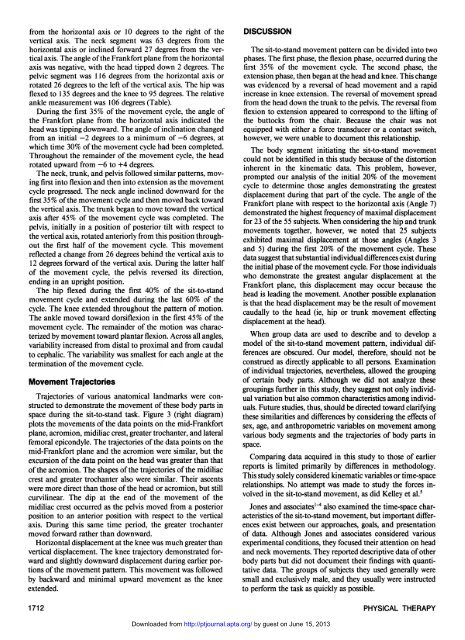
Trajec<strong>to</strong>ries of various ana<strong>to</strong>mical landmarks were constructed<br />
<strong>to</strong> demonstrate the movement of these body parts in<br />
space during the sit-<strong>to</strong>-stand task. Figure 3 (right diagram)<br />
plots the movements of the data points on the mid-Frankfort<br />
plane, acromion, midiliac crest, greater trochanter, and lateral<br />
femoral epicondyle. The trajec<strong>to</strong>ries of the data points on the<br />
mid-Frankfort plane and the acromion were similar, but the<br />
excursion of the data point on the head was greater than that<br />
of the acromion. The shapes of the trajec<strong>to</strong>ries of the midiliac<br />
crest and greater trochanter also were similar. Their ascents<br />
were more direct than those of the head or acromion, but still<br />
curvilinear. The dip at the end of the movement of the<br />
midiliac crest occurred as the pelvis moved from a posterior<br />
position <strong>to</strong> an anterior position with respect <strong>to</strong> the vertical<br />
axis. During this same time period, the greater trochanter<br />
moved forward rather than downward.<br />
Horizontal displacement at the knee was much greater than<br />
vertical displacement. The knee trajec<strong>to</strong>ry demonstrated forward<br />
and slightly downward displacement during earlier portions<br />
of the movement pattern. This movement was followed<br />
by backward and minimal upward movement as the knee<br />
extended.<br />
DISCUSSION<br />
The sit-<strong>to</strong>-stand movement pattern can be divided in<strong>to</strong> two<br />
phases. The first phase, the flexion phase, occurred during the<br />
first 35% of the movement cycle. The second phase, the<br />
extension phase, then began at the head and knee. This change<br />
was evidenced by a reversal of head movement and a rapid<br />
increase in knee extension. The reversal of movement spread<br />
from the head down the trunk <strong>to</strong> the pelvis. The reversal from<br />
flexion <strong>to</strong> extension appeared <strong>to</strong> correspond <strong>to</strong> the lifting of<br />
the but<strong>to</strong>cks from the chair. Because the chair was not<br />
equipped with either a force transducer or a contact switch,<br />
however, we were unable <strong>to</strong> document this relationship.<br />
The body segment initiating the sit-<strong>to</strong>-stand movement<br />
could not be identified in this study because of the dis<strong>to</strong>rtion<br />
inherent in the kinematic data. This problem, however,<br />
prompted our analysis of the initial 20% of the movement<br />
cycle <strong>to</strong> determine those angles demonstrating the greatest<br />
displacement during that part of the cycle. The angle of the<br />
Frankfort plane with respect <strong>to</strong> the horizontal axis (Angle 7)<br />
demonstrated the highest frequency of maximal displacement<br />
for 23 of the 55 subjects. When considering the hip and trunk<br />
movements <strong>to</strong>gether, however, we noted that 25 subjects<br />
exhibited maximal displacement at those angles (Angles 3<br />
and 5) during the first 20% of the movement cycle. These<br />
data suggest that substantial individual differences exist during<br />
the initial phase of the movement cycle. For those individuals<br />
who demonstrate the greatest angular displacement at the<br />
Frankfort plane, this displacement may occur because the<br />
head is leading the movement. Another possible explanation<br />
is that the head displacement may be the result of movement<br />
caudally <strong>to</strong> the head (ie, hip or trunk movement effecting<br />
displacement at the head).<br />
When group data are used <strong>to</strong> describe and <strong>to</strong> develop a<br />
model of the sit-<strong>to</strong>-stand movement pattern, individual differences<br />
are obscured. Our model, therefore, should not be<br />
construed as directly applicable <strong>to</strong> all persons. Examination<br />
of individual trajec<strong>to</strong>ries, nevertheless, allowed the grouping<br />
of certain body parts. Although we did not analyze these<br />
groupings further in this study, they suggest not only individual<br />
variation but also common characteristics among individuals.<br />
Future studies, thus, should be directed <strong>to</strong>ward clarifying<br />
these similarities and differences by considering the effects of<br />
sex, age, and anthropometric variables on movement among<br />
various body segments and the trajec<strong>to</strong>ries of body parts in<br />
space.<br />
Comparing data acquired in this study <strong>to</strong> those of earlier<br />
reports is limited primarily by differences in methodology.<br />
This study solely considered kinematic variables or time-space<br />
relationships. No attempt was made <strong>to</strong> study the forces involved<br />
in the sit-<strong>to</strong>-stand movement, as did Kelley et al. 5<br />
Jones and associates 1-4 also examined the time-space characteristics<br />
of the sit-<strong>to</strong>-stand movement, but important differences<br />
exist between our approaches, goals, and presentation<br />
of data. Although Jones and associates considered various<br />
experimental conditions, they focused their attention on head<br />
and neck movements. They reported descriptive data of other<br />
body parts but did not document their findings with quantitative<br />
data. The groups of subjects they used generally were<br />
small and exclusively male, and they usually were instructed<br />
<strong>to</strong> perform the task as quickly as possible.<br />
1712 PHYSICAL THERAPY<br />
Downloaded from<br />
http://ptjournal.apta.org/ by guest on June 15, 2013