

Popliteomeniscal Fascicles: Anatomic Considerations Using MR ...
Popliteomeniscal Fascicles: Anatomic Considerations Using MR ...
Popliteomeniscal Fascicles: Anatomic Considerations Using MR ...

Create successful ePaper yourself
Turn your PDF publications into a flip-book with our unique Google optimized e-Paper software.
Peduto et al.<br />
<strong>MR</strong> Arthrography of <strong>Popliteomeniscal</strong> <strong>Fascicles</strong><br />
Musculoskeletal Imaging<br />
Original Research<br />
Anthony J. Peduto 1,2<br />
Alison Nguyen 1<br />
Debra J. Trudell 1<br />
Donald L. Resnick 1<br />
Peduto AJ, Nguyen A, Trudell DJ, Resnick DL<br />
Keywords: anatomy, fascicles, knee, <strong>MR</strong>I,<br />
popliteomeniscal<br />
DOI:10.2214/AJR.07.2643<br />
Received May 29, 2007; accepted after revision<br />
July 10, 2007.<br />
1<br />
Department of Radiology, Veterans Affairs Healthcare<br />
System, San Diego, CA.<br />
2 Department of Radiology, Westmead Hospital, Darcy<br />
Rd., Westmead, Sydney, New South Wales, Australia<br />
2145. Address correspondence to A. J. Peduto.<br />
AJR 2008; 190:442–448<br />
0361–803X/08/1902–442<br />
© American Roentgen Ray Society<br />
Musculoskeletal Imaging • Original Research<br />
<strong>Popliteomeniscal</strong> <strong>Fascicles</strong>:<br />
<strong>Anatomic</strong> <strong>Considerations</strong> <strong>Using</strong><br />
<strong>MR</strong> Arthrography in Cadavers<br />
OBJECTIVE. This study was performed to examine the normal <strong>MR</strong> arthrographic anatomy<br />
of the popliteomeniscal fascicles with specific reference to the number of popliteomeniscal<br />
fascicles, thickness and course of the fascicles, and presence of other posterior attachments<br />
from the medial aponeurosis of the popliteus musculotendinous region.<br />
MaTERIals and METhOds. Multiplanar 1.5-T <strong>MR</strong> arthrography of 10 cadaveric<br />
knees was performed using a quadrature knee coil. Specimens were frozen and sectioned in<br />
the sagittal (n = 4), axial (n = 3), and coronal (n = 3) planes. <strong>MR</strong> images and anatomic specimens<br />
were correlated by two musculoskeletal radiologists.<br />
REsUlTs. Three popliteomeniscal fascicles were identified on <strong>MR</strong> arthrography: anteroinferior<br />
and posterosuperior fascicles in all 10 knees and posteroinferior fascicles in four<br />
of the knees. The posterosuperior popliteomeniscal fascicle was uniform in thickness, and the<br />
anteroinferior popliteomeniscal fascicle was variable in thickness. The anteroinferior popliteomeniscal<br />
fascicle formed a conjoined fibular attachment with the popliteofibular ligament.<br />
A medial aponeurotic extension from the popliteus musculotendinous region gave rise to the<br />
posteroinferior popliteomeniscal fascicle, which extended upward and attached to the inferomedial<br />
aspect of the posterior horn of the lateral meniscus. Additional attachments from the<br />
medial aponeurosis of the popliteus musculotendinous region to the posterior cruciate ligament,<br />
posterior capsule, oblique popliteal ligament, and posterior meniscofemoral ligament<br />
of Wrisberg were seen.<br />
COnClUsIOn. Three popliteomeniscal fascicles were identified on <strong>MR</strong> arthrographic images.<br />
The popliteus muscle–tendon unit forms robust attachments in the superior, inferior, medial,<br />
and lateral oblique aspects, highlighting its importance in posterolateral stability of the knee.<br />
B<br />
etter understanding of the clinical<br />
significance of injuries to the<br />
posterolateral corner of the knee<br />
has led to an increasing focus on<br />
clinical evaluation, treatment, and <strong>MR</strong>I of<br />
this region. Unrecognized injuries to the<br />
posterolateral corner have been cited as an<br />
important factor in postsurgical failure after<br />
cruciate ligament reconstruction and in chronic<br />
instability and degenerative changes after<br />
knee trauma [1, 2]. Within the posterolateral<br />
corner of the knee, the functional and structural<br />
relations among the lateral meniscus,<br />
popliteus muscle and tendon attachments, and<br />
the popliteomeniscal fascicles have received<br />
considerable emphasis [3–7].<br />
The proximal intraarticular insertion of the<br />
popliteus tendon is situated within a shallow<br />
concavity in the lateral aspect of the femur<br />
designated the popliteal sulcus. The tendon<br />
descends in an inferoposterior helicoid man-<br />
ner to the posterolateral corner of the knee. As<br />
it passes the posterior horn of the lateral meniscus,<br />
the popliteus tendon becomes extraarticular.<br />
The popliteomeniscal fascicles are<br />
posterolateral meniscocapsular extensions<br />
that blend inferiorly into the popliteus musculotendinous<br />
region and allow the tendon to<br />
pass from an intraarticular to an extraarticular<br />
compartment while maintaining the compartmental<br />
integrity of the knee joint. The popliteomeniscal<br />
fascicles are considered functionally<br />
important stabilizers of the lateral meniscus,<br />
working in conjunction with the popliteus<br />
musculotendinous unit to prevent excessive<br />
lateral meniscal movement and possible entrapment<br />
[8–10]. Injuries to the popliteomeniscal<br />
fascicles are commonly underrecognized<br />
both clinically and on imaging studies<br />
and are reported [3, 9] to occur in association<br />
with acute anterior cruciate ligament tears in<br />
as many as 25% of patients. Isolated tears of<br />
442 AJR:190, February 2008
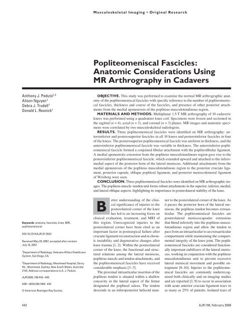
Fig. 1—Drawing shows superolateral view<br />
of posterolateral corner of knee with femur<br />
and superficial fibular attachments removed.<br />
Arrangement between popliteus muscle (8)<br />
and tendon (9) (cut proximally and reflected)<br />
and anteroinferior (11) and posterosuperior (10)<br />
popliteomeniscal fascicles is apparent. Inferolateral<br />
portion of anteroinferior popliteomeniscal fascicle<br />
(11) forms common fibular styloid attachment<br />
with anterior arm of popliteofibular ligament<br />
(6). 1 = anterior cruciate ligament, 2 = posterior<br />
cruciate ligament, 3 = lateral meniscus, 4 = anterior<br />
meniscofemoral ligament of Humphry, 5 = posterior<br />
meniscofemoral ligament of Wrisberg, 7 = posterior<br />
arm of popliteofibular ligament, 12 = fibula. (Reprinted<br />
with permission from Stäubli HU, Birrer S. The<br />
popliteus tendon and its fascicles at the popliteal<br />
hiatus: gross anatomy and functional arthroscopic<br />
evaluation with and without anterior cruciate<br />
ligament deficiency. Arthroscopy 1990; 6:209–220 [3])<br />
the popliteomeniscal fascicles can be symptomatic<br />
and manifest as localized posterolateral<br />
pain and locking of the knee joint [8, 10,<br />
11]. Some authors [11] have referred to this<br />
disorder as hypermobile lateral meniscus and<br />
describe specific clinical examination techniques<br />
that can help in the diagnosis.<br />
Although there is debate about the number<br />
of popliteomeniscal fascicles, most studies<br />
have described at least two: an anteroinferior<br />
fascicle and a posterosuperior fascicle [3, 7,<br />
12, 13]. The anteroinferior popliteomeniscal<br />
fascicle originates from the lateral aspect of<br />
the body of the lateral meniscus, courses in<br />
an inferoposterior direction to form the floor<br />
of the popliteal hiatus, and then blends with<br />
the musculotendinous portion of the popli-<br />
<strong>MR</strong> Arthrography of <strong>Popliteomeniscal</strong> <strong>Fascicles</strong><br />
teus muscle. The lateral portion of the anteroinferior<br />
popliteomeniscal fascicle takes<br />
an inferoposterior course and fuses with the<br />
popliteofibular ligament to form a conjoined<br />
attachment at the fibular styloid process. The<br />
origin of the posterosuperior popliteomeniscal<br />
fascicle is the posterosuperior margin of<br />
the posterior horn of the lateral meniscus<br />
medial to the popliteus tendon. This fascicle<br />
forms the roof of the popliteal hiatus.<br />
The posterosuperior popliteomeniscal fascicle<br />
has a posterior course and attaches to the<br />
posterior joint capsule, which fuses with the<br />
musculotendinous portion of the popliteus<br />
tendon (Fig. 1).<br />
The presence of a third popliteomeniscal<br />
fascicle, known as the posteroinferior popliteomeniscal<br />
fascicle, is controversial. This<br />
fascicle is reported to be located medial to<br />
the popliteal hiatus [11, 14–16]. Last [17] in<br />
1950 described a broad and robust aponeurotic<br />
extension from the medial aspect of the<br />
musculotendinous region of the popliteus<br />
muscle that had a prominent attachment to<br />
the inferior margin of the posterior horn of<br />
the lateral meniscus. Terry and LaPrade [15]<br />
and Ullrich et al. [16] also described the medial<br />
aponeurotic extension and designated<br />
the attachment to the inferior margin of the<br />
posterior horn of the lateral meniscus the<br />
posteroinferior popliteomeniscal fascicle.<br />
This fascicle passes upward from the medial<br />
aponeurosis of the popliteus muscle and inserts<br />
on the inferior margin of the posterior<br />
horn of the lateral meniscus near the origin<br />
of the posterior meniscofemoral ligament of<br />
Wrisberg. Feipel et al. [14] found the posteroinferior<br />
popliteomeniscal fascicle present in<br />
17% of 42 dissections of embalmed knee<br />
specimens. Most other reports of the posteroinferior<br />
popliteomeniscal fascicle do not<br />
state its frequency.<br />
The medial aponeurosis of the popliteus<br />
musculotendinous region has been reported<br />
to have additional medial attachments to the<br />
inferolateral aspect of the posterior cruciate<br />
ligament, the posterior capsule, and an<br />
infero lateral connection with the oblique<br />
popliteal ligament [14, 16]. A variant of the<br />
posterior meniscofemoral ligament of Wrisberg<br />
has been reported in which the origin is<br />
the medial aponeurosis of the popliteus musculotendinous<br />
region rather than the posterior<br />
horn of the lateral meniscus [14].<br />
This study was performed to examine the<br />
normal <strong>MR</strong> arthrographic imaging anatomy<br />
of the popliteomeniscal fascicles with specific<br />
reference to the number of popliteomeniscal<br />
fascicles, the thickness and course of the fascicles,<br />
and the presence of other posterior attachments<br />
from the medial aponeurosis of the<br />
popliteus musculotendinous region.<br />
Materials and Methods<br />
After institutional review board approval was<br />
obtained, 10 fresh unembalmed frozen cadaveric<br />
knee specimens were obtained from the<br />
department of anatomy donor program at our<br />
institution. Specimens were from five men and<br />
five women whose average age at death was 85<br />
years (range, 73–93 years).<br />
<strong>MR</strong> Arthrography<br />
Before <strong>MR</strong>I, specimens were allowed to thaw<br />
to room temperature for 24 hours, after which<br />
arthrography was performed under fluoroscopic<br />
guidance. Approximately 55–60 mL of a solution<br />
containing 1 mL of gadopentetate dimeglumine<br />
(Magnevist, Bayer Schering Pharma) diluted in<br />
250 mL of equal parts saline solution and iohexol<br />
(Omnipaque 350, GE Healthcare) was injected<br />
into each knee joint via a lateral suprapatellar<br />
approach with an 18-gauge needle. T1-weighted<br />
spin-echo imaging was performed on a 1.5-T<br />
<strong>MR</strong>I system (Signa LX Horizon, software version<br />
8.3, GE Healthcare) with a quadrature knee coil<br />
in the orthogonal sagittal, axial, and coronal<br />
planes. The following <strong>MR</strong>I parameters were used:<br />
TR/TE, 900/22; bandwidth, 16 kHz; matrix size,<br />
512 × 256; field of view, 12 × 12 cm; slice<br />
thickness, 2.5-mm; interslice gap, 0.5 mm; single<br />
acquisition; imaging time, approximately 5<br />
minutes for each sequence.<br />
Specimen Sectioning and Photography<br />
After <strong>MR</strong>I, the knee specimens were placed in a<br />
freezer (Forma Bio-Freezer, Forma Scientific) and<br />
deep frozen to −40°C. The frozen knee specimens<br />
were sectioned with a band saw into 3-mm slices<br />
in the sagittal (n = 4), axial (n = 3), and coronal<br />
(n = 3) planes. After debris was rinsed from the<br />
surface of the specimens, the sections were<br />
thawed, floodlit, and photographed with a digital<br />
camera (Coolpix 990, Nikon).<br />
Image Interpretation<br />
<strong>MR</strong> arthrographic images and specimen<br />
photographs were simultaneously reviewed by two<br />
musculoskeletal radiologists working in consensus.<br />
Identification and location of the anteroinferior,<br />
posterosuperior, and postero inferior popliteomeniscal<br />
fascicles and the medial aponeurosis with<br />
its medial attachments were based on gross<br />
anatomic descriptions obtained from the literature<br />
[3, 12, 14–16]. The number of popliteomeniscal<br />
fascicles visualized and their location on <strong>MR</strong><br />
AJR:190, February 2008 443
arthrographic images were recorded for each<br />
specimen. The reviewers inspected <strong>MR</strong> arthrographic<br />
images obtained through the popliteal<br />
hiatus, with the anteroinferior popliteomeniscal<br />
fascicle originating from the lateral surface of the<br />
body of the lateral meniscus and forming the floor<br />
and lateral wall of the hiatus, and the posterosuperior<br />
A<br />
Peduto et al.<br />
popliteomeniscal fascicle originating from the<br />
superior edge of the posterior horn of the lateral<br />
meniscus and forming the roof and medial wall of<br />
the hiatus. <strong>MR</strong> arthrographic images obtained<br />
medial to the popliteal hiatus were inspected for the<br />
presence of a medial aponeurotic extension from the<br />
musculotendinous junction of the popliteus muscle,<br />
C<br />
d<br />
Fig. 2—Anteroinferior popliteomeniscal fascicle and popliteofibular ligament of cadaver specimens. A and B<br />
are matching <strong>MR</strong>I and anatomic sections from one cadaver; C and D are matching <strong>MR</strong>I and anatomic sections<br />
from another cadaver.<br />
A–D, Sagittal T1-weighted <strong>MR</strong> arthrographic images with corresponding cadaveric sections show<br />
anteroinferior popliteomeniscal fascicle extending in posteroinferior course from lateral aspect of lateral<br />
meniscus (LM) and to blend with popliteus tendon. Conjoined attachment of anteroinferior popliteomeniscal<br />
fascicle and popliteofibular ligament (asterisk) at styloid process of fibula (f) is evident. Variable appearance<br />
of anteroinferior popliteomeniscal fascicle (arrows) is thin and membrane-like in A and B and thick in C and D.<br />
POP = popliteus tendon.<br />
which has been reported [15–17] to send an<br />
attachment to the inferior edge of the posterior horn<br />
of the lateral meniscus. This attachment is designated<br />
the posteroinferior popliteomeniscal fascicle and is<br />
immediately beneath the origin of the meniscofemoral<br />
ligament of Wrisberg.<br />
Electronic calipers were used to measure the<br />
thickness of each popliteomeniscal fascicle on<br />
<strong>MR</strong> arthrographic images. The fascicles were<br />
categorized as thin (≤ 1 mm), intermediate (1–2<br />
mm), or thick (≥ 2 mm). The following anatomic<br />
features were recorded: presence of a popliteofibular<br />
ligament and its relation to the anteroinferior<br />
popliteomeniscal fascicle, presence of a medial<br />
aponeurotic extension from the popliteus<br />
musculotendinous unit, and presence of medial<br />
attachments from the medial aponeurosis to the<br />
posterior cruciate ligament, posterior joint capsule,<br />
oblique popliteal ligament, and the posterior<br />
meniscofemoral ligament of Wrisberg.<br />
Results<br />
The anteroinferior and posterosuperior popli-<br />
teomeniscal fascicles were identified with <strong>MR</strong><br />
arthrography in all 10 specimens. Together<br />
the anteroinferior and posterosuperior popliteomeniscal<br />
fascicles formed a meniscocapsular<br />
sheath enveloping the popliteal tendon as it<br />
passed through the popliteal hiatus and became<br />
extraarticular in location (Fig. 1).<br />
The anteroinferior popliteomeniscal fascicle<br />
(Fig. 2) extended in an inferoposterior direction<br />
from its attachment at the lateral aspect<br />
of the body of the lateral meniscus and formed<br />
the lateral wall and floor of the popliteal hiatus.<br />
The thickness of the anteroinferior popliteomeniscal<br />
fascicle was variable. In five of<br />
10 specimens, this fascicle was categorized<br />
as thick, in three as intermediate, and in two<br />
as thin. The anteroinferior popliteomeniscal<br />
fascicle curved in the inferior direction adjacent<br />
to the posteromedial aspect of the fibular<br />
styloid process and blended with the popliteofibular<br />
ligament to form a conjoined fibular<br />
attachment, which was found in eight of 10<br />
specimens (Fig. 2). In the more medial aspect<br />
the anteroinferior popliteomeniscal fascicle<br />
formed the floor of the popliteal hiatus and<br />
fused with the deep musculotendinous portion<br />
of the popliteus complex. The popliteofibular<br />
ligament was seen on <strong>MR</strong> arthro graphic<br />
images of nine of 10 specimens and had a robust<br />
attachment to the posteromedial aspect<br />
of the fibular styloid process (Fig. 2). Only a<br />
single attachment site of the popliteofibular<br />
ligament was discernible.<br />
The posterosuperior popliteomeniscal fascicle<br />
was in a medial position in relation to<br />
444 AJR:190, February 2008<br />
B
<strong>MR</strong> Arthrography of <strong>Popliteomeniscal</strong> <strong>Fascicles</strong><br />
A B<br />
C<br />
d E F<br />
G<br />
Fig. 3—<strong>Popliteomeniscal</strong> fascicular attachments of cadaver specimen.<br />
A–G, Series of sagittal T1-weighted <strong>MR</strong> arthrographic images of lateral meniscus extending from lateral to<br />
medial shows three popliteomeniscal fascicular attachments. Anteroinferior popliteomeniscal fascicle (AI-<br />
PMF) is thinner than posterosuperior popliteomeniscal fascicle (PS-PMF) in this knee. The posteroinferior<br />
popliteomeniscal fascicle (PI-PMF) extends upward and in medial direction from medial aponeurotic extension<br />
(arrowheads, F and G) of popliteus musculotendinous region and attaches to inferior margin of posterior horn of<br />
lateral meniscus immediately below posterior meniscofemoral ligament of Wrisberg (curved arrow in G).<br />
AJR:190, February 2008 445
the popliteus tendon at the level of the<br />
popliteal hiatus and extended in a posterior<br />
direction from the posterosuperior corner of<br />
the posterior horn of the lateral meniscus<br />
(Fig. 3) to the posterior joint capsule imme-<br />
Peduto et al.<br />
diately above the diverging popliteus tendon.<br />
In this region the popliteus tendon widened<br />
and formed a broad aponeurotic attachment<br />
with the posterior capsule, anchoring the<br />
posterior horn of the lateral meniscus to the<br />
popliteus muscle via the posterosuperior<br />
popliteomeniscal fascicle and capsule. The<br />
posterosuperior popliteomeniscal fascicle<br />
A B<br />
Fig. 4—Attachment of medial aponeurosis to posterior cruciate ligament.<br />
A, Axial T1-weighted <strong>MR</strong> arthrographic image shows inferomedial extension from medial aponeurosis to<br />
inferolateral aspect (arrows) of posterior cruciate ligament (PCL).<br />
B, Axial section of different specimen from A with traction on medial aponeurotic extension of popliteus<br />
muscle–tendon unit shows attachment to posterior cruciate ligament (arrows). POP = popliteus tendon.<br />
A<br />
C<br />
was uniform in thickness in all 10 of the<br />
specimens and was categorized as thick.<br />
A broad medial aponeurotic expansion from<br />
the medial aspect of the musculotendinous<br />
region of the popliteus tendon was identified<br />
in all 10 specimens (Fig. 3). From the medial<br />
aponeurosis an attachment to the inferior margin<br />
of the posterior horn of lateral meniscus<br />
was seen that corresponded to the anatomic<br />
descriptions by Terry and LaPrade [15] of<br />
the third, or posteroinferior, popliteomeniscal<br />
fascicle. The posteroinferior popliteomeniscal<br />
fascicle was seen on <strong>MR</strong> arthrographic images<br />
of four of 10 knee specimens (Fig. 3).<br />
All four posteroinferior popliteomeniscal<br />
fascicles identified were categorized as thick<br />
on measurement.<br />
Additional attachments of the medial<br />
aponeurosis were seen. An attachment to the<br />
posterior joint capsule was seen on <strong>MR</strong> arthrographic<br />
images of eight of the 10 knees.<br />
A deeper extension to the inferolateral aspect<br />
of the posterior cruciate ligament was found<br />
in seven of the 10 specimens (Fig. 4). A focal<br />
thickening of the medial aponeurosis coursed<br />
upward, where it joined the oblique popliteal<br />
ligament to form an inferior connection between<br />
the medial aponeurosis and the oblique<br />
popliteal ligament in seven of the 10 specimens<br />
(Fig. 5). In one knee in which both the<br />
anterior and posterior meniscofemoral ligaments<br />
were present, the medial aponeurotic<br />
extension from the popliteus musculotendinous<br />
region extended medially to form the<br />
posterior meniscofemoral ligament of Wrisberg<br />
(Fig. 5). In this knee, the anterior meniscofemoral<br />
ligament of Humphry had a<br />
normal attachment to the posterior horn of<br />
the lateral meniscus.<br />
discussion<br />
There has been increasing interest in the<br />
meniscocapsular attachments of the popliteus<br />
Fig. 5—Medial aponeurosis attachments of<br />
cadaver specimen. Series of axial T1-weighted <strong>MR</strong><br />
arthrographic images from inferior (A) to superior (D)<br />
aspects. PCL = posterior cruciate ligament.<br />
A, <strong>MR</strong> arthrographic image shows relation between<br />
popliteus tendon (POP) and medial aponeurosis<br />
(arrowheads). Medial attachments to posterior<br />
capsule (large arrow) and ligament of Wrisberg (small<br />
arrow) extend from medial aponeurosis. LM = lateral<br />
meniscus.<br />
B–D, Successive superior <strong>MR</strong> arthrographic images<br />
show Wrisberg extension (short arrows) of medial<br />
aponeurosis can be followed upward. Upward<br />
extension of medial aponeurosis (arrowheads)<br />
forms inferior connection with oblique popliteal<br />
ligament (long arrows, D). Asterisk (B and C) indicates<br />
meniscofemoral ligament of Humphry.<br />
446 AJR:190, February 2008<br />
B<br />
d
muscle–tendon complex. These attachments<br />
not only are important in allowing the tendon<br />
to pass through the joint capsule to assume an<br />
extraarticular location but also act in concert<br />
with the popliteus complex to retract the lateral<br />
meniscus from the joint during knee flexion<br />
to prevent excessive meniscal shearing<br />
forces and entrapment [8–10]. A number of<br />
studies [3, 9] have shown a relatively high<br />
prevalence of disruption of the popliteomeniscal<br />
fascicle at arthroscopic surgery on patients<br />
with anterior cruciate ligament tears.<br />
In this series, the anteroinferior and posterosuperior<br />
popliteomeniscal fascicles were<br />
seen on all <strong>MR</strong> arthrographic studies. In comparison,<br />
Feipel et al. [14] found the anteroinferior<br />
popliteomeniscal fascicle in 83% and the<br />
posterosuperior popliteomeniscal fascicle in<br />
90% of dissections of 42 embalmed knee<br />
specimens. Terry and LaPrade [15] and Stäubli<br />
and Birrer [3] described the presence of<br />
these fascicles in their studies of 30 and 14<br />
fresh cadavers, but they did not discuss how<br />
frequently the fascicles were seen in the specimens.<br />
In arthroscopic studies [3, 15, 18], the<br />
anteroinferior and posterosuperior popliteomeniscal<br />
fascicles have been reported to be<br />
present in nearly all patients examined. Tria et<br />
al. [19], unlike most other investigators, found<br />
fascicular attachments to the lateral meniscus<br />
in only 22 of 40 knee dissections.<br />
In our study, unlike the posterosuperior<br />
popliteomeniscal fascicle, which had uniform<br />
thickness, the anteroinferior popliteomeniscal<br />
fascicle had variable thickness, ranging from a<br />
thin membrane-like structure to a much more<br />
robust structure. Bozkurt et al. [20] described<br />
a lateral meniscofibular ligament that appeared<br />
to correspond to the anatomic description of<br />
the anteroinferior popliteomeniscal fascicle in<br />
all 50 specimens examined by microdissection<br />
and transillumination. We found the lateral<br />
portion of the anteroinferior popliteomeniscal<br />
fascicle passed downward and in a lateral<br />
direction to form a conjoined attachment<br />
with the popliteofibular ligament at the fibular<br />
styloid process, resulting in a connection between<br />
the lateral aspect of the body of the lateral<br />
meniscus and the styloid process of the<br />
fibula that matched the description of a meniscofibular<br />
ligament by Bozkurt et al.<br />
Terry and LaPrade [15] described a third<br />
popliteomeniscal fascicle designated the<br />
posteroinferior popliteomeniscal fascicle,<br />
which extended from the medial aponeurotic<br />
extension of the popliteus tendon to attach to<br />
the inferior margin of the posterior horn of<br />
the lateral meniscus. The posteroinferior<br />
<strong>MR</strong> Arthrography of <strong>Popliteomeniscal</strong> <strong>Fascicles</strong><br />
popliteomeniscal fascicle was seen in 40%<br />
of the knees in our study compared with 17%<br />
of those studied by Feipel et al. [14]. Ullrich<br />
et al. [16] found a third popliteomeniscal fascicle<br />
in their dissections of 13 fresh knees,<br />
but the frequency of the finding of a posteroinferior<br />
popliteomeniscal fascicle was not<br />
stated. Inconsistencies in descriptions of the<br />
third popliteomeniscal fascicle and doubts<br />
about its existence may relate to studies concentrated<br />
solely on the popliteal hiatus region<br />
without consideration of the more medially<br />
located capsular aponeurotic extension from<br />
the popliteus tendon and its complex posteromedial<br />
attachments. Last [17], in a report<br />
on the popliteus complex in 1950, described<br />
the broad medial aponeurotic extension from<br />
the medial portion of the popliteus muscle<br />
with a prominent attachment to the inferior<br />
margin of the posterior horn of the lateral<br />
meniscus. This description corresponds to<br />
other descriptions of the posteroinferior<br />
popliteomeniscal fascicle.<br />
The medial aponeurotic extension of the<br />
popliteus muscle appears to be an important<br />
structural element of the popliteus complex. In<br />
addition to blending with the posterior capsule,<br />
this extension forms an inferior connection<br />
with the popliteal oblique ligament, sends attachments<br />
to the posterior cruciate ligament<br />
and posterior horn of the lateral meniscus<br />
(posteroinferior popliteomeniscal fascicle),<br />
and in some individuals gives origin to a variant<br />
of the ligament of Wrisberg. Thus the popliteus<br />
muscle–tendon complex has attachments<br />
that form a robust-appearing cruciate arrangement:<br />
a superior attachment to the femur at the<br />
popliteal sulcus, an inferior triangular attachment<br />
of the main muscle bulk to the posterior<br />
aspect of the tibia, a robust inferolateral attachment<br />
to the fibular styloid process via the<br />
popliteofibular ligament, and several complex<br />
superomedial attachments to the joint capsule,<br />
lateral meniscus, oblique popliteal ligament,<br />
and ligament of Wrisberg. The importance of<br />
the popliteus muscle–tendon unit is highlighted<br />
by these robust-appearing attachments and<br />
by study findings [16, 21, 22] of dynamic and<br />
static functions that include balancing and<br />
controlling neutral tibial rotation, acting as a<br />
principal dorsolateral knee stabilizer, and preventing<br />
lateral meniscal entrapment during<br />
knee flexion by retraction of the meniscus<br />
via popliteomeniscal fascicle attachments.<br />
Limitations of this study included the relatively<br />
small number of specimens, allowing<br />
only limited comment on the frequency of<br />
variations in the attachments of the popliteus<br />
complex. In addition, the cadavers were those<br />
of elderly persons (average age at death, 85<br />
years). In most specimens, moderate degenerative<br />
joint disease was present, with variable<br />
areas of articular surface wear and meniscal<br />
degeneration or tearing. These changes might<br />
have affected visualization of structures on<br />
<strong>MR</strong> arthrographic images. Ligament, capsular,<br />
and fascicular degenerative changes might<br />
also have contributed to variability in the appearance<br />
of these structures with resultant<br />
adaptive thickening or attenuation. No history<br />
of knee injury or surgery was evident in any of<br />
the knee specimens, but untreated or unreported<br />
injury cannot be excluded.<br />
Arthrographic fluid in the joints provided<br />
excellent joint distention and optimized visualization<br />
of the popliteomeniscal fascicles,<br />
but this technique is not part of routine <strong>MR</strong>I<br />
of the knee. In the absence of substantial<br />
joint effusion or hemarthrosis, it is unlikely<br />
that visualization of the popliteomeniscal<br />
fascicles will be as optimal in nonarthrographic<br />
studies of the knee. Hemarthrosis is<br />
a common finding in patients with acute tear<br />
of the anterior cruciate ligament, who have<br />
been found to be at particular risk of popliteomeniscal<br />
fascicle tears [3, 9]. Sakai et al.<br />
[7] used an optimized oblique coronal plane<br />
in combination with nonarthrographic <strong>MR</strong>I<br />
and found the anteroinferior popliteomeniscal<br />
fascicle in 94.1% and the posterosuperior<br />
popliteomeniscal fascicle in 88.2% of subjects.<br />
We used all three orthogonal <strong>MR</strong> arthrographic<br />
imaging planes in analysis and<br />
did not assess the visibility of the popliteomeniscal<br />
fascicles in individual imaging<br />
planes. Our impression, however, was that<br />
the popliteomeniscal fascicles were best seen<br />
on sagittal <strong>MR</strong> arthrographic images.<br />
We studied the normal <strong>MR</strong>I anatomic features<br />
of the popliteomeniscal fascicles and<br />
found three fascicles. The third, or posteroinferior,<br />
popliteomeniscal fascicle was located<br />
medial in relation to the popliteal hiatus and<br />
arose from a medial aponeurotic extension of<br />
the popliteus musculotendinous region, which<br />
had additional capsular, oblique popliteal ligament,<br />
posterior cruciate, and ligament of<br />
Wrisberg attachments. These extensive attachments,<br />
combined with femoral, tibial, and<br />
fibular attachments, highlight the important<br />
role of the popliteus muscle–tendon unit in the<br />
posterolateral corner of the knee.<br />
References<br />
1. Fleming RE Jr, Blatz DJ, McCarroll JR. Posterior<br />
problems in the knee: posterior cruciate insuffi-<br />
AJR:190, February 2008 447
ciency and posterolateral rotatory insufficiency.<br />
Am J Sports Med 1981; 9:107–113<br />
2. Hughston JC, Jacobson KE. Chronic posterolateral<br />
rotatory instability of the knee. J Bone Joint<br />
Surg Am 1985; 67:351–359<br />
3. Stäubli HU, Birrer S. The popliteus tendon and its<br />
fascicles at the popliteal hiatus: gross anatomy<br />
and functional arthroscopic evaluation with and<br />
without anterior cruciate ligament deficiency. Arthroscopy<br />
1990; 6:209–220<br />
4. De Smet AA, Asinger DA, Johnson RL. Abnormal<br />
superior popliteomeniscal fascicle and posterior<br />
pericapsular edema: indirect <strong>MR</strong> imaging signs<br />
of a lateral meniscal tear. AJR 2001; 176:63–66<br />
5. Blankenbaker DG, De Smet AA, Smith JD. Usefulness<br />
of two indirect <strong>MR</strong> imaging signs to diagnose<br />
lateral meniscal tears. AJR 2002; 178:579–582<br />
6. Johnson RL, De Smet AA. <strong>MR</strong> visualization of<br />
the popliteomeniscal fascicles. Skeletal Radiol<br />
1999; 28:561–566<br />
7. Sakai H, Sasho T, Wada Y, Sano S, Morita F,<br />
Moriya H. <strong>MR</strong>I of the popliteomeniscal fasciculi.<br />
AJR 2006; 186:460–466<br />
8. Simonian PT, Sussmann PS, Wickiewicz Tl, et al.<br />
<strong>Popliteomeniscal</strong> fasciculi and the unstable lateral<br />
meniscus: clinical correlation and magnetic resonance<br />
diagnosis. Arthroscopy 1997; 13:590–596<br />
Peduto et al.<br />
9. LaPrade RF. Arthroscopic evaluation of the lateral<br />
compartment of knees with grade 3 posterolateral<br />
knee complex injuries. Am J Sports Med<br />
1997; 25:596–602<br />
10. Simonian PT, Sussmann PS, van Trommel M,<br />
Wickiewicz TL, Warren RF. <strong>Popliteomeniscal</strong><br />
fasciculi and lateral meniscal stability. Am J<br />
Sports Med 1997; 25:849–853<br />
11. LaPrade RF, Konowalchuk BK. <strong>Popliteomeniscal</strong><br />
fascicle tears causing symptomatic lateral compartment<br />
knee pain: diagnosis by the figure-4 test<br />
and treatment by open repair. Am J Sports Med<br />
2005; 33:1231–1236<br />
12. Diamantopoulos A, Tokis A, Tzurbakis M, Patsopoulos<br />
I, Georgoulis A. The posterolateral corner<br />
of the knee: evaluation under microsurgical dissection.<br />
Arthroscopy 2005; 21:826–833<br />
13. Munshi M, Pretterklieber ML, Kwak S, Antonio<br />
GE, Trudell DJ, Resnick D. <strong>MR</strong> imaging, <strong>MR</strong> arthrography,<br />
and specimen correlation of the posterolateral<br />
corner of the knee: an anatomic study.<br />
AJR 2003; 180:1095–1101<br />
14. Feipel V, Simonnet ML, Rooze M. The proximal<br />
attachments of the popliteus muscle: a quantitative<br />
study and clinical significance. Surg Radiol<br />
Anat 2003; 25:58–63<br />
15. Terry GC, LaPrade RF. The posterolateral aspect<br />
of the knee: anatomy and surgical approach. Am J<br />
Sports Med 1996; 24:732–739<br />
16. Ullrich K, Krudwig WK, Witzel U. Posterolateral<br />
aspect and stability of the knee joint. Part 1. Anatomy<br />
and function of the popliteus muscle–tendon<br />
unit: an anatomical and biomechanical study. Knee<br />
Surg Sports Traumatol Arthrosc 2002; 10:86–90<br />
17. Last RJ. The popliteus muscle and the lateral meniscus:<br />
with a note on the attachment of the medial<br />
meniscus. J Bone Joint Surg Br 1950; 32:93–99<br />
18. Patel D. Proximal approaches to arthroscopic surgery<br />
of the knee. Am J Sports Med 1981; 9:296–303<br />
19. Tria AJ, Johnson CD, Zawadsky JP. The popliteus<br />
tendon. J Bone Joint Surg Am 1989; 71:714–716<br />
20. Bozkurt M, Elhan A, Tekdemir I, Tonuk E. An<br />
anatomical study of the meniscofibular ligament.<br />
Knee Surg Sports Traumatol Arthrosc 2004;<br />
12:429–433<br />
21. Moorman CT 3rd, LaPrade RF. Anatomy and biomechanics<br />
of the posterolateral corner of the<br />
knee. J Knee Surg 2005; 18:137–145<br />
22. LaPrade RF, Ly TV, Wentorf FA, Engebretsen L.<br />
The posterolateral attachments of the knee: a qualitative<br />
and quantitative morphologic analysis of<br />
the fibular collateral ligament, popliteus tendon,<br />
popliteofibular ligament, and lateral gastrocnemius<br />
tendon. Am J Sports Med 2003; 31:854–860<br />
448 AJR:190, February 2008