

Popliteomeniscal Fascicles: Anatomic Considerations Using MR ...
Popliteomeniscal Fascicles: Anatomic Considerations Using MR ...
Popliteomeniscal Fascicles: Anatomic Considerations Using MR ...

Create successful ePaper yourself
Turn your PDF publications into a flip-book with our unique Google optimized e-Paper software.
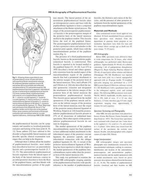
Fig. 1—Drawing shows superolateral view<br />
of posterolateral corner of knee with femur<br />
and superficial fibular attachments removed.<br />
Arrangement between popliteus muscle (8)<br />
and tendon (9) (cut proximally and reflected)<br />
and anteroinferior (11) and posterosuperior (10)<br />
popliteomeniscal fascicles is apparent. Inferolateral<br />
portion of anteroinferior popliteomeniscal fascicle<br />
(11) forms common fibular styloid attachment<br />
with anterior arm of popliteofibular ligament<br />
(6). 1 = anterior cruciate ligament, 2 = posterior<br />
cruciate ligament, 3 = lateral meniscus, 4 = anterior<br />
meniscofemoral ligament of Humphry, 5 = posterior<br />
meniscofemoral ligament of Wrisberg, 7 = posterior<br />
arm of popliteofibular ligament, 12 = fibula. (Reprinted<br />
with permission from Stäubli HU, Birrer S. The<br />
popliteus tendon and its fascicles at the popliteal<br />
hiatus: gross anatomy and functional arthroscopic<br />
evaluation with and without anterior cruciate<br />
ligament deficiency. Arthroscopy 1990; 6:209–220 [3])<br />
the popliteomeniscal fascicles can be symptomatic<br />
and manifest as localized posterolateral<br />
pain and locking of the knee joint [8, 10,<br />
11]. Some authors [11] have referred to this<br />
disorder as hypermobile lateral meniscus and<br />
describe specific clinical examination techniques<br />
that can help in the diagnosis.<br />
Although there is debate about the number<br />
of popliteomeniscal fascicles, most studies<br />
have described at least two: an anteroinferior<br />
fascicle and a posterosuperior fascicle [3, 7,<br />
12, 13]. The anteroinferior popliteomeniscal<br />
fascicle originates from the lateral aspect of<br />
the body of the lateral meniscus, courses in<br />
an inferoposterior direction to form the floor<br />
of the popliteal hiatus, and then blends with<br />
the musculotendinous portion of the popli-<br />
<strong>MR</strong> Arthrography of <strong>Popliteomeniscal</strong> <strong>Fascicles</strong><br />
teus muscle. The lateral portion of the anteroinferior<br />
popliteomeniscal fascicle takes<br />
an inferoposterior course and fuses with the<br />
popliteofibular ligament to form a conjoined<br />
attachment at the fibular styloid process. The<br />
origin of the posterosuperior popliteomeniscal<br />
fascicle is the posterosuperior margin of<br />
the posterior horn of the lateral meniscus<br />
medial to the popliteus tendon. This fascicle<br />
forms the roof of the popliteal hiatus.<br />
The posterosuperior popliteomeniscal fascicle<br />
has a posterior course and attaches to the<br />
posterior joint capsule, which fuses with the<br />
musculotendinous portion of the popliteus<br />
tendon (Fig. 1).<br />
The presence of a third popliteomeniscal<br />
fascicle, known as the posteroinferior popliteomeniscal<br />
fascicle, is controversial. This<br />
fascicle is reported to be located medial to<br />
the popliteal hiatus [11, 14–16]. Last [17] in<br />
1950 described a broad and robust aponeurotic<br />
extension from the medial aspect of the<br />
musculotendinous region of the popliteus<br />
muscle that had a prominent attachment to<br />
the inferior margin of the posterior horn of<br />
the lateral meniscus. Terry and LaPrade [15]<br />
and Ullrich et al. [16] also described the medial<br />
aponeurotic extension and designated<br />
the attachment to the inferior margin of the<br />
posterior horn of the lateral meniscus the<br />
posteroinferior popliteomeniscal fascicle.<br />
This fascicle passes upward from the medial<br />
aponeurosis of the popliteus muscle and inserts<br />
on the inferior margin of the posterior<br />
horn of the lateral meniscus near the origin<br />
of the posterior meniscofemoral ligament of<br />
Wrisberg. Feipel et al. [14] found the posteroinferior<br />
popliteomeniscal fascicle present in<br />
17% of 42 dissections of embalmed knee<br />
specimens. Most other reports of the posteroinferior<br />
popliteomeniscal fascicle do not<br />
state its frequency.<br />
The medial aponeurosis of the popliteus<br />
musculotendinous region has been reported<br />
to have additional medial attachments to the<br />
inferolateral aspect of the posterior cruciate<br />
ligament, the posterior capsule, and an<br />
infero lateral connection with the oblique<br />
popliteal ligament [14, 16]. A variant of the<br />
posterior meniscofemoral ligament of Wrisberg<br />
has been reported in which the origin is<br />
the medial aponeurosis of the popliteus musculotendinous<br />
region rather than the posterior<br />
horn of the lateral meniscus [14].<br />
This study was performed to examine the<br />
normal <strong>MR</strong> arthrographic imaging anatomy<br />
of the popliteomeniscal fascicles with specific<br />
reference to the number of popliteomeniscal<br />
fascicles, the thickness and course of the fascicles,<br />
and the presence of other posterior attachments<br />
from the medial aponeurosis of the<br />
popliteus musculotendinous region.<br />
Materials and Methods<br />
After institutional review board approval was<br />
obtained, 10 fresh unembalmed frozen cadaveric<br />
knee specimens were obtained from the<br />
department of anatomy donor program at our<br />
institution. Specimens were from five men and<br />
five women whose average age at death was 85<br />
years (range, 73–93 years).<br />
<strong>MR</strong> Arthrography<br />
Before <strong>MR</strong>I, specimens were allowed to thaw<br />
to room temperature for 24 hours, after which<br />
arthrography was performed under fluoroscopic<br />
guidance. Approximately 55–60 mL of a solution<br />
containing 1 mL of gadopentetate dimeglumine<br />
(Magnevist, Bayer Schering Pharma) diluted in<br />
250 mL of equal parts saline solution and iohexol<br />
(Omnipaque 350, GE Healthcare) was injected<br />
into each knee joint via a lateral suprapatellar<br />
approach with an 18-gauge needle. T1-weighted<br />
spin-echo imaging was performed on a 1.5-T<br />
<strong>MR</strong>I system (Signa LX Horizon, software version<br />
8.3, GE Healthcare) with a quadrature knee coil<br />
in the orthogonal sagittal, axial, and coronal<br />
planes. The following <strong>MR</strong>I parameters were used:<br />
TR/TE, 900/22; bandwidth, 16 kHz; matrix size,<br />
512 × 256; field of view, 12 × 12 cm; slice<br />
thickness, 2.5-mm; interslice gap, 0.5 mm; single<br />
acquisition; imaging time, approximately 5<br />
minutes for each sequence.<br />
Specimen Sectioning and Photography<br />
After <strong>MR</strong>I, the knee specimens were placed in a<br />
freezer (Forma Bio-Freezer, Forma Scientific) and<br />
deep frozen to −40°C. The frozen knee specimens<br />
were sectioned with a band saw into 3-mm slices<br />
in the sagittal (n = 4), axial (n = 3), and coronal<br />
(n = 3) planes. After debris was rinsed from the<br />
surface of the specimens, the sections were<br />
thawed, floodlit, and photographed with a digital<br />
camera (Coolpix 990, Nikon).<br />
Image Interpretation<br />
<strong>MR</strong> arthrographic images and specimen<br />
photographs were simultaneously reviewed by two<br />
musculoskeletal radiologists working in consensus.<br />
Identification and location of the anteroinferior,<br />
posterosuperior, and postero inferior popliteomeniscal<br />
fascicles and the medial aponeurosis with<br />
its medial attachments were based on gross<br />
anatomic descriptions obtained from the literature<br />
[3, 12, 14–16]. The number of popliteomeniscal<br />
fascicles visualized and their location on <strong>MR</strong><br />
AJR:190, February 2008 443