Microdeletion Syndromes Detected by FISH - Kamla-Raj Enterprises
Microdeletion Syndromes Detected by FISH - Kamla-Raj Enterprises
Microdeletion Syndromes Detected by FISH - Kamla-Raj Enterprises

Create successful ePaper yourself
Turn your PDF publications into a flip-book with our unique Google optimized e-Paper software.
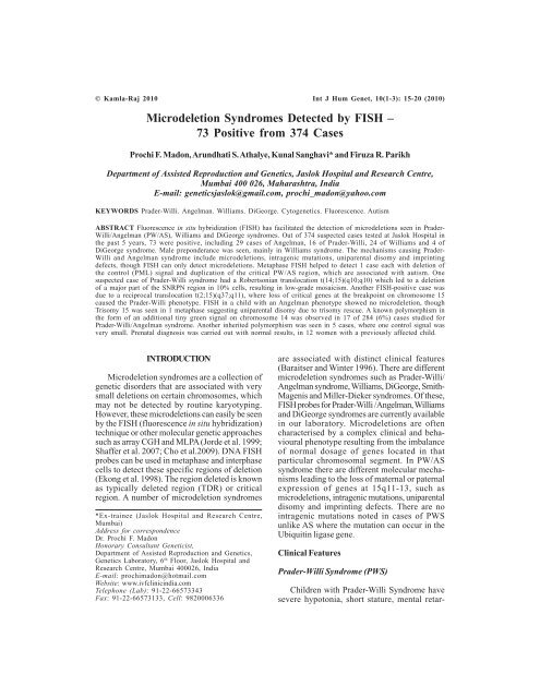
© <strong>Kamla</strong>-<strong>Raj</strong> 2010 Int J Hum Genet, 10(1-3): 15-20 (2010)<br />
<strong>Microdeletion</strong> <strong>Syndromes</strong> <strong>Detected</strong> <strong>by</strong> <strong>FISH</strong> –<br />
73 Positive from 374 Cases<br />
Prochi F. Madon, Arundhati S. Athalye, Kunal Sanghavi* and Firuza R. Parikh<br />
Department of Assisted Reproduction and Genetics, Jaslok Hospital and Research Centre,<br />
Mumbai 400 026, Maharashtra, India<br />
E-mail: geneticsjaslok@gmail.com, prochi_madon@yahoo.com<br />
KEYWORDS Prader-Willi. Angelman. Williams. DiGeorge. Cytogenetics. Fluorescence. Autism<br />
ABSTRACT Fluorescence in situ hybridization (<strong>FISH</strong>) has facilitated the detection of microdeletions seen in Prader-<br />
Willi/Angelman (PW/AS), Williams and DiGeorge syndromes. Out of 374 suspected cases tested at Jaslok Hospital in<br />
the past 5 years, 73 were positive, including 29 cases of Angelman, 16 of Prader-Willi, 24 of Williams and 4 of<br />
DiGeorge syndrome. Male preponderance was seen, mainly in Williams syndrome. The mechanisms causing Prader-<br />
Willi and Angelman syndrome include microdeletions, intragenic mutations, uniparental disomy and imprinting<br />
defects, though <strong>FISH</strong> can only detect microdeletions. Metaphase <strong>FISH</strong> helped to detect 1 case each with deletion of<br />
the control (PML) signal and duplication of the critical PW/AS region, which are associated with autism. One<br />
suspected case of Prader-Willi syndrome had a Robertsonian translocation t(14;15)(q10;q10) which led to a deletion<br />
of a major part of the SNRPN region in 10% cells, resulting in low-grade mosaicism. Another <strong>FISH</strong>-positive case was<br />
due to a reciprocal translocation t(2;15)(q37;q11), where loss of critical genes at the breakpoint on chromosome 15<br />
caused the Prader-Willi phenotype. <strong>FISH</strong> in a child with an Angelman phenotype showed no microdeletion, though<br />
Trisomy 15 was seen in 1 metaphase suggesting uniparental disomy due to trisomy rescue. A known polymorphism in<br />
the form of an additional tiny green signal on chromosome 14 was observed in 17 of 284 (6%) cases studied for<br />
Prader-Willi/Angelman syndrome. Another inherited polymorphism was seen in 5 cases, where one control signal was<br />
very small. Prenatal diagnosis was carried out with normal results, in 12 women with a previously affected child.<br />
INTRODUCTION<br />
<strong>Microdeletion</strong> syndromes are a collection of<br />
genetic disorders that are associated with very<br />
small deletions on certain chromosomes, which<br />
may not be detected <strong>by</strong> routine karyotyping.<br />
However, these microdeletions can easily be seen<br />
<strong>by</strong> the <strong>FISH</strong> (fluorescence in situ hybridization)<br />
technique or other molecular genetic approaches<br />
such as array CGH and MLPA (Jorde et al. 1999;<br />
Shaffer et al. 2007; Cho et al.2009). DNA <strong>FISH</strong><br />
probes can be used in metaphase and interphase<br />
cells to detect these specific regions of deletion<br />
(Ekong et al. 1998). The region deleted is known<br />
as typically deleted region (TDR) or critical<br />
region. A number of microdeletion syndromes<br />
*Ex-trainee (Jaslok Hospital and Research Centre,<br />
Mumbai)<br />
Address for correspondence<br />
Dr. Prochi F. Madon<br />
Honorary Consultant Geneticist,<br />
Department of Assisted Reproduction and Genetics,<br />
Genetics Laboratory, 6 th Floor, Jaslok Hospital and<br />
Research Centre, Mumbai 400026, India<br />
E-mail: prochimadon@hotmail.com<br />
Website: www.ivfclinicindia.com<br />
Telephone (Lab): 91-22-66573343<br />
Fax: 91-22-66573133, Cell: 9820006336<br />
are associated with distinct clinical features<br />
(Baraitser and Winter 1996). There are different<br />
microdeletion syndromes such as Prader-Willi/<br />
Angelman syndrome, Williams, DiGeorge, Smith-<br />
Magenis and Miller-Dieker syndromes. Of these,<br />
<strong>FISH</strong> probes for Prader-Willi /Angelman, Williams<br />
and DiGeorge syndromes are currently available<br />
in our laboratory. <strong>Microdeletion</strong>s are often<br />
characterised <strong>by</strong> a complex clinical and behavioural<br />
phenotype resulting from the imbalance<br />
of normal dosage of genes located in that<br />
particular chromosomal segment. In PW/AS<br />
syndrome there are different molecular mechanisms<br />
leading to the loss of maternal or paternal<br />
expression of genes at 15q11-13, such as<br />
microdeletions, intragenic mutations, uniparental<br />
disomy and imprinting defects. There are no<br />
intragenic mutations noted in cases of PWS<br />
unlike AS where the mutation can occur in the<br />
Ubiquitin ligase gene.<br />
Clinical Features<br />
Prader-Willi Syndrome (PWS)<br />
Children with Prader-Willi Syndrome have<br />
severe hypotonia, short stature, mental retar-
16<br />
dation, obesity with increasing age, hypogonadism,<br />
small hands and feet, fair hair and skin.<br />
These children have a habit of excessive skin<br />
picking. (Robinson et al. 1991; Cassidy et al.<br />
1992).<br />
Angelman Syndrome (AS)<br />
This is also known as Happy Puppet syndrome.<br />
The children in general have a happy<br />
predisposition with outbursts of laughter often<br />
accompanied <strong>by</strong> frequent hand flapping. These<br />
children have mental retardation with microcephaly,<br />
jerky movements affecting the trunk and<br />
upper limbs, ataxia, unsteady and wide-based<br />
gait, wide mouth with constant dribbling and<br />
prominent chin (Boyd et al. 1988; Clayton-Smith<br />
1992).<br />
In a majority of cases, both the above clinically<br />
different syndromes are caused <strong>by</strong> the same<br />
microdeletion +/- 4 Mb on chromosome 15 at the<br />
15q11-13 region. Because of imprinting, the<br />
presence of a microdeletion on the paternal<br />
chromosome 15 leads to Prader-Willi syndrome,<br />
while a microdeletion on the maternal chromosome<br />
15 causes Angelman syndrome (Wagstaff<br />
et al. 1992). Besides micordeletions and<br />
imprinting, uniparental disomy and intragenic<br />
mutations can also cause the two syndromes<br />
(Malcolm et al. 1991).<br />
Williams Syndrome (Williams-Beuren<br />
Syndrome) - (WS)<br />
This is caused <strong>by</strong> a microdeletion on<br />
chromosome 7 at the Elastin locus 7q11 (Ewart et<br />
al. 1993). The facial features consist of a medial<br />
eyebrow flare, a stellate iris pattern, flat nose<br />
bridge and long smooth philtrum with widely<br />
spaced teeth. The characteristic heart defect is a<br />
supravalvular aortic stenosis (Burn 1986;<br />
Halladie-Smith and Karas 1988; Jones 1990).<br />
DiGeorge Syndrome (Velocardiofacial<br />
Syndrome or CATCH 22) – (DS)<br />
The chromosomal microdeletion is at 22q11.<br />
The clinical features include hypoplasia, hypoparathyroidism<br />
and cardiac malformations. Dysmorphic<br />
features include hypertelorism, low-set<br />
ears and micrognathia. The most common cardiac<br />
defects include interrupted aortic arch, often with<br />
VSD (Ventricular Septal Defect) and a persistent<br />
PROCHI F. MADON, ARUNDHATI S. ATHALYE ET AL.<br />
truncus arteriosus (Driscoll et al. 1992; Laena-<br />
Cox et al. 1996; Scambler et al. 1992). It is also<br />
called CATCH 22 because it describes the<br />
abnormal findings of cardiac anomalies, abnormal<br />
facies, thymic hypoplasia, cleft palate and<br />
hypocalcemia due to a deletion on chromosome<br />
22.<br />
<strong>FISH</strong> Probes and Interpretation<br />
<strong>Microdeletion</strong> probes have control signals<br />
on the same chromosome, which can clearly be<br />
seen on metaphases. For Williams and DiGeorge<br />
syndromes, the control signals are labeled in<br />
green (G) and the critical region with orange (O)<br />
in the present study. Therefore a normal cell will<br />
show a 2G2O pattern whereas a cell with the<br />
deletion will show a 2G1O pattern (Fig. 1a, b).<br />
The SNRPN probe for detection of PW/AS has<br />
got two internal controls, a large proximal green<br />
signal (CEP 15) and a smaller orange signal (PML)<br />
towards the distal end. The critical region<br />
(SNRPN) has a small orange signal and lies<br />
between the two control signals on chromosome<br />
15, adjacent to the green signal. With this probe,<br />
normal cells show a 2G4O signal pattern whereas<br />
cells with the microdeletion show a 2G3O signal<br />
pattern (Baumer et al.1999) (Fig. 1c, d). <strong>FISH</strong><br />
signals on metaphases should be checked with<br />
this probe to avoid false positive results due to<br />
deletion of the control orange signal. The D15S11<br />
probe for PW/AS is used to double check such<br />
cases as it has only 1 internal control region in<br />
green.<br />
MATERIAL AND METHODS<br />
Over the past 5 years, a total of 374 blood<br />
samples of patients referred <strong>by</strong> pediatricians<br />
across the country were tested for microdeletions<br />
at Jaslok Hospital. PHA stimulated 72 hour whole<br />
blood cultures were set up to obtain metaphases<br />
using standard techniques. Fixed WBC pellets<br />
of blood cultures were also accepted from other<br />
laboratories. <strong>FISH</strong> was carried out using Vysis<br />
(Abbott) probes <strong>by</strong> codenaturation of the probe<br />
with the test sample at 73 o C for 5 minutes, followed<br />
<strong>by</strong> overnight hybridization at 37 o C. After<br />
washing as per the manufacturer’s protocol, the<br />
slides were mounted in the counter-stain DAPI<br />
and observed under a Zeiss fluorescent microscope.<br />
The images were captured and processed<br />
with Metasystems isis software. About 100-200
MICRODELETION SYNDROMES DETECTED BY <strong>FISH</strong> 17<br />
interphase nuclei and 5-10 metaphases were<br />
usually analyzed for detection of microdeletions<br />
<strong>by</strong> <strong>FISH</strong>. Currently, the following Vysis (Abbott)<br />
microdeletion probes are used in our laboratory:<br />
PWS/AS- LSI SNRPN (orange) / PML<br />
(orange)/ CEP 15 (green) dual color DNA probe<br />
and LSI D15S11(orange) /CEP 15 (green) probe<br />
WS - LSI ELN (orange)/ LSI D7S486, D7S522<br />
(green) dual color DNA probe<br />
DS - LSI TUPLE 1 (orange)/ LSI ARSA (green)<br />
dual color DNA probe<br />
RESULTS<br />
Out of 374 samples tested, 73(20%) were<br />
found to be positive for various microdeletions<br />
(Table 1). Among the 73 positive cases, 29(40%)<br />
had Angelman syndrome, 16(22%) had Prader-<br />
Willi syndrome, 24(33%) had Williams syndrome<br />
and 4(5%) had DiGeorge syndrome. There was a<br />
male preponderance in DiGeorge syndrome (3/4<br />
cases). Out of the suspected cases tested<br />
syndrome-wise, the percentage of positive cases<br />
detected <strong>by</strong> <strong>FISH</strong> was 36% (24/67) for Williams<br />
syndrome, 18% (29 of 163) for Angelman, 17%<br />
(4/23) for DiGeorge and 13% (16/121) for Prader-<br />
Willi syndrome. A few interesting cases are<br />
described below.<br />
Case 1 (BF219): A male child suspected to<br />
have Prader-Willi syndrome was incidentally<br />
found to have a Robertsonian translocation,<br />
while observing <strong>FISH</strong> signals on metaphases with<br />
the SNRPN probe. This was later confirmed to be<br />
t(14;15)(q10;q10) on karyotyping. <strong>FISH</strong> showed<br />
that of the 2 green (control) signals at 15p11.2,<br />
the one on the normal 15 was of the regular size<br />
Table 1: Distribution of positive cases<br />
and the other on the translocated chromosome<br />
was very small in 90% cells (Fig. 2a,b). In 10%<br />
cells, the green signal and the orange signal<br />
(critical region) adjoining it on the translocated<br />
chromosome were barely visible, probably<br />
because of a partial deletion of this region due to<br />
the translocation. This suggested low-grade<br />
mosaicism for the microdeletion (Table 2).<br />
Case 2 (BF438): An 8 year old male child<br />
with a Prader-Willi phenotype was found to have<br />
a reciprocal translocation t(2;15)(q37;q11) on<br />
karyotyping in another laboratory. <strong>FISH</strong> in our<br />
laboratory showed the presence of the<br />
microdeletion in 95% cells, though this was<br />
caused <strong>by</strong> loss of genes in the critical SNRPN<br />
region where the breakpoint on chromosome 15<br />
was located. The control CEP 15 green signal<br />
was missing in all cells, because of the translocation<br />
(Fig. 2c,d).<br />
Case 3 (BF198): A 3 year old male child with<br />
a phenotype of Angelman syndrome did not<br />
show the microdeletion <strong>by</strong> <strong>FISH</strong>. However,<br />
Trisomy 15 was clearly seen in 1 metaphase <strong>by</strong><br />
<strong>FISH</strong>. Therefore, this was probably a case of AS<br />
due to uniparental disomy (UPD) caused <strong>by</strong><br />
trisomy rescue, resulting in loss of the maternal<br />
homologue and paternal disomy (Fig. 2e).<br />
Case 4 (BF300): In a child with autism, <strong>FISH</strong><br />
analysis on metaphases with the SNRPN probe<br />
showed partial deletion of the distal control<br />
orange (PML) signal, where the fourth orange<br />
was barely visible (Fig. 2f).<br />
In another child, duplication/amplification of<br />
the critical PW/AS region was seen in some cells,<br />
instead of a deletion. A known polymorphism in<br />
the form of an extra small green signal on chromo-<br />
Syndrome Suspected Positive Positive Positive %+ve out of % +ve out of<br />
cases cases females males total (73) suspected<br />
positive cases cases for each<br />
syndrome<br />
Angelman 163 29 12 17 40 18<br />
Prader-Willi 121 16 6 10 22 13<br />
Williams 67 24 8 16 33 36<br />
DiGeorge 23 4 1 3 5 17<br />
Total 374 73 27 46 100 20<br />
Table 2: Variation in signals seen using the SNRPN probe in Case 1<br />
Green Orange No. of Interpretation<br />
Locus CEP 15 PML/SNRPN cells<br />
Signals per cell 1 big 1 small 4 180 No deletion of the SNRPN region (90%)<br />
1 big 1 small 3+1(small) 20 Partial deletion of the SNRPN region (10%)
18<br />
some 14 was observed in 17 of 284 (6%) cases<br />
studied for Prader-Willi/ Angelman syndrome<br />
(Fig. 2g). Another rare polymorphism was seen<br />
in 5 cases, where one green signal was of the<br />
regular size while the other was much smaller<br />
(Fig. 2h). On studying some parents, it was<br />
observed that these are normal polymorphisms<br />
inherited from one of the parents. Prenatal<br />
diagnosis was carried out with normal results, in<br />
12 women with a previously affected child.<br />
DISCUSSION<br />
Apparently balanced Robertsonian translo-<br />
PROCHI F. MADON, ARUNDHATI S. ATHALYE ET AL.<br />
Fig. 1. <strong>FISH</strong> on interphase cells using probes with a single control signal (a,b) and 2 control signals<br />
(c,d). 1a: 2G2O (Normal); 1b: 2G1O (Deletion); 1c: 2G4O (Normal); 1d: 2G3O (Deletion).<br />
Fig. 2 a,b: Case 1. Arrow indicates t(14;15) with a partially deleted (small) green signal.<br />
2 c,d: Case 2. Arrow indicates t(2;15) with a del. of CEP 15 & SNRPN (2c) /deletion CEP 15 only (2d).<br />
2e: Case 3. Trisomy 15 w/o SNRPN deletion. UPD.<br />
2f: Case 4. Deletion of distal PML signal in autism.<br />
2g: Additional small green signal on chr. 14.<br />
2h: Variation (inherited) in size of CEP 15 signal.<br />
cations can cause phenotypic abnormalities in<br />
3-4% cases (Groupe de Cytogeneticiens Francias<br />
1989). A de novo microdeletion at 14q32 on an<br />
inherited 45,XX,t(dic)(14;21)(pl1;pl1) translocation<br />
was reported, implicating that the<br />
translocation was responsible for the subsequent<br />
de novo structural anomaly (Bonthron et al.<br />
1993). Case 1 in the present study showed a<br />
microdeletion close to the breakpoint of the<br />
Robertsonian translocation t(14;15)(q10;q10) in<br />
10% cells, suggesting that this was also a<br />
secondary event, though it could even have been<br />
due to a variable breakpoint in the adjacent<br />
SNRPN (15q11.2) region. Both uniparental
MICRODELETION SYNDROMES DETECTED BY <strong>FISH</strong> 19<br />
disomy and a small de novo deletion on an<br />
inherited t(6;15) were observed in one family,<br />
causing Prader-Willi syndrome in one cousin and<br />
Angelman sundrome in another cousin (Smeets<br />
et al. 1992). Case 2 in the present study had a<br />
different reciprocal translocation t(2;15), resulting<br />
in the Prader-Willi phenotype. Uniparental<br />
disomy (UPD) occurs in 24% of PWS patients as<br />
compared to 3-5% of AS and is most likely to be<br />
due to trisomy 15 rescue, suggested <strong>by</strong><br />
observation of trisomy 15 mosaicism in patients<br />
with unusual PWS manifestations. If the cause<br />
is uniparental disomy, it will not be detected <strong>by</strong><br />
<strong>FISH</strong> analysis as was seen in Case 3 in the present<br />
study. Imprinting defects are found in 2 % of the<br />
AS cases and in less than 1% of the PWS cases<br />
(Vogels and Fryns 2004). Absence of all or a part<br />
of the PML gene, such as a 1 megabase deletion<br />
in 15q22-q23 was identified in a patient with<br />
autism, developmental delay and mild<br />
dysmorphism (Smith et al. 2000), similar to Case 4<br />
in our study. This could have been mistaken as a<br />
positive case of PW/AS if only interphase cells<br />
were scored. The polymorphism with an extra<br />
small green signal at the centromeric region on<br />
one homologue of chromosome 14 is present in<br />
10-15% cases (Vysis SNRPN probe pamphlet),<br />
and was seen in 6% cases in our study from the<br />
Indian population.<br />
An Indian study of chromosome 22<br />
microdeletions in isolated congenital heart<br />
disease (Gawde et al. 2006) showed the<br />
microdeletion in 6/105 (5.71%) patients. In the<br />
present study, chromosome 22 microdeletions<br />
were seen in 4/23 (17%) cases.<br />
Duplications as compared to deletions<br />
produce less serious complications (Thomas et<br />
al. 2006). In approximately 1% cases of autism,<br />
duplication of the 15q11-13 region has been<br />
reported (Peters et al 2004; Battaglia 2005;<br />
Koochek et al. 2006). In the present study,<br />
duplication/amplification of the critical PW/AS<br />
region was seen in some cells in one patient.<br />
Reciprocal duplication in a case of Williams<br />
syndrome was shown to be associated with<br />
severe delay in expressive speech (Somerville et<br />
al. 2005).<br />
Recently, preimplantation genetic diagnosis<br />
(PGD) using <strong>FISH</strong> on 1-2 blastomeres biopsied<br />
from embryos obtained <strong>by</strong> IVF-ICSI has been<br />
successfully used to detect a microdeletion in<br />
women predisposed to cancer demonstrating that<br />
<strong>FISH</strong>-based PGD is a straightforward approach<br />
to detect microdeletions in single blastomeres<br />
(Vanneste et al. 2009). The facility of <strong>FISH</strong>-based<br />
PGD is available in our Department at Jaslok<br />
Hospital.<br />
CONCLUSION<br />
Although there is no specific treatment<br />
available so far for microdeletion syndromes,<br />
early diagnosis with the use of <strong>FISH</strong> probes,<br />
accurate interpretation and genetic counseling<br />
would certainly help detect these microdeletion<br />
syndromes at an early stage and help prevent its<br />
recurrence in the family through prenatal<br />
diagnosis or PGD.<br />
RECOMMENDATIONS<br />
Utmost care and expertise is required while<br />
performing <strong>FISH</strong> analysis and interpreting results.<br />
It is always better to analyze both interphase as<br />
well as metaphase cells. If <strong>FISH</strong> for PW/AS using<br />
the SNRPN probe is carried out only on<br />
interphase cells, there is a possibility of getting a<br />
false positive result, if there is a partial deletion<br />
of the distal 15q region (PML) where the control<br />
orange signal is situated. On metaphases, the<br />
orange signal of the critical region is clearly seen<br />
adjacent to the green control signal. In doubtful<br />
cases, <strong>FISH</strong> can be repeated using a probe such<br />
as D15S11, which has only one internal control.<br />
ACKNOWLEDGEMENT<br />
We wish to thank the referring doctors,<br />
especially Dr. Vrajesh Udani, Dr. Anaita Udwadia-<br />
Hegde, Dr. Vibha Krishnamurthy, Dr. Hema<br />
Purandarey, Dr. Zarine Patel, Dr. P.G. Samdani, Dr.<br />
K.P. Mehta, Dr. M. Malkani, Dr. Fazal Nabi, Dr.<br />
A.B. Mehta, Dr. Hemant Thaker, Dr. Bharat Dalvi,<br />
Dr. N.B. Kumta, Dr. Archana Kher, Dr. Shilpa<br />
Kulkarni, Dr. Viraj Sanghi, Dr. Ashwin Sainani,<br />
Dr. Prachi Pawar, Dr. M.R. Lokeshwar, Dr. Vaman<br />
Khadilkar, Dr. Tushar Maniar, Dr. Nalini Shah, Dr.<br />
Prakash Gambhir, Dr. Koumudi Godbole, Dr.<br />
Mrinalini Moghe, Dr. Sreelata Nair, Dr. Salil<br />
Vaniawala, Dr. Prashant Naik, Dr. R.H. Ramadwar,<br />
Dr. Dhaval Mody, Dr. Shakuntala Parab, Dr.<br />
Archana Juneja, Dr. Palia, Dr. Pooja Ramchandran,<br />
Dr. Bani Ganguly, Dr. Manjeet Mehta, Dr. Shyam<br />
Shroff, Dr. Neetu Desai and others.<br />
Technical expertise of our staff, Vijay Bandkar,<br />
Mahadev Kawle, Rupesh Sanap, Vasant Dhumal,
20<br />
Prashant Padyal, Suresh Dhumal, Mangesh<br />
Sanap and Mahendra Sute is highly appreciated.<br />
REFERENCES<br />
Baraitser M, Winter RM 1996. Color Atlas of Congenital<br />
Malformation <strong>Syndromes</strong>. Spain: Times Mirror<br />
International Publishers Limited.<br />
Battaglia A 2005.The inv dup(15) or idic(15) syndrome:<br />
a clinically recognizable neurogenetic disorder. Brain<br />
Dev, 27: 365-369.<br />
Baumer A, Balmer D, Schinzel A 1999. Screening for<br />
UBE3A gene mutations in a group of Angelman<br />
syndrome patients selected according to non-stringent<br />
clinical criteria. Hum Genet, 105: 598-602.<br />
Bonthron DT, Smith SJ, Fantes J, Gosden CM 1993. De<br />
novo microdeletion on an inherited Robertsonian<br />
translocation chromosome: A cause for dysmorphism<br />
in the apparently balanced translocation<br />
carrier. Am J Hum Genet, 53: 629-637.<br />
Boyd SG, Harden A, Patton MA 1988. The EEG in early<br />
diagnosis of the Angelman (happy puppet)<br />
syndrome. Eur J Pediatr, 147: 508-513.<br />
Burn J 1986. Syndrome of the month: Williams syndrome.<br />
J Med Gen, 23: 389-395.<br />
Cassidy SB, Lai L-W, Erickson RP, Magnuson L, Thomas<br />
E, Gendron R, Herrmann J 1992. Trisomy 15 with<br />
the loss of the paternal 15 as a cause of Prader-<br />
Willi syndrome due to maternal disomy. Am J Hum<br />
Genet, 51: 701-708.<br />
Cho EH, Park BY, Cho JH, Kang YS 2009. Comparing<br />
two diagnostic laboratory tests for several microdeletions<br />
causing mental retardation syndromes:<br />
multiplex ligation-dependent amplification vs<br />
fluorescent in situ hybridization. Korean J Lab Med,<br />
29: 71-76.<br />
Clayton-Smith J 1992. Angleman’s syndrome. Arch Dis<br />
Child, 67: 889-890.<br />
Driscoll DA, Budarf ML, Emanuel BS 1992. A genetic<br />
etiology for DiGeorge syndrome: consistent<br />
deletions and microdeletion of 22q11. Am J Hum<br />
Genet, 50: 924-933.<br />
Ekong R, Wolfe J 1998. Advances in fluorescence in situ<br />
hybridization. Curr Opin Biotechnol, 9: 19-24.<br />
Ewart AK, Morris CA, Atkinson D, Jin W, Sternes K,<br />
et al. 1993. Hemizygosity at the elastin locus in a<br />
developmental disorder, Williams syndrome. Nature<br />
Genetics, 5: 11-16.<br />
Gawde H, Patel ZM, Khatkhatey MI, D’Souza A, Babu S,<br />
Adhia R, Kerkar P 2006. Chromosome 22<br />
microdeletion <strong>by</strong> F.I.S.H. in isolated congenital heart<br />
disease. Indian J Pediatr, 73: 885-888.<br />
Groupe de Cytogeneticiens Francias 1989. Robertsonian<br />
translocations and abnormal phenotypes. Ann<br />
Genet, 32: 5-9.<br />
Halladie-Smith KA, Karas S 1988. Cardiac anomalies in<br />
Williams-Beuren syndrome. Arch Dis Child, 63: 809-<br />
813.<br />
Jones KL 1990. Williams syndrome: a historical perspective<br />
of its evolution, natural history and etiology.<br />
Am J Med Genet Suppl, 6: 89-96.<br />
Jorde LB, Carey JC, Bamshad MJ, White RL 2000.<br />
Medical Genetics. USA: Mos<strong>by</strong>, Inc.<br />
Koochek M, Harvard C, Hildebrand MJ, Van Allen M,<br />
PROCHI F. MADON, ARUNDHATI S. ATHALYE ET AL.<br />
Wingert H, et al. 2006. 15q duplication associated<br />
with autism in a multiplex family with a familial<br />
cryptic translocation t(14;15)(q11.2;q11.3)<br />
detected using array CGH. Clinical Genet, 69(2):<br />
124-134.<br />
Laena-Cox J, Pangkanon S, Eanet KR, Curtin MS,<br />
Wulfsberg EA 1996. Familial DiGeorge/velocardiofacial<br />
syndrome with deletions of chromosome<br />
area 22q11.2:report of five families with a review<br />
of literature. Am J Med Genet, 65: 309-316.<br />
Peters SU, Beaudet al, Madduri N, Bacino CA 2004.<br />
Autism in Angelman syndrome: implications for<br />
autism research. Clinical Genetics, 66: 530-536.<br />
Robinson WP, Bottani A, Yagang X, Balakrishnan J,<br />
Binkert F et al. 1991. Molecular cytogenetic and<br />
clinical investigations of Prader-Willi syndrome<br />
patients. Am J Hum Genet, 49: 1219-1234.<br />
Scambler PJ, Kelly D, Lindsay, Williamson R, Goldberg<br />
R 1992. Velo-cardio-facial syndrome associated with<br />
chromosome 22 deletions encompassing the<br />
DiGeorge locus. Lancet, 1: 1138-1139.<br />
Shaffer LG, Bejjani BA, Torchia B, Kirkpatrick S,<br />
Croppinger J, Ballif BC 2007. The identification<br />
of microdeletion syndromes and other chromosome<br />
abnormalities: cytogenetic methods of the past,<br />
new technologies for the future. Am J Med Genet C<br />
Semin Med Genet, 145C(4): 335-345.<br />
Smeets DF, Hamel BC, Nelen MR, Smeets HJ, Bollen JH<br />
et al. 1992. Prader-Willi Syndrome and Angelman<br />
syndrome in cousins from a family with a<br />
translocation between chromosomes 6 and 15. N<br />
Engl J Med, 326: 807-811.<br />
Smith M, Filipek PA, Wu C, Bocian M, Hakim S, Modahl<br />
C, Spence A. 2000. Analysis of a 1 megabase deletion<br />
in 15q22-q23 in an autistic patient: Identification<br />
of candidate genes for autism and of homologous<br />
DNA segments in 15q22-q23 and 15q11-q13. Am J<br />
Med Genet (Neuropsychiatr Genet), 96: 765-770.<br />
Somerville MJ, Mervis CB, Young EJ, Seo EU, Campo<br />
M, Bamforth S, Peregrine E, LooW, Lilley M,<br />
Morris C, Scherer S, Osborne L 2005. Severe<br />
expressive-language delay related to duplication of<br />
the William-Beuren locus. NJEM, 353: 1694-1701.<br />
Thomas NS, Durkie M, Potts G, Sandford R, Van Zyl B,<br />
Youings S, Dennis NR, Jacobs P 2006. Parental and<br />
chromosomal origins of microdeletion and<br />
duplication syndromes involving 7q11.23, 15q11q13<br />
and 22q11. Eur J Hum Genet, 14: 831-837.<br />
Vanneste E, Melotte C, Debrock S, D’Hooghe T, Brems<br />
H, Fryns JP, Legius E, Vermeesch JR 2009. Preimplantation<br />
genetic diagnosis using fluorescent in<br />
situ hybridization for cancer predisposition syndromes<br />
caused <strong>by</strong> microdeletions. Hum Reprod, 0:<br />
dep034v1-7.<br />
Vogels A, Fryns JP 2004. <strong>Microdeletion</strong>s and Molecular<br />
Genetics. Atlas Genet Cytogenet Oncol Haematol,<br />
February 2004 http://AtlasGeneticsOncology.org/<br />
Educ/<strong>Microdeletion</strong>ID30059ES.html (Accessed on<br />
09. 01. 2010).<br />
Wagstaff J, Knoll JHM, Glatt KA, Shugart YY, Sommer<br />
A, et al. 1992. Maternal but not paternal transmission<br />
of 15q11-13-linked nondeletion Angelman<br />
syndrome leads to phenotype expression. Nature<br />
Genet, 1: 291-294.