CH16 Cytoskeleton.pdf - finedrafts
CH16 Cytoskeleton.pdf - finedrafts
CH16 Cytoskeleton.pdf - finedrafts

Create successful ePaper yourself
Turn your PDF publications into a flip-book with our unique Google optimized e-Paper software.
1006 Chapter 16:The <strong>Cytoskeleton</strong><br />
u.5 um<br />
functional plectin gene die within a few days of birth, with blistered skin and<br />
abnormal skeletal and heart muscles. Thus, although plectin may not be necessary<br />
for the initial formation and assembly of intermediate filaments, its crosslinking<br />
action is required to provide cells with the strength they need to withstand<br />
the mechanical stresses<br />
inherent to vertebrate life.<br />
cross-linking Proteins with Distinct properties organize Different<br />
Assemblies of Actin Filaments<br />
Actin filaments in animal cells are organized into two types of arrays: bundles<br />
and weblike (gel-like) networks (Figure lHz). As described earlier, these different<br />
structures are initiated by the action of distinct nucleating proteins: the<br />
long straight filaments produced by formins make bundles and the ARp complex<br />
makes webs. The actin filament cross-linking proteins that help to stabilize<br />
and maintain these distinct structures are divided into tvvo classes: bundling<br />
proteins and gel-forming proteins. Bundling proteins cross-link actin filaments<br />
into a parallel array, while gel-forming proteins hold two actin filaments<br />
together at a large angle to each other, thereby creating a looser meshwork. Both<br />
Each type of bundling protein also determines which other molecules can<br />
interact with an actin filament. Myosin II (discussed later) is the motor protein<br />
in stress fibers and other contractile arrays that enables them to contract. The<br />
contractile bundle<br />
gel-like network<br />
100 nm<br />
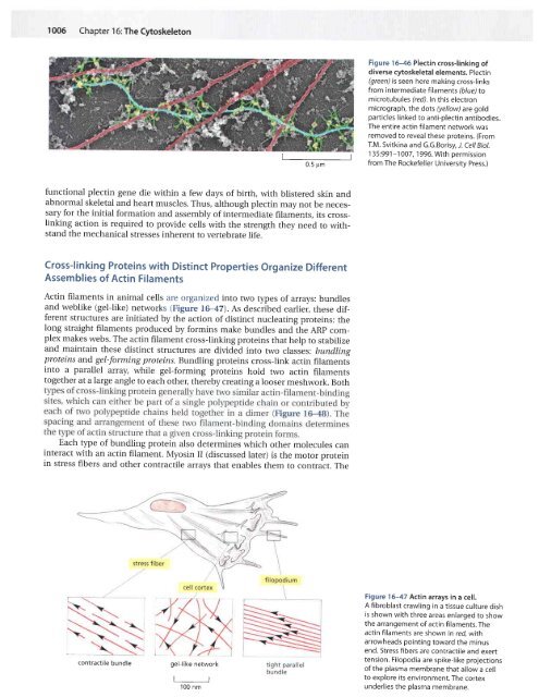
Figure 16-46 Plectin cross-linking of<br />
diverse cytoskeletal elements. Plectin<br />
(green) is seen here making cross-links<br />
from intermediate filaments (blue) to<br />
microtubules ( red). ln this electron<br />
micrograph, the dots (yellow) are gold<br />
particles linked to anti-plectin antibodies.<br />
The entire actin filament network was<br />
removed to reveal these proteins. (From<br />
T.M. Svitkina and G.G.Borisy, J. Cell Biol.<br />
1 35:991-1 007, 1 996. With permission<br />
from The Rockefeller University Press.)<br />
Figure 16-47 Actin arrays in a cell.<br />
A fibroblast crawling in a tissue culture dish<br />
is shown with three areas enlarged to show<br />
the arrangement of actin filaments. The<br />
actin fifaments are shown in red, with<br />
arrowheads pointing toward the minus<br />
end. Stress fibers are contractile and exert<br />
tension. Filopodia are spike-like projections<br />
of the plasma membrane that allow a cell<br />
to explore its environment. The cortex<br />
underlies the plasma membrane.