CH16 Cytoskeleton.pdf - finedrafts
CH16 Cytoskeleton.pdf - finedrafts
CH16 Cytoskeleton.pdf - finedrafts

You also want an ePaper? Increase the reach of your titles
YUMPU automatically turns print PDFs into web optimized ePapers that Google loves.
1030 Chapter 16:The <strong>Cytoskeleton</strong><br />
,".<br />
10<br />
"t<br />
tropon in<br />
comPlex tropomvosin<br />
rc T<br />
+ ca2*<br />
ca2*<br />
myosin-binding site exposed<br />
by Ca'*-mediated tropomyosin<br />
movement<br />
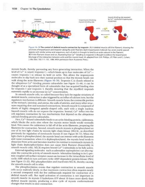
Figure 16-78 The control of skeletal muscle contraction by troponin. (A) A skeletal muscle cell thin filament, showing the<br />
positions of tropomyosin and troponin along the actin filament. Each tropomyosin molecule has seven evenly spaced<br />
regions with similar amino acid sequences, each of which is thought to bind to an actin subunit in the filament.<br />
(B) A thin filament shown end-on, illustrating how Ca2+ (binding to troponin) is thought to relieve the tropomyosin<br />
blockage of the interaction between actin and the myosin head. (A, adapted from G.N. Phillips, J.P. Fillers and C. Cohen,<br />
J. Mol. Biol. 192:111-131,1986. With permission from Academic Press.)<br />
myosin heads, thereby preventing any force-generating interaction. \Mhen the<br />
level of Ca2* is raised, troponin C-which binds up to four molecules of Caz+causes<br />
troponin I to release its hold on actin. This allows the tropomyosin<br />
molecules to slip back into their normal position so that the myosin heads can<br />
walk along the actin filaments (Figure f 6-78). Troponin C is closely related to<br />
the ubiquitous Caz*-binding protein calmodulin (see Figure lS=44); it can be<br />
thought of as a specialized form of calmodulin that has acquired binding sites<br />
for troponin I and troponin ! thereby ensuring that the myofibril responds<br />
extremely rapidly to an increase in Ca2+ concentration.<br />
In smooth muscle cells, so-called because they lack the regular striations of<br />
skeletal muscle, contraction is also triggered by an influx of calcium ions, but the<br />
regulatory mechanism is different. Smooth muscle forms the contractile portion<br />
of the stomach, intestine, and uterus, the walls of arteries, and many other structures<br />
requiring slow and sustained contractions. smooth muscle is composed of<br />
sheets of highly elongated spindle-shaped cells, each with a single nucleus.<br />
Smooth muscle cells do not express the troponins. Instead, Caz* influx into the<br />
cell regulates contraction by two mechanisms that depend on the ubiquitous<br />
calcium binding protein calmodulin.<br />
First, Ca2+-bound calmodulin binds to an actin-binding protein, caldesmon,<br />
which blocks the actin sites where the myosin motor heads would normally<br />
bind. This causes the caldesmon to fall off of the actin filaments, preparing the<br />
filaments for contraction. Second, smooth muscle myosin is phosphorylated on<br />
one of its two light chains by myosin light chain kinase (MLCK.), as described<br />
previously for regulation of nonmuscle myosin II (see Figure 16-72).lVhen the<br />
light chain is phosphorylated, the myosin head can interact with actin filaments<br />
and cause contraction; when it is dephosphorylated, the myosin head tends to<br />
dissociate from actin and becomes inactive (in contrast to nonmuscle myosin II,<br />
light chain dephosphorylation does not cause thick filament disassembly in<br />
smooth muscle cells). MLCK requires bound ca2*/calmodulin to be fully active.<br />
External signaling molecules such as adrenaline (epinephrine) can also regulate<br />
the contractile activity of smooth muscle. Adrenaline binding to its G-protein-coupled<br />
cell surface receptor causes an increase in the intracellular level of<br />
cyclic AMf; which in turn activates cyclic-AMP-dependent protein kinase (pKA)<br />
(see Figure l5-35). PKA phosphorylates and inactivates MLCK, thereby causing<br />
the smooth muscle cell to relax.<br />
The phosphorylation events that regulate contraction in smooth muscle<br />
cells occur realtively slowly, so that maximum contraction often requires nearly<br />
a second (compared with the few milliseconds required for contraction of a<br />
skeletal muscle cell). But rapid activation of contraction is not important in<br />
smooth muscle: its myosin II hydrolyzes ArP about l0 times more slowly than<br />
skeletal muscle myosin, producing a slow cycle of myosin conformational<br />
changes that results in slow contraction.