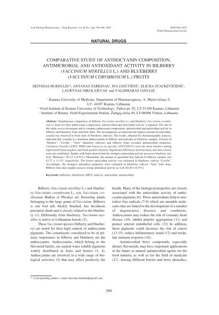
Comparative study of anthocyanin composition, antimicrobial
Comparative study of anthocyanin composition, antimicrobial
Comparative study of anthocyanin composition, antimicrobial

Create successful ePaper yourself
Turn your PDF publications into a flip-book with our unique Google optimized e-Paper software.
Acta Poloniae Pharmaceutica ñ Drug Research, Vol. 66 No. 4 pp. 399ñ408, 2009 ISSN 0001-6837<br />
Polish Pharmaceutical Society<br />
Bilberry (Vaccinium myrtillus L.) and blueberry<br />
(Vaccinium corymbosum L., syn. Vaccinium covilleanum<br />
Butkus et Pliszka) are flowering plants<br />
belonging to the large genus <strong>of</strong> Vaccinium. Bilberry<br />
is one foot tall, thickly brached, has deciduous<br />
perennial shrub and is closely related to the blueberry<br />
(1). Differently from blueberry, Vaccinium myrtillus<br />
is native to Lithuanian forests (2).<br />
These Vaccinium species (bilberry and blueberry)<br />
are noted for their high content <strong>of</strong> <strong>anthocyanin</strong><br />
pigments. The constituents considered to be <strong>of</strong> primary<br />
importance in bilberry and blueberry are the<br />
<strong>anthocyanin</strong>s. Anthocyanins, which belong to the<br />
flavonoid family, are natural water soluble pigments<br />
widely distributed in fruits and berries (3, 4).<br />
Anthocyanins are involved in a wide range <strong>of</strong> biological<br />
activities (5) that may affect positively the<br />
NATURAL DRUGS<br />
COMPARATIVE STUDY OF ANTHOCYANIN COMPOSITION,<br />
ANTIMICROBIAL AND ANTIOXIDANT ACTIVITY IN BILBERRY<br />
(VACCINIUM MYRTILLUS L.) AND BLUEBERRY<br />
(VACCINIUM CORYMBOSUM L.) FRUITS<br />
DEIVIDAS BURDULIS 1 *, ANTANAS äARKINAS 2 , INA JASUTIENË 2 , ELICIJA STACKEVI»IENË 3 ,<br />
LAURYNAS NIKOLAJEVAS 1 and VALDIMARAS JANULIS 1<br />
1 Kaunas University <strong>of</strong> Medicine, Department <strong>of</strong> Pharmacognosy, A. MickeviËiaus 9,<br />
LT- 44307 Kaunas, Lithuania<br />
2 Food Institute <strong>of</strong> Kaunas University <strong>of</strong> Technology, Taikos pr. 92, LT-51180 Kaunas, Lithuania<br />
3 Institute <strong>of</strong> Botany, Field Experimental Station, Þaliøjø eþerø 49, LT-08406 Vilnius, Lithuania<br />
Abstract: Simultaneous comparison <strong>of</strong> bilberry (Vaccinium myrtillus L.) and blueberry (Vaccinium corymbosum<br />
L) fruits for their <strong>anthocyanin</strong> <strong>composition</strong>, <strong>antimicrobial</strong> and antioxidant activity is reported. The aim <strong>of</strong><br />
this <strong>study</strong> was to investigate and to compare <strong>anthocyanin</strong> <strong>composition</strong>, <strong>antimicrobial</strong> and antioxidant activity in<br />
bilberry and blueberry fruits and their skins. The investigations revealed that the highest amount <strong>of</strong> total <strong>anthocyanin</strong>s<br />
was observed in fruits skins <strong>of</strong> blueberry cultivars. The results, obtained by chromatographic analysis,<br />
indicated that cyanidin is a dominant anthocyanidin in bilberry and malvidin in blueberry samples. Extracts <strong>of</strong><br />
ìHerbertî, ìCovilleî, ìToroî blueberry cultivars and bilberry fruits revealed <strong>antimicrobial</strong> properties.<br />
Citrobacter freundii (ATCC 8090) and Enterococcus faecalis (ATCC29212) were the most sensitive among<br />
eight tested Gram-negative and Gram-positive bacteria. Significant differences between berry and skin extracts<br />
were not established. Studies with fruits showed that the strongest antioxidant activity possesses blueberry cultivar<br />
ìBerkeleyî (82.13 ± 0.51%). Meanwhile, the amount <strong>of</strong> quenched free radicals in bilberry samples was<br />
63.72 ± 1.11%, respectively. The lowest antioxidant activity was estimated in blueberry cultivar ìCovilleî.<br />
Accordingly, the strongest antiradical properties were estimated in blueberry cultivar ìAmaî fruit skins.<br />
Bilberry fruit skin samples possess strong antiradical activity as well (82.69 ± 0.37%).<br />
Keywords: bilberries, blueberries, HPLC analysis, antioxidant, <strong>antimicrobial</strong><br />
* Corresponding author: e-mail: deividas.burdulis@gmail.com<br />
399<br />
health. Many <strong>of</strong> the biological properties are closely<br />
associated with the antioxidant activity <strong>of</strong> <strong>anthocyanin</strong><br />
pigments (6). These antioxidants help to neutralize<br />
free radicals (7-9) which are unstable molecules<br />
that are linked to the development <strong>of</strong> a number<br />
<strong>of</strong> degenerative diseases and conditions.<br />
Anthocyanins may reduce the risk <strong>of</strong> coronary heart<br />
disease (10), inhibit platelet aggregation (11) and<br />
protect arterial endothelial cells (12) In addition,<br />
these pigments could decrease the risk <strong>of</strong> cancer<br />
(13-15), reduce inflammatory insult (12) and modulate<br />
immune response (16).<br />
Antimicrobial berry compounds, especially<br />
dietary flavonoids, may have important applications<br />
in the future as natural <strong>antimicrobial</strong> agents for the<br />
food industry as well as for the field <strong>of</strong> medicine<br />
(17). Among different berries and berry phenolics,
400 DEIVIDAS BURDULIS et al.<br />
cranberry, cloudberry, raspberry, strawberry and bilberry<br />
especially possess considerable <strong>antimicrobial</strong><br />
effects against, e.g. Salmonella and Staphylococcus<br />
(18). Several mechanisms <strong>of</strong> action in the growth<br />
inhibition <strong>of</strong> bacteria are involved, such as destabilization<br />
<strong>of</strong> cytoplasmic membrane, permeabilization<br />
<strong>of</strong> plasma membrane, inhibition <strong>of</strong> extracellular<br />
microbial enzymes, direct actions on microbial<br />
metabolism and deprivation <strong>of</strong> the substrates<br />
required for microbial growth. Antimicrobial activity<br />
<strong>of</strong> berries may also be related to antiadherence <strong>of</strong><br />
bacteria to epithelial cells, which is a prerequisite for<br />
colonization and infection <strong>of</strong> many pathogens. It<br />
was found that myricetin inhibited the growth <strong>of</strong> all<br />
lactic acid bacteria derived from the human gastrointestinal<br />
tract flora but did not affect the<br />
Salmonella strain (19). Extracts from common<br />
Finnish berries inhibited the growth <strong>of</strong> Gram-negative,<br />
but not Gram-positive bacteria. Other authors<br />
reported that there was no correlation between<br />
Gram-positive or Gram-negative bacterial status and<br />
susceptibility to the berries (20).<br />
The aim <strong>of</strong> our <strong>study</strong> was to compare total<br />
<strong>anthocyanin</strong> and anthocyanidin content and their<br />
<strong>composition</strong>, <strong>antimicrobial</strong> and antioxidant activity<br />
in the bilberry and blueberry fruits and skins.<br />
MATERIALS AND METHODS<br />
Raw materials and extraction procedures<br />
Wild bilberry (Vaccinium myrtillus) fruit samples<br />
were collected randomly in natural sampling<br />
sites in the territory <strong>of</strong> Lithuania and blueberry<br />
(Vaccinium corymbosum) were collected in the institute<br />
<strong>of</strong> botany in Vilnius. Fresh, ripe fruit samples<br />
were frozen and stored in the freezer at -19 ± 1 O C<br />
until use.<br />
Approximately 50 g <strong>of</strong> frozen berries were<br />
crushed in a pounder to produce a thick puree. A 5 g<br />
(accurate weight) aliquot was placed into a 100 mL<br />
flat-bottom flask and 95 mL <strong>of</strong> methanol was added.<br />
An ultrasonic bath BioSonic UC 100 (Coltene/<br />
Whaledent, Mahwah, NJ, USA) was used for sonication<br />
with occasional swirling. Ten min period <strong>of</strong><br />
sonication was applied for extraction procedure<br />
(21). Three replicates <strong>of</strong> samples were prepared for<br />
all analytes.<br />
Chemicals<br />
All solvents, reagents and standards, used in<br />
this <strong>study</strong>, were <strong>of</strong> analytical grade. Acetonitrile<br />
(HPLC grade) and orthophosphoric acid (85% purity,<br />
HPLC grade) were purchased from Sigma ñ<br />
Aldrich GmbH, (Buchs, Switzerland). The Folin-<br />
Ciocalteu reagent, sodium carbonate and 2,2diphenyl-1-picrylhydrazyl<br />
radical (DPPH) (95%)<br />
were purchased from Sigma-Aldrich Chemie<br />
GmbH, (Steinheim, Germany). Methanol was provided<br />
by Roth (Karlsruhe, Germany). Hydrochloric<br />
acid (35%) was purchased from Lachema<br />
(Neratovice, Czech Republic). Anthocyanidin standards<br />
(cyanidin chloride, delphinidin chloride, petunidin<br />
chloride, peonidin chloride and malvidin chloride)<br />
were <strong>of</strong> HPLC purity grade and purchased<br />
from Roth (Karlsruhe, Germany) and Chromadex<br />
(Santa Ana, USA). Water was deionized and filtered<br />
through a Millipore filter system (Millipore, USA)<br />
before use.<br />
Total phenolics measurement<br />
The amount <strong>of</strong> total phenolics in the berry<br />
extracts was determined with the Folin-Ciocalteu<br />
reagent according to the method <strong>of</strong> Slinkard and<br />
Singleton (22) using gallic acid as a standard. The<br />
reagent was prepared by diluting a stock solution<br />
(Sigma-Aldrich Chemie GmbH, Steinheim, Germany)<br />
with distilled water (1:10, v/v). Samples (1.0<br />
mL, two replicates) were introduced into test<br />
cuvettes, and then 5.0 mL <strong>of</strong> Folin-Ciocalteuís phenol<br />
reagent, and 4.0 mL <strong>of</strong> Na 2CO 3 (7.5%) were<br />
added. The absorbance <strong>of</strong> all samples was measured<br />
at 765 nm using the Genesys10 UV/Vis spectrophotometer<br />
(Thermo Spectronic, Rochester, USA) after<br />
incubation at 20 O C for 1.0 h.<br />
Total <strong>anthocyanin</strong>s measurement<br />
Total <strong>anthocyanin</strong> contents <strong>of</strong> berry extracts<br />
were measured using the spectrophotometric<br />
method. The samples were determined using a<br />
model Unicam Helios ALPHA spectrophotometer<br />
(Unicam, Cambridge, UK). The extract was filtrered<br />
through paper filter into a 100.0 mL volumetric<br />
flask and diluted with appropriate extractant to<br />
100.0 mL. A 50-fold dilution <strong>of</strong> this solution in a<br />
0.1% v/v <strong>of</strong> hydrochloric acid in methanol was prepared.<br />
The absorbance <strong>of</strong> the solution was measured<br />
at 528 nm wavelength, using a 0.1% v/v solution <strong>of</strong><br />
hydrochloric acid in methanol as a blank. The results<br />
are expressed as a percentage <strong>of</strong> cyanidin 3-glucoside<br />
equivalents (23).<br />
Acid hydrolysis and chromatographic analysis<br />
Twenty five mL <strong>of</strong> methanolic extract was<br />
mixed with 8.5 mL <strong>of</strong> concentrated hydrochloric<br />
acid. The mixture was heated under reflux condenser<br />
for 2 h. After acid hydrolysis, the <strong>anthocyanin</strong><br />
content was converted to five major anthocyanidins.<br />
The hydrolyzed samples were passed through 0.22
µm pore size membrane filters (Carl Roth GmbH,<br />
Karlsruhe, Germany) prior to HPLC injection (24).<br />
Commercially available anthocyanidin standards<br />
were dissolved together in methanol to form a standard<br />
mixture <strong>of</strong> cyanidin chloride, delphinidin chloride,<br />
petunidin chloride, peonidin chloride, and malvidin<br />
chloride. The standard mixture was diluted in<br />
methanol to generate a five-point calibration curve for<br />
five anthocyanidin standard compounds separately.<br />
The peak areas <strong>of</strong> the anthocyanidins in fruit extracts<br />
were within the linear range <strong>of</strong> the calibration curves.<br />
The experiments were performed with Waters 2690<br />
Alliance HPLC system, equipped with Waters 2487<br />
Dual Absorbance Detector (UV/Vis), and Waters 996<br />
Photodiode Array (PDA) Detector (Waters<br />
Corporation, Milford, MA, USA), coupled to a personal<br />
computer with Waters Millennium 2000w chromatographic<br />
manager system (Waters Corporation,<br />
Milford, MA, USA) for data storage and processing.<br />
Separation <strong>of</strong> anthocyanidins was carried out using a<br />
Supelco Hypersil ODS (C18) column (100 ◊ 3 mm<br />
i.d.; particle size 3 mm), guarded with a Hypersil<br />
ODS (10 ◊ 4.6 mm i.d.; particle size 5 mm) guard column<br />
(Supelco, Bellefonte USA). The column was<br />
kept at ambient temperature. The flow rate was kept<br />
constant at 1.0 mL/min for a total run time <strong>of</strong> 45 min.<br />
The mobile phases were: A ñ acetonitrile and B ñ 4%<br />
aqueous ortophosphoric acid (v/v). The system was<br />
run with a gradient program: 0ñ37 min, 7ñ25% A,<br />
37ñ40 min, 25ñ7% A and 40ñ45 min 7% A. There<br />
was a 5 min post run at initial conditions for the column<br />
equilibration. The injection volume for all standard<br />
solutions and bilberry extracts was 10 µL. The<br />
UV/Vis detector was set at 520 nm, PDA 200-800 nm<br />
wavelength. Identification <strong>of</strong> anthocyanidins was<br />
achieved by comparing their retention times and UV-<br />
Vis spectra, obtained with a PDA detector, with those<br />
<strong>of</strong> the standards (21).<br />
Antimicrobial test<br />
Assessment <strong>of</strong> the <strong>antimicrobial</strong> activity <strong>of</strong> the<br />
extracts was performed on the following Gram-positive<br />
bacterial test cultures Listeria monocytogenes<br />
(ATCC 19117), Staphylococcus aureus (ATCC<br />
25923), Bacillus subtilis ( ATCC 6633) (spores and<br />
vegetative cells), Enterococcus faecalis (ATCC<br />
29212), and also on the Gram-negative bacterial test<br />
cultures Escherichia coli (ATCC 25922),<br />
Pseudomonas aeruginosa (ATCC 27853),<br />
Citrobacter freundii (ATCC 8090), and Salmonella<br />
typhimurium (ATCC 14028). Eight strains <strong>of</strong> yeasts<br />
were also used: Debaryomyces hansenii,<br />
Trichosporon cutaneum, Kluyveromyces marxianus<br />
var. lactis, Sacharomyces cerevisiae, Candida para-<br />
<strong>Comparative</strong> <strong>study</strong> <strong>of</strong> <strong>anthocyanin</strong> <strong>composition</strong>, <strong>antimicrobial</strong>... 401<br />
psilosis, Torulaspora delbrueckii, Pichia kluyveri,<br />
and Rhodotorula rubra.<br />
The <strong>antimicrobial</strong> properties were evaluated by<br />
the agar well diffusion method. The bacteria were<br />
grown in peptone-soy bouillon (LAB 04, LAB M)<br />
for 24 h at 37 O C. After cultivation, test culture cells<br />
were mixed using a minishaker MS 1 (Wilmington.<br />
USA) and the cell suspensions were adjusted<br />
according to McFarland No. 0.5 standard (25). The<br />
suspension <strong>of</strong> bacteria cells was introduced into the<br />
dissolved media and cooled to 47 O C; 20 mL was<br />
pipetted into a 90 mm diameter Petri plate. Wells<br />
eight-millimeters in diameter were pushed in the<br />
agar and filled with 50 µL <strong>of</strong> the ethanol extracts <strong>of</strong><br />
berry or berry skin. The plates were incubated<br />
overnight at 37 O C.<br />
The yeasts were grown on a potato dextrose<br />
agar slant (LAB 98, LAB M) for 48 h at 25 O C. After<br />
cultivation the yeast cells were washed with saline<br />
and the cell suspensions were treated as above. The<br />
media containing the yeast cells and oils were incubated<br />
overnight at 25 O C. A 50 mL volume <strong>of</strong> ethanol<br />
extract <strong>of</strong> berry or berry cakes was added to the agar<br />
wells using an automatic pipettor. Ethanol was used<br />
as a control in blank samples for bacteria and yeast<br />
at the same conditions. Ceftazidime/clavulanic acid<br />
30/10 µg sensi-disc (Becton, Dickinson and<br />
Company, USA) was used as positive control. After<br />
incubation the inhibition zones were measured with<br />
callipers to an accuracy <strong>of</strong> 0.1 mm and the effect<br />
was calculated as a mean <strong>of</strong> three replicate tests.<br />
Antioxidant activity assay<br />
The antioxidant activities <strong>of</strong> the extracts were<br />
determined using 2,2-diphenyl-1-picrylhydrazyl<br />
(DPPH) method (26) to determine free radical-scavenging<br />
potential <strong>of</strong> each sample. The antiradical<br />
activity was calculated as a percentage <strong>of</strong> DPPH<br />
decoloration versus control (methanolic solution).<br />
Fifty µL <strong>of</strong> prepared extract was added to 2 mL <strong>of</strong><br />
DPPH methanolic solution (6 ◊ 10 -5 M). The reaction<br />
was carried out at ambient temperature.<br />
Changes in the absorbance at 515 nm due to the<br />
scavenging <strong>of</strong> DPPH radical were measured with a<br />
spectrophotometer every 10 s for a period <strong>of</strong> 30 min,<br />
to attain reaction equilibrium (27). Antioxidant<br />
activity <strong>of</strong> berry extracts was expressed by percentage<br />
<strong>of</strong> inactivated DPPH reagent (28):<br />
A b ñ A a<br />
DDPH = ñññññññ × 100<br />
A b<br />
where Ab = absorbtion <strong>of</strong> blank aliquot (t = 0 min.),<br />
Aa = absorbtion <strong>of</strong> solution with analyzed extract<br />
(after 30 min).
402 DEIVIDAS BURDULIS et al.<br />
Figure 1. Total <strong>anthocyanin</strong>s content in bilberry and blueberry cultivars, berry and berry skins<br />
Figure 2. Representative chromatograms <strong>of</strong> anthocyanidins, a ñ bilberry, b ñ blueberry, 1 ñ delphinidin, 2 ñ cyanidin, 3 ñ petunidin, 4 ñ<br />
peonidin, 5 ñ malvidin.<br />
Statistical data analysis<br />
All <strong>of</strong> the assays were performed in triplicate.<br />
The mean values and standard deviations were<br />
obtained using the SPSS (Chicago, JAV) computer<br />
package.<br />
RESULTS AND DISCUSSION<br />
Anthocyanin and anthocyanidin <strong>composition</strong><br />
For the quantification <strong>of</strong> total <strong>anthocyanin</strong>s<br />
content in bilberry and blueberry fruits, the spectrophotometric<br />
assay, descripted in the European<br />
Pharmacopoeia, was performed. Spectrophotometric<br />
method is widely used for standardization <strong>of</strong><br />
natural raw material. Total <strong>anthocyanin</strong> content in<br />
bilberry and blueberry fruits and their skins is presented<br />
in Figure 1. The highest amount <strong>of</strong> <strong>anthocyanin</strong>s<br />
was observed in fruits skins <strong>of</strong> blueberry<br />
cultivars. The highest content <strong>of</strong> <strong>anthocyanin</strong>s in<br />
blueberry skins was found in ìCovilleî cultivar<br />
skins (1.77%), the lowest ñ in ìToroî cultivar fruits<br />
skins. Though, identical amounts <strong>of</strong> total <strong>anthocyanin</strong>s<br />
were determined in ìHeermaî cultivar and<br />
bilberry skins samples (1.13 %). However the high-
Figure 3. Radical scavenging activity dependence on time<br />
Table 1. Anthocyanidin contents in bilberry and blueberry cultivars berry<br />
est content <strong>of</strong> <strong>anthocyanin</strong>s in fruits was found in<br />
bilberry fruits (0.6%) compared with blueberry<br />
fruits. The higher amount <strong>of</strong> <strong>anthocyanin</strong>s in blueberry<br />
fruits was found in ìBerkeleyî and<br />
ìNorthlandî cultivars berries. The lowest one ñ in<br />
ìToroî cultivar berries (0.12%).<br />
Acid mediated hydrolysis was performed and<br />
five major anthocyanidins (delphinidin, cyanidin,<br />
petunidin, peonidin and malvidin) were estimated<br />
by high performance liquid chromatography<br />
(HPLC) in bilberry and blueberry extracts, respectively.<br />
Numerous techniques are used for separation<br />
and quantification <strong>of</strong> <strong>anthocyanin</strong>s. HPLC is a<br />
method <strong>of</strong> choice for the analysis <strong>of</strong> <strong>anthocyanin</strong>s<br />
for it ensures efficient and simple identification,<br />
<strong>Comparative</strong> <strong>study</strong> <strong>of</strong> <strong>anthocyanin</strong> <strong>composition</strong>, <strong>antimicrobial</strong>... 403<br />
Sample raw material<br />
Anthocyanidin contents in berry (%)<br />
Delphinidin Cyanidin Petunidin Peonidin Malvidin Sum<br />
Bilberry 0.12 0.25 0.10 0.05 0.06 0.58<br />
Berkeley 0.04 0.04 0.05 0.01 0.05 0.17<br />
Heerma 0.02 0.01 0.03 0.002 0.02 0.09<br />
Northland 0.05 0.02 0.06 0.01 0.07 0.20<br />
Coville 0.01 0.01 0.02 0.002 0.04 0.08<br />
Herbert 0.01 0.002 0.02 0.002 0.03 0.07<br />
Toro 0.03 0.01 0.04 0.002 0.05 0.13<br />
Mean 0.04 0.05 0.05 0.01 0.05<br />
Min. 0.01 0.002 0.02 0.002 0.02<br />
Max. 0.12 0.25 0.10 0.05 0.07<br />
SD 0.03 0.08 0.02 0.02 0.02<br />
separation and quantification <strong>of</strong> individual anthocyanidins<br />
in bilberry and blueberry fruits and their<br />
products. The developed HPLC method, used in this<br />
<strong>study</strong> was optimized, validated and published previously<br />
(21). Representative HPLC chromatograms <strong>of</strong><br />
bilberry and blueberry extracts are shown in Figure<br />
2. Chromatographic analysis revealed that qualitative<br />
<strong>composition</strong> <strong>of</strong> anthocyanidins in bilberry and<br />
blueberry fruits and their skins is homogeneous.<br />
Quantitative results obtained for the amount <strong>of</strong> individual<br />
anthocyanidins in bilberry and blueberry<br />
fruits are presented in Table 1 and those in fruit<br />
skins in Table 2. The HPLC method was applied and<br />
variation <strong>of</strong> anthocyanidin <strong>composition</strong> in bilberry<br />
and blueberry extracts was estimated. The obtained
404 DEIVIDAS BURDULIS et al.<br />
Table 2. Anthocyanidin contens in bilberry and blueberry cultivars berry skins<br />
Sample raw material<br />
Anthocyanidin contents in berry (%)<br />
Delphinidin Cyanidin Petunidin Peonidin Malvidin Sum<br />
Bilberry 0.23 0.49 0.18 0.09 0.10 1.09<br />
Berkeley 0.24 0.24 0.31 0.07 0.37 1.24<br />
Heerma 0.24 0.24 0.31 0.07 0.37 1.24<br />
Northland 0.24 0.24 0.31 0.07 0.37 1.24<br />
Coville 0.24 0.24 0.31 0.07 0.37 1.24<br />
Herbert 0.11 0.04 0.17 0.01 0.30 0.63<br />
Toro 0.23 0.12 0.12 0.03 0.48 0.99<br />
Mean 0.29 0.18 0.31 0.05 0.42<br />
Min. 0.11 0.04 0.12 0.01 0.10<br />
Max. 0.49 0.49 0.59 0.09 0.77<br />
SD 0.11 0.13 0.15 0.02 0.19<br />
Table 3. The <strong>antimicrobial</strong> influence <strong>of</strong> 50 mL berry extracts on Gram-negative test cultures<br />
Sample raw<br />
Inhibition zone size, mm<br />
material E. coli P. aeruginosa S. typhimurium C. freundii<br />
ìHerbertî berry 13.66 ± 0.47 14.00 ± 0.00 20.00 ± 0.00 21.66 ± 1.24<br />
ìCovilleî berry 14.66 ± 0.47 12.66 ± 0.47 18.00 ± 0.00 21.66 ± 0.47<br />
ìToroî berry 15.66 ± 0.47 12.66 ± 0.47 15.00 ± 0.00 21.66 ± 0.94<br />
ìHerbertî skin 13.66 ± 0.47 14.66 ± 0.47 16.33 ± 0.47 22.66 ± 0.47<br />
ìCovilleî skin 13.33 ± 0.47 13.00 ± 0.00 16.00 ± 0.00 23.00 ± 0.00<br />
ìToroî skin 13.66 ± 0.47 14.33 ± 0.47 14.00 ± 0.00 25.00 ± 0.00<br />
Bilberry skin 17.33 ± 0.47 14.66 ± 1.90 11.00 ± 0.01 Not investigated<br />
Bilberry berry<br />
Ceftazidime/<br />
15.39 ± 1.24 12.30 ± 0.40 13.33 ± 1.24 Not investigated<br />
Clavulanic acid<br />
30/10 µg<br />
38.0 ± 0.00 36.10 ± 0.47 34.30 ± 0.06 34.00 ± 0.00<br />
results indicated that cyanidin is a dominant anthocyanidin<br />
in bilberry and malvidin in blueberry samples.<br />
It was determined that bigger amounts <strong>of</strong><br />
anthocyanidins are accumulated in skins <strong>of</strong> blueberry<br />
and bilberry fruits than in entire fruits. The<br />
biggest amounts <strong>of</strong> anthocyanidins were estimated<br />
in blueberry cultivars skins, where total athocyanidins<br />
reached 2% (ìCovilleî cultivar), whereas the<br />
smallest amount <strong>of</strong> total anthocyanidins was determined<br />
in ìHerbertî cultivar skins (0.63%), however,<br />
differently from skins, the biggest amounts <strong>of</strong> anthocyanidins<br />
were found in bilberry fruit samples. The<br />
amounts <strong>of</strong> total anthocyanidins in bilberry fruits<br />
exceeded 0.5% and the smallest amounts <strong>of</strong> total<br />
anthocyanidins were assessed in blueberry<br />
ìHerbertî cultivar (0.07%). Variation <strong>of</strong> individual<br />
anthocyanidin <strong>composition</strong> in bilberry and blueberry<br />
cultivar fruits and fruit skins was evaluated as<br />
well. It was found that the biggest amounts <strong>of</strong> individual<br />
anthocyanidins (delphinidin, cianidin, petunidin,<br />
peonidin) are accumulated in bilberry fruits,<br />
except <strong>of</strong> malvidin. The biggest amounts <strong>of</strong> malvidin<br />
were estimated in blueberry ìNorthlandî cultivar<br />
fruits (0.07%).<br />
The highest content <strong>of</strong> delphinidin, petunidin<br />
and malvidin (0.49%, 0.59% and 0.77%, respectively)<br />
were determined in ìCovilleî cultivar fruit skins,<br />
however, the biggest amounts <strong>of</strong> cyanidin and peoni-
din were assayed in bilberry skin samples. The lowest<br />
content <strong>of</strong> all individual anthocyanidins was<br />
determined in ìHerbertî cultivar fruit skins.<br />
Antimicrobial assay<br />
Investigation <strong>of</strong> <strong>antimicrobial</strong> properties <strong>of</strong><br />
berry extracts by the diffusion to agar method<br />
showed that among Gram-negative test cultures C.<br />
<strong>Comparative</strong> <strong>study</strong> <strong>of</strong> <strong>anthocyanin</strong> <strong>composition</strong>, <strong>antimicrobial</strong>... 405<br />
Table 4. The <strong>antimicrobial</strong> influence <strong>of</strong> 50 mL blueberry extracts on Gram-positive test cultures<br />
Sample raw<br />
Inhibition zone size, mm<br />
material B. subtilis<br />
vegetative cells<br />
B. subtilis<br />
spores<br />
L. monocytogenes S. aureus E. faecalis<br />
ëHerbertí berry 17.33 ± 0.47 17.66 ± 0.47 19.00 ± 0.00 18.66 ± 0.47 26.00 ± 0.00<br />
ëCovilleí berry 15.00 ± 0.00 13.66 ± 0.47 17.66 ± 0.47 18.00 ± 0.81 26.00 ± 0.00<br />
ëToroí berry 19.33 ± 0.47 14.33 ± 0.47 20.33 ± 0.47 17.00 ± 0.00 30.66 ± 0.94<br />
ëHerbertí skin 17.00 ± 0.00 13.33 ± 0.47 18.00 ± 0.00 15.66 ± 0.47 22.66 ± 0.47<br />
ëCovilleí skin 14.66 ± 0.47 13.33 ± 0.47 19.00 ± 0.00 16.00 ± 0.00 23.00 ± 0.00<br />
ëToroí skin 17.66 ± 0.47 14.00 ± 0.00 22.33 ± 0.47 17.66 ± 0.47 25.00 ± 0.00<br />
Bilberry skin 18.00 ± 0.00 12.00 ± 0.00 15.00 ± 0.00 13.00 ± 0.00 Not investigated<br />
Bilberry berry<br />
Ceftazidime/<br />
18.00 ± 0.00 14.66 ± 0.47 18.00 ± 1.60 15.00 ± 0.81 Not investigated<br />
Clavulanic acid<br />
30/10µg<br />
31.00 ± 0.00 31.00 ± 0.00 32.00±0.00 27.00 ± 0.00 36.00 ± 0.00<br />
Table 5. The <strong>antimicrobial</strong> influence <strong>of</strong> 50 mL blueberry extracts on yeast test cultures<br />
Sample Inhibition zone size, mm<br />
raw Debaryomyces Trichosporon Kliuveromyces Torulaspora Saccharomyces Candida Pichia Rhodotorula<br />
material<br />
ìHerbertî<br />
hansenii cataneum marxianus<br />
var. lactis<br />
delbrueckii cerevisiae parapsilosis kluyveri rubra<br />
berry<br />
ìCovilleî<br />
0 0 0 0 0 0 0 0<br />
berry<br />
ìToroî<br />
0 0 0 9.33 ± 0.47 10.00 ± 0.0 0 0 0<br />
berry<br />
ìHerbertî<br />
0 0 0 0 0 0 0 0<br />
skin<br />
ìCovilleî<br />
0 0 0 13.33 ± 0.47 17.00 ± 0.00 0 0 0<br />
skin<br />
ìToroî<br />
0 0 0 13.00 ± 0.00 17.00 ± 0.00 0 0 0<br />
skin<br />
Bilberry<br />
0 0 0 10.33 ± 0.81 16.33 ± 0.81 0 0 0<br />
skin<br />
Bilberry<br />
0 0 0 13.00 ± 0.00 11.50 ± 0.07 12.00 ± 0.47 13.00 ± 0.00 0<br />
berry 0 0 0 11.05 ± 0.33 15.00 ± 0.00 9.66 ± 0.47 9.66 ± 0.47 0<br />
freundii were the most sensitive ñ average zones <strong>of</strong><br />
inhibition were 21.66 mm for skin, and 23.55 mm<br />
for berry extracts (Table 3). E. coli showed largest<br />
resistance to berry extracts; the average zone <strong>of</strong> inhibition<br />
was 13.6 mm for all investigated samples.<br />
Though phenolic compounds and <strong>anthocyanin</strong> content<br />
was higher in the extracts <strong>of</strong> skin, significant<br />
differences between extracts <strong>of</strong> berries and berry
406 DEIVIDAS BURDULIS et al.<br />
Table 6. Antioxidant activity <strong>of</strong> bilberry and blueberry cultivars<br />
Radical ñ scavenging activity (%)<br />
Berry Berry skins<br />
Bilberry<br />
Blueberry cultivars<br />
63.72 ± 1.11 82.69 ± 0.37<br />
Berkeley 82.13 ± 0.51 87.25 ± 0.23<br />
Heerma 50.40 ± 0.57 92.10 ± 1.10<br />
Ama 81.65 ± 0.68 93.15 ± 0.11<br />
Northland 53.34 ± 0.72 76.68 ± 3.15<br />
Coville 51.30 ± 0.72 59.88 ± 0.45<br />
Herbert 76.22 ± 0.36 82.19 ± 0.34<br />
Toro 71.27 ± 0.23 77.73 ± 0.24<br />
Each value is the mean ± standart devation <strong>of</strong> 3 replicate assays<br />
skin were not established. All investigated cultivars<br />
showed the same <strong>antimicrobial</strong> activity. The extracts<br />
<strong>of</strong> bilberry and bilberry skin had the strongest influence<br />
on E. coli. Unfortunately, the <strong>antimicrobial</strong><br />
influence to C. freundii was not investigated.<br />
The ethanol extracts <strong>of</strong> berry and berry skins<br />
showed inhibitory effects on growing <strong>of</strong> Gram-positive<br />
test cultures L. monocytogenes and B. subtilis<br />
(Table 4). More sensitive was L. monocytogenes test<br />
culture. After comparison <strong>of</strong> <strong>antimicrobial</strong> influence<br />
on spores and vegetative cells, it was found that B.<br />
subtilis vegetative cells were more sensitive and<br />
made large inhibition zones (16.83 mm). When<br />
spores graduate to the vegetative form or during germination<br />
process, B. subtilis spores made transparent<br />
inhibitory zones with the average diameter <strong>of</strong><br />
14.38 mm. The test cultures in coccus form showed<br />
sensitivity to the blueberry extracts <strong>of</strong> investigated<br />
cultivars: S. aureus and E. faecalis made large inhibition<br />
zone in diameter 15.66 ñ 18.66 mm and 22.66<br />
ñ 30.66 mm, respectively. The differences between<br />
extracts <strong>of</strong> the investigated blueberry cultivars and<br />
bilberry were not established. All investigated test<br />
cultures were more sensitive than the<br />
ceftazidime/clavulanic acid 30/10 µg sensi-disc.<br />
The eight yeast species Debaryomyces<br />
hansenii, Trichosporon cutaneum, Kluyveromyces<br />
marxianus var. Laktis, Sacharomyces cerevisiae,<br />
Candida parapsilosis, Torulaspora delbrueckii,<br />
Pichia kluyveri, and Rhodotorula rubra showed<br />
complete resistance to the blueberry extracts (Table<br />
5). Most <strong>of</strong> inhibition zones were 0.00 mm. Minimal<br />
sensitivity was demonstrated only by Torulaspora<br />
delbrueckii and Saccharomyces cerevisiae. Similar<br />
results were obtained after investigation <strong>of</strong> black<br />
currant, cranberry and bilberry extracts (29).<br />
Minimal sensitivity showed only the bilberry<br />
extract. Such resistance is explained by the wide<br />
occurrence <strong>of</strong> yeasts on the plants and berries.<br />
Antioxidant activity<br />
Anthocyanins are efficient free radical scavengers,<br />
due to their ability to inactivate free radicals.<br />
Photometric DPPH bleaching method was applied<br />
for measuring radical scavenging activity <strong>of</strong> bilberry<br />
and blueberry extracts. This method is unsophisticated,<br />
cheap and sufficiently accurate and shows<br />
the radical scavenging activities <strong>of</strong> berry extracts,<br />
contributed by both <strong>anthocyanin</strong>s and other compounds.<br />
However, as it is well known and confirmed<br />
by our <strong>study</strong>, berries contain a large amount <strong>of</strong> phenolic<br />
compounds (Table 3), which has antioxidant<br />
activity as well. Since the blueberry and bilberry<br />
extracts are very complex, it is difficult to distinguish<br />
which compounds contribute the most to<br />
antioxidant activity. The bilberry and blueberry<br />
fruits, besides <strong>anthocyanin</strong>s, are rich in flavonoids<br />
(catechin, epikatechin, myrcetin, quercetin, and<br />
kempferol), phenolic acids, chlorogenic acid and<br />
ascorbic acid, which possess antiradical properties<br />
as well (30, 31).<br />
Anthocyanins and other polyphenolics, major<br />
contributors to antioxidant activity in berries, have<br />
similar patterns <strong>of</strong> behavior in response to time. The<br />
DPPH radical inactivation dependence from time<br />
was evaluated. With an increase <strong>of</strong> time the amount<br />
<strong>of</strong> quenched free radical was increased proportionally.<br />
It was found that equilibrium, when amount <strong>of</strong><br />
inactivated free radical is stable, was attained after<br />
30 min. The analysis was carried out with bilberry<br />
and blueberry fruits and fruit skins. The free radical<br />
inactivation dependence on time is presented in<br />
Figure 3. The antioxidant activity was assayed for<br />
bilberry and blueberry (ìAmaî, ìBerkeleyî,
ìCovilleî, ìHeermaî, ìHerbertî, ìToroî cultivars)<br />
fruits and fruit skins. On the basis <strong>of</strong> the obtained<br />
data, bilberry and blueberry skins were distinguished<br />
by stronger antioxidant activity. This may<br />
be due to the estimated higher content <strong>of</strong> <strong>anthocyanin</strong>s<br />
and phenolic compounds in the investigated<br />
samples. Our studies with fruits revealed that the<br />
strongest antioxidant activity possess blueberry cultivar<br />
ìBerkeleyî, in which the amount <strong>of</strong> quenched<br />
free radicals was 82.13 ± 0.51%. Meanwhile, the<br />
amount <strong>of</strong> quenched free radicals in bilberry samples<br />
was 63.72 ± 1.11%, respectively. The lowest<br />
antioxidant activity was estimated in blueberry cultivar<br />
ìCovilleî (51.30 ± 0.72%). Accordingly, the<br />
strongest antiradical properties were estimated in<br />
blueberry cultivar ìAmaî fruit skins where the<br />
amount <strong>of</strong> quenched free radical was 93.15 ± 0.11%.<br />
The bilberry fruit samples possess strong antiradical<br />
activity as well (82.69 ± 0.37%). The lowest antioxidant<br />
activity was found in blueberry cultivar<br />
ìCovilleî fruit skins (59.88 ± 0.45%). The amounts<br />
(in percentage) <strong>of</strong> the inactivated DPPH radical are<br />
presented in Table 6.<br />
CONCLUSIONS<br />
Our <strong>study</strong> revealed that the highest amount <strong>of</strong><br />
total <strong>anthocyanin</strong>s was observed in fruits skins <strong>of</strong><br />
blueberry cultivars. The highest content <strong>of</strong> <strong>anthocyanin</strong>s<br />
in blueberry skins was found in ìCovilleî<br />
cultivar skins (1.77%), the lowest ñ in ìToroî cultivar<br />
fruits skins.<br />
Chromatographic results indicated that cyanidin<br />
is a dominant anthocyanidin in bilberry and malvidin<br />
in blueberry samples. It was determined that<br />
bigger amounts <strong>of</strong> anthocyanidins are accumulated<br />
in skins <strong>of</strong> blueberry and bilberry fruits than in<br />
entire fruits.<br />
The extracts <strong>of</strong> ìHerbertî, ìCovilleî, ìToroî<br />
blueberry cultivars and bilberry revealed <strong>antimicrobial</strong><br />
properties. Citrobacter freundii (ATCC 8090)<br />
and Enterococcus faecalis (ATCC29212) were the<br />
most sensitive among eight tested Gram-negative<br />
and Gram-positive bacteria: inhibitory zones were<br />
22.6 and 25.6 mm, respectively. Significant differences<br />
between berry and skin extracts were not<br />
established. The eight yeast species showed resistance<br />
to the blueberry and bilberry extracts.<br />
Our studies with fruits revealed that the<br />
strongest antioxidant activity posseses blueberry<br />
cultivar ìBerkeleyî (82.13 ± 0.51%). Meanwhile,<br />
the amount <strong>of</strong> quenched free radical in bilberry samples<br />
was 63.72 ± 1.11%, respectively. The lowest<br />
antioxidant activity was estimated in blueberry cul-<br />
<strong>Comparative</strong> <strong>study</strong> <strong>of</strong> <strong>anthocyanin</strong> <strong>composition</strong>, <strong>antimicrobial</strong>... 407<br />
tivar ìCovilleî whereas the strongest antradical<br />
properties were estimated in blueberry cultivar<br />
ìAmaî fruit skins. The amount <strong>of</strong> quenched free<br />
radical was 93.15 ± 0.11%. The bilberry fruit skin<br />
samples possess strong antiradical activity as well<br />
(82.69 ± 0.37%).<br />
Acknowledgment<br />
We thank the scientific foundation <strong>of</strong> Kaunas<br />
University <strong>of</strong> Medicine for supporting this <strong>study</strong>.<br />
REFERENCES<br />
1. American Herbal Pharmacopoeia. p. 1, Santa<br />
Cruz, CA: HAP 2001.<br />
2. StackeviËienë E., Butkus V., Butkienë Z., Eds.:<br />
Large cranberry, highbush and lowbush blueberry<br />
and lingonberry introduction in Lithuania,<br />
p. 58, Lithuanian Academy <strong>of</strong> Sciences<br />
Publishers, Vilnius 1997.<br />
3. Skrede G., Wrolstad R.: Flavanoids from Beries<br />
and Grapes. In Functional Foods: Biochemical<br />
and Processing Aspects, vol. 2, Shi J., Mazza<br />
G., Le Maguer M., Boca R. Eds., p. 71, CRC<br />
Press, Boca Raton 2002.<br />
4. Mazza G., Minaiti E. Eds. Anthocyanins in<br />
fruits, vegetable, and grains. p. 362, CRC Press,<br />
Boca Raton 1993.<br />
5. Kong J.M., Chia L.S., Goh N.K., et al.:<br />
Phytochemistry 64, 923 (2003).<br />
6. Prior R.L., Cao G., Martin A., S<strong>of</strong>ic E., et al.: J.<br />
Agric. Food Chem. 46, 2686 (1998).<br />
7. Prior, R.L.: Am. J. Clin. Nutr. 78, 570 (2003).<br />
8. Ichiyanagi T., Hatano Y., Matsuo S., et al.:<br />
Chem. Pharm. Bull. 52, 1312 (2004).<br />
9. Zheng W., Wang S.Y.: J. Agric. Food Chem.<br />
51, 502 (2003).<br />
10. Reed J.: Crit. Rev. Food Sci. Nutr. 42, 301<br />
(2002).<br />
11. Marozzoni P., Magistretti M. J.: Fitoterapia 61,<br />
13 (1990).<br />
12. Yuodim K.A., McDonald J., Kalt W., et al.: J.<br />
Nutr. Biochem. 13, 282 (2002).<br />
13. Lazze M.C., Savio M., Pizzala R., et al.:<br />
Carcinogenesis 25, 1427 (2004).<br />
14. Martin S., Giannone G., Andriantsitohaina R.,<br />
et al.: Br. J. Pharmacol. 139, 1095 (2003).<br />
15. Katsube N., Iwashita K., Tsushida T., et al.: J.<br />
Agric. Food Chem. 51, 68 (2003).<br />
16. Wang J., Mazza G.: J. Agric. Food Chem. 50,<br />
4183 (2002).<br />
17. Puupponen-Pimi‰ R., Nohynek L., Alakomi, H.L.,<br />
et al.: Appl. Microbiol. Biotechnol. 67, 8 (2005).
408 DEIVIDAS BURDULIS et al.<br />
18. Puupponen-Pimi‰ R., Nohynek L., Hartmann-<br />
Schmidlin S., et al.: J. Appl. Microbiol. 98, 991<br />
(2005).<br />
19. Puupponen-Pimi‰ R., Nohynek L., Meier C., et<br />
al.: J. Appl. Microbiol. 90, 494 (2001).<br />
20. Cavanagh H.M.A., Hipwell M., Wilkinson J.<br />
M.: J. Med. Food 6, 57 (2003).<br />
21. Burdulis D., Janulis V., Milaöius A., et al.: J.<br />
Liq. Chromatogr. Relat. Technol. 31, 850<br />
(2008).<br />
22. Slinkard K., Singleton V. L.: Am. J. Enol. Vitic.<br />
28, 49 (1977).<br />
23. European Pharmacopoeia. 4 th Ed., p. 1099,<br />
Council <strong>of</strong> Europe, Strasbourg 2005.<br />
24. Burdulis D., Ivanauskas L., Jakötas V., et al.:<br />
Medicina 43, 568 (2007) (in Lithuanian).<br />
25. Hood J. R., Wilkinson J. M., Cavanagh H. M.<br />
A. J. Essen. Oil Res. 15, 428 (2003).<br />
26. Gadow A., Joubert E., Hausmann C.F.: J. Agric.<br />
Food Chem. 45, 632 (1997).<br />
27. Burda S., Oleszek W.: J. Agric. Food Chem. 49,<br />
2774 (2001).<br />
28. Bandonienë D, MarkoviË MË, Venskutonis Pr.,<br />
et al.: Eur. Food Res. Technol. 214, 143 (2002).<br />
29. äarkinas A., JasutienÎ I., Viökelis P.:<br />
SodininkystÎ ir darþininkystë 24, 100 (2005) (in<br />
Lithuanian).<br />
30. Sellappan S., Akoh C.C., Krewer G.: J. Agric.<br />
Food Chem. 50, 2432 (2002).<br />
31. Moyer R.A., Hummer K.E., Finn C.E., et al.: J.<br />
Agric. Food Chem. 50, 519 (2002).<br />
Received: 23. 09. 2008