new postcranial remains of smilodon populator lund, 1842
new postcranial remains of smilodon populator lund, 1842
new postcranial remains of smilodon populator lund, 1842

You also want an ePaper? Increase the reach of your titles
YUMPU automatically turns print PDFs into web optimized ePapers that Google loves.
202<br />
12) or right (MZSP-PV 10, 18, 19) ribs. The capitulum has two<br />
articular surfaces separated by a shallow notch, and the<br />
tuberculum includes the articular portion and a small lateral<br />
projection. The surface between tuberculum and capitulum<br />
is more excavated in MZSP-PV 10 and MZSP-PV 18. In the<br />
former, the caudal portion <strong>of</strong> that surface bears a tuberosity<br />
dorsal to a roughened area, while in the latter that portion<br />
possesses a number <strong>of</strong> foramina. The posterior angle is<br />
preserved in specimens MZSP-PV 9 and MZSP-PV 11.<br />
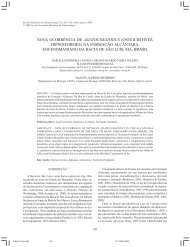
Ulna<br />
The partial right ulna (MZSP-PV 05) is broken at the distal<br />
portion <strong>of</strong> the interosseus ligament scar, so that about 70% <strong>of</strong><br />
the bone is preserved (Figure 3). Also fragmented are the lateral<br />
surface <strong>of</strong> the olecranon, the tip <strong>of</strong> the coronoid process, and<br />
the cranial portion <strong>of</strong> the articular surface for the radius. Distal<br />
REVISTA BRASILEIRA DE PALEONTOLOGIA,11(3), 2008<br />
to the latter, a flattened area in the diaphysis represents the<br />
origin <strong>of</strong> the supinator muscle. A prominent tubercle is seen<br />
distal to the coronoid process. Lateral to this a large nutrient<br />
foramen is seen, whereas other foramina spread over the<br />
epiphysis and around the proximal portion <strong>of</strong> the sigmoid cavity.<br />
Radius<br />
The well preserved left radius (MZSP-PV 04) lacks only<br />
the proximal epiphysis and a small medial portion <strong>of</strong> the distal<br />
articulation (Figure 3), precluding the observation <strong>of</strong> grooves<br />
for mm. extensor ossis metacarpi pollicis and extensor<br />
communis digitorum. A wide nutrient foramen perforates the<br />
medial surface <strong>of</strong> the proximal-most part <strong>of</strong> the shaft, distal to<br />
which is located the tubercle for m. biceps brachialis. The<br />
middle part <strong>of</strong> the shaft is laterally flattened for the insertion<br />
<strong>of</strong> m. supinator. As pointed out by Merriam & Stock (1932),<br />
PROVAS<br />
Figure 3. Smilodon <strong>populator</strong> from Abismo Iguatemi. A-C, MZSP-PV 04, left radius in cranial (A), caudal (B) and distal (C) views; D-F,<br />
MZSP-PV 05, right ulna in medial (D), cranial (E) and caudal (F) views. Abbreviations: ar, articulation for the radius; cp, coronoid<br />
process; mb, tubercle for m. biceps brachialis; ms, flatness for the m. supinator; ms’, origin <strong>of</strong> m. supinator; sc, sigmoid cavity. Scale<br />
bars = 100 mm.