Molecular assembly of a squaraine dye with cationic surfactant and ...
Molecular assembly of a squaraine dye with cationic surfactant and ...
Molecular assembly of a squaraine dye with cationic surfactant and ...

Create successful ePaper yourself
Turn your PDF publications into a flip-book with our unique Google optimized e-Paper software.
The current <strong>assembly</strong> design utilized 333 m MCTABt<strong>of</strong>orm<br />
just sufficient micelles, which were used as the building blocks<br />
for further <strong>assembly</strong> <strong>with</strong> the ATP <strong>and</strong> SQ. In other words, the<br />
strategy provided the optimum condition to incorporate the ATP<br />
<strong>and</strong> SQ into the desirable <strong>assembly</strong>, while avoiding the interruption<br />
<strong>of</strong> the SQ aggregate structure by excessive CTAB.<br />
Excitation spectra obtained by monitoring the emission at<br />
645 <strong>and</strong> 676 nm revealed maxima at 630 <strong>and</strong> 643 nm (Fig. 6),<br />
respectively. In the excitation spectra, a shoulder at ~585 nm<br />
was also visible, which was attributed to the H-aggregate (in<br />
consistency <strong>with</strong> its absorption at 569 nm). The emission b<strong>and</strong> at<br />
the longer wavelength (676 nm), therefore, indicated the formation<br />
<strong>of</strong> a new species, which was likely to be the J-aggregate from<br />
SQ on the basis <strong>of</strong> the spectral red-shift. 21 Although the control<br />
experiment by using excess CTAB also exhibited an emission at<br />
676 nm, a similar signal intensity at ~676 nm required the use<br />
<strong>of</strong> a higher concentration <strong>of</strong> CTAB (in comparison <strong>with</strong> ATP)<br />
(Fig. 5). In summary, the fluorescence at 645 nm was attributed to<br />
SQ monomer <strong>and</strong> the emissison at 676 nm to its J-aggregate.<br />
Fig. 6 Excitation spectra <strong>of</strong> SQ (5 mM) in the presence <strong>of</strong> CTAB (333 mM)<br />
<strong>and</strong> ATP (133 mM).<br />
Dynamic light scattering (DLS) techniques were employed<br />
to probe the size <strong>of</strong> the self-assembled structures in solution<br />
quantitatively. CTAB (333 mM) exhibited average solvodynamic<br />
diameters <strong>of</strong> 220 nm, corresponding to its micelle formation<br />
(Fig. 7). The solution <strong>of</strong> SQ (5 mM) revealed a broad distribution <strong>of</strong><br />
species (<strong>with</strong> average size <strong>of</strong> about 430 nm), attributed to H- <strong>and</strong> Jaggregates<br />
<strong>of</strong> various sizes. The solution <strong>of</strong> CTAB <strong>and</strong> SQ revealed<br />
aspeciesat~400 nm (similar to the aggregate <strong>of</strong> SQ alone),<br />
Fig. 7 Solvodynamic diameters <strong>of</strong> CTAB (333 mM) aggregates, CTAB<br />
<strong>with</strong> SQ, <strong>and</strong> CTAB <strong>with</strong> SQ <strong>and</strong> ATP in water determined by dynamic<br />
light scattering.<br />
in addition to a significantly larger particle at ~3000 nm. Lack<br />
<strong>of</strong> characteristic sizes from either SQ or SQ–CTAB in solutions<br />
suggested that the well-defined <strong>assembly</strong> was not occurring in the<br />
absence <strong>of</strong> ATP. Interestingly, addition <strong>of</strong> ATP (to the solution<br />
including CTAB <strong>and</strong> SQ) led to a defined nanostructure <strong>with</strong> a<br />
diameter <strong>of</strong> about 570 nm, clearly indicating that a self-<strong>assembly</strong><br />
process took place in the solution. A significant increase in<br />
the dimension <strong>of</strong> the CTAB–SQ–ATP <strong>assembly</strong> (570 nm), in<br />
comparison <strong>with</strong> that <strong>of</strong> CTAB micelle (~220 nm), supported<br />
the assumption that the ATP <strong>and</strong> SQ were interacting <strong>with</strong><br />
CTAB micelle at its outer perimeter. Light scattering data also<br />
revealed a second species (size at ~5200 nm) for CTAB–SQ–ATP<br />
<strong>assembly</strong>, suggesting that the SQ molecule could link multiple<br />
CTAB micelles during the <strong>assembly</strong> process.<br />
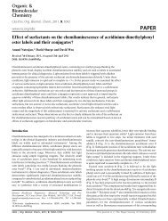
Atomic force microscopy (AMF) further confirmed the interaction<br />
pattern. The CTAB micelle was detected to be slightly less<br />
than 0.1 mm (Fig. 8). Addition <strong>of</strong> SQ to CTAB did not alter<br />
the micelle size. From the CTAB+SQ+ATP sample, however, two<br />
distinctive sizes were observed. One species had a dimension <strong>of</strong><br />
about 0.1–0.15 nm, in consistency <strong>with</strong> the proposed <strong>assembly</strong><br />
pattern. A second species <strong>with</strong> significant larger size (~0.4–1 nm)<br />
was also observed, in agreement <strong>with</strong> the two distinct species<br />
Fig. 8 AFM images <strong>of</strong> films spin-cast from the CTAB, CTAB+SQ,<strong>and</strong>CTAB+SQ+ATP solutions. The image <strong>of</strong> CTAB+SQ+ATP contains the single<br />
species <strong>of</strong> dimension ~0.1 mm (small arrow) <strong>and</strong> connected species (thick arrow).<br />
This journal is © The Royal Society <strong>of</strong> Chemistry 2011 Org. Biomol. Chem., 2011, 9, 2878–2884 | 2881