Molecular assembly of a squaraine dye with cationic surfactant and ...
Molecular assembly of a squaraine dye with cationic surfactant and ...
Molecular assembly of a squaraine dye with cationic surfactant and ...

You also want an ePaper? Increase the reach of your titles
YUMPU automatically turns print PDFs into web optimized ePapers that Google loves.
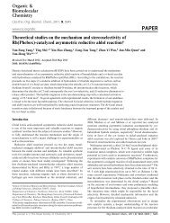
Organic &<br />
Biomolecular<br />
Chemistry<br />
Cite this: Org. Biomol. Chem., 2011, 9, 2878<br />
www.rsc.org/obc PAPER<br />
<strong>Molecular</strong> <strong>assembly</strong> <strong>of</strong> a <strong>squaraine</strong> <strong>dye</strong> <strong>with</strong> <strong>cationic</strong> <strong>surfactant</strong> <strong>and</strong><br />
nucleotides: its impact on aggregation <strong>and</strong> fluorescence response†<br />
Yongqian Xu, a Andrey Malkovskiy, b Qiuming Wang c <strong>and</strong> Yi Pang* a<br />
Received 22nd November 2010, Accepted 1st February 2011<br />
DOI: 10.1039/c0ob01061h<br />
A novel fluorescent system has been assembled by using ATP, <strong>surfactant</strong>, <strong>and</strong> a <strong>squaraine</strong> <strong>dye</strong> in an<br />
aqueous buffer solution. In the system, a <strong>cationic</strong> <strong>surfactant</strong> such as cetyl trimethyl ammonium<br />
bromide (CTAB) forms a sphere-like micelle, whose positive charge at the surface <strong>of</strong> the micelle attracts<br />
the negatively charged ATP to form a unique organized nanostructure. Such an organized system is<br />
shown to interact <strong>with</strong> the <strong>squaraine</strong> <strong>dye</strong> (SQ) to perturb its aggregate structure, thereby generating the<br />
optical response. The nanostructure <strong>of</strong> the <strong>assembly</strong> has been characterized by dynamic light-scattering<br />
(DLS) <strong>and</strong> atomic force microscopy (AFM). The unique feature <strong>of</strong> the developed sensing system is that<br />
the analytes ATP form part <strong>of</strong> the <strong>assembly</strong> structure. The system utilizes forces such as electrostatic<br />
interaction <strong>and</strong> p–p stacking <strong>of</strong> the aromatic segment <strong>of</strong> ATP <strong>and</strong> SQ to achieve the selective detection<br />
<strong>of</strong> ATP.<br />
Introduction<br />
The design <strong>and</strong> construction <strong>of</strong> chemical sensors that selectively<br />
recognize anions has received ever-increasing attention in recent<br />
years, because anions are ubiquitous in biological systems. 1<br />
Among various anions, detection <strong>of</strong> phosphates, pyrophosphate<br />
(PPi), <strong>and</strong> nucleotides is <strong>of</strong> special interest. 2 As a multifunctional<br />
nucleotide, adenosine triphosphate (ATP) functions as an energy<br />
shuttle to power nearly all forms <strong>of</strong> cellular work, from the<br />
generation <strong>of</strong> light by fireflies to the movement <strong>of</strong> muscle cells.<br />
It also provides an energy source for biosynthetic reaction,<br />
consumption <strong>of</strong> many enzymes <strong>and</strong> cell division. 3 Adenosine<br />
diphosphate (ADP) is another nucleotide, which is the product <strong>of</strong><br />
ATP dephosphorylation by ATPases <strong>and</strong> can be converted back<br />
to ATP by ATP synthases. The conversion <strong>of</strong> these two molecules<br />
plays a critical role in supplying energy for many processes <strong>of</strong><br />
life. 4 Thus, selective detection <strong>of</strong> ATP from ATP/ADP in aqueous<br />
media has emerged as an important area <strong>of</strong> research. Although<br />
several studies have employed colorimetric <strong>and</strong> fluorescent probes,<br />
electrostatic <strong>and</strong> hydrogen bonding interactions to recognize ATP, 5<br />
it remains a challenge to find new approaches to simplify ATP<br />
detection while keeping higher selectivity over other nucleoside<br />
triphosphates such as GTP. Fluorescent probes as a conventional<br />
<strong>and</strong> highly sensitive analytical method have been used to detect<br />
aDepartment <strong>of</strong> Chemistry & Maurice Morton Institute <strong>of</strong> Polymer Science,<br />
The University <strong>of</strong> Akron, Akron, OH, 44325. E-mail: yp5@uakron.edu<br />
bDepartment <strong>of</strong> Polymer Science, The University <strong>of</strong> Akron, Akron, OH,<br />
44325<br />
cDepartment <strong>of</strong> Chemical <strong>and</strong> Biomolecular Engineering, The University <strong>of</strong><br />
Akron, Akron, OH, 44325<br />
† Electronic supplementary information (ESI) available: Synthetic procedures,<br />
additional spectroscopic data. See DOI: 10.1039/c0ob01061h<br />
ATP, however, few <strong>of</strong> these sensors show turn-on emission upon<br />
ATP binding in aqueous medium over PPi. 5e–g<br />
Squaraines form a class <strong>of</strong> novel <strong>dye</strong>s possessing sharp <strong>and</strong><br />
intense absorption <strong>and</strong> fluorescence in the red to near-infrared<br />
region. 6 In the solution, <strong>squaraine</strong> <strong>dye</strong>s can be spontaneously<br />
assembled <strong>with</strong> chromophores in a parallel-oriented fashion (Haggregates),<br />
or in a head-to-tail arrangement (J-aggregates) by<br />
varying the solvents or adding ionic species. The J-aggregates<br />
give red-shifted absorption b<strong>and</strong>s (as compared <strong>with</strong> monomer<br />
absorption), <strong>and</strong> H-aggregates give blue shifted absorption b<strong>and</strong>s.<br />
The different form <strong>of</strong> the aggregate also affects the emission<br />
properties, <strong>with</strong> H-aggregates usually being poor emitters while<br />
J-aggregates <strong>of</strong>ten give efficient luminescence. 7,8 The distinction<br />
in optical characteristics between J- <strong>and</strong> H-aggregates can be<br />
used as an effective means to monitor the molecular <strong>assembly</strong>.<br />
Although ATP has been demonstrated to be a building block<br />
to aid the H-aggregate formation <strong>of</strong> a cyanine <strong>dye</strong>, 9 no study<br />
has been found utilizing the electrostatic interaction (between the<br />
negatively charged ATP <strong>and</strong> positively charged <strong>squaraine</strong> <strong>dye</strong>)<br />
to influence the aggregate structures <strong>of</strong> <strong>squaraine</strong> <strong>dye</strong> (SQ) in<br />
aqueous solution. An interesting question is whether such an<br />
aggregate structure change, if induced, could lead to a useful<br />
spectroscopic response to detect ATP.<br />
<strong>Molecular</strong> self-<strong>assembly</strong> <strong>of</strong>fers an excellent tool to construct, via<br />
a “bottom-up” approach, diverse supermolecular architectures in<br />
which the organization <strong>of</strong> individual molecules can be controlled<br />
by proper selection <strong>of</strong> different self-assembling components. 10<br />
For example, <strong>surfactant</strong>s are well known to self-assemble in<br />
aqueous solutions to give organized nanostructures (e.g., micelles<br />
<strong>and</strong> vesicles). 11 Utilization <strong>of</strong> <strong>surfactant</strong>s in the chemical<br />
sensor ensemble has led to detection <strong>of</strong> several transition-metal<br />
cations, 12 proteins 13 <strong>and</strong> other analytes such as Cl - , pH, Ba 2+ <strong>and</strong><br />
2878 | Org. Biomol. Chem., 2011, 9, 2878–2884 This journal is © The Royal Society <strong>of</strong> Chemistry 2011
Scheme 1 Structures <strong>of</strong> SQ, ATP <strong>and</strong> schematic illustration <strong>of</strong> the working micellar system. In the SQ structure, the counter ion CF 3SO 3 - is not shown<br />
for clarity.<br />
lipophilicity. 14 The current strategy is limited to encapsulating a<br />
fluorophore in the micelle, whose fluorescence is then quenched by<br />
an analyte (e.g., by using heavy atom effect or electron transfer).<br />
The well-defined nanostructure <strong>of</strong> the <strong>surfactant</strong> micelle raises<br />
the possibility that it can be used as an <strong>assembly</strong> building block.<br />
To demonstrate its feasibility, we have examined a ternary system<br />
that consists <strong>of</strong> ATP, <strong>squaraine</strong> <strong>dye</strong>, <strong>and</strong> <strong>cationic</strong> <strong>surfactant</strong> (cetyl<br />
trimethyl ammonium bromide (CTAB)) as illustrated in Scheme 1.<br />
The basis for our hypothesis is as follows: in the system, CTAB<br />
can form a sphere-like micelle, 15 whose positive charge at the<br />
surface <strong>of</strong> the sphere attracts the ATP to form a unique organized<br />
nanostructure through electrostatic interaction. It is known that<br />
<strong>squaraine</strong> <strong>dye</strong> tends to form dimers or H-aggregates through p–p<br />
stacking, owing to its poor solubility in aqueous media. 7 Upon<br />
interaction <strong>with</strong> ATP at the outer surface <strong>of</strong> the nanostructured<br />
sphere, the <strong>squaraine</strong> <strong>dye</strong>s in the H-aggregate form are found to<br />
be changed to the J-aggregate form (head-to-tail arrangement), as<br />
a consequence <strong>of</strong> electrostatic interaction between the <strong>squaraine</strong><br />
<strong>and</strong> the phosphate group <strong>of</strong> ATP. 16 In this study, we report the<br />
formation <strong>of</strong> a nanostructured <strong>assembly</strong> by using CTAB, ATP<br />
<strong>and</strong> <strong>squaraine</strong> SQ. The optical responses <strong>of</strong> SQ, induced by the<br />
<strong>assembly</strong> structure, exhibit potential for desirable ATP detection<br />
in aqueous solution.<br />
Results <strong>and</strong> discussion<br />
The SQ <strong>dye</strong> was synthesized as reported previously. 17 UV–vis<br />
absorption spectra <strong>of</strong> SQ in various organic solvents gives one<br />
b<strong>and</strong> at about 635 nm <strong>with</strong> similar absorbance, attributed to the<br />
monomeric form (Fig. 1). Solvent polarity only slightly affects the<br />
absorption peak, <strong>with</strong> l max = 646 nm in the nonpolar toluene <strong>and</strong><br />
l max = 631 nm in methanol. To assess the feasibility <strong>of</strong> detecting<br />
ATP at physiological pH values, the sensing ability <strong>of</strong> SQ toward<br />
ATP was examined in an aqueous solution containing phosphate<br />
buffer (10 mM, pH 7.20) <strong>and</strong> 1% EtOH. Poor water solubility<br />
requires us to first dissolve SQ <strong>dye</strong>inEtOH,whichwasthen<br />
dilutedinwater.<br />
Fig. 1 UV–vis absorption spectra <strong>of</strong> SQ (5 mM) in different solvents.<br />
In the aqueous buffer solution, SQ showed characteristic<br />
absorption maxima at 547, 569 <strong>and</strong> 627 nm (Fig. 2a). The<br />
absorption b<strong>and</strong> at 627 nm was assigned to the monomeric species.<br />
On the basis <strong>of</strong> the observed spectral blue shift, the b<strong>and</strong>s at 547<br />
<strong>and</strong> 569 nm were attributed to the H-aggregates (in the dimer<br />
This journal is © The Royal Society <strong>of</strong> Chemistry 2011 Org. Biomol. Chem., 2011, 9, 2878–2884 | 2879
Fig. 2 Absorption change (a) <strong>and</strong> fluorescence response (b) <strong>of</strong> SQ (5 mM) in phosphate buffer (10 mM, pH 7.2) upon addition <strong>of</strong> CTAB (0–333 mM),<br />
followed by ATP (0–133 mM). The emission spectra were acquired when the solutions were excited at 620 nm.<br />
or oligomeric form). 7,8 The absorption intensity <strong>of</strong> these three<br />
peaks increased slightly <strong>with</strong> increasing CTAB concentration (Fig.<br />
S2, ESI†). After the CTAB concentration in the solution reached<br />
0.33 mM, ATP was added. When the ATP concentration was<br />
increased, the aggregate absorption peaks at 547 <strong>and</strong> 569 nm<br />
weakened, while the monomeric absorption at 627 nm went up.<br />
No change in the absorption spectra was observed upon addition<br />
<strong>of</strong> other ions, including AMP, PPi, phosphate, adenosine, CO 3 2- ,<br />
AcO - ,SO 4 2- ,NO3 - ,Cl - ,Br - ,I - ,Na + ,K + ,Ca 2+ ,Mg 2+ . The specific<br />
optical response from ATP, which was visible (Fig. 3), indicated<br />
that the SQ could be used as colorimetric probe for ATP in<br />
aqueous buffer solution. Addition <strong>of</strong> ATP also induced a shoulder<br />
at ~665 nm, suggesting the formation <strong>of</strong> a new chemical species-<br />
“SQ-<strong>surfactant</strong>” complex, 18 although formation <strong>of</strong> J-aggregate<br />
remained a possibility.<br />
Fig. 3 Color changes induced in SQ (5 mM) in the presence <strong>of</strong> CTAB<br />
(333 mM) in 3 mL phosphate buffer (10 mM, pH 7.2) upon addition <strong>of</strong><br />
different anions <strong>and</strong> adenosine (0.2 mM).<br />
In order to underst<strong>and</strong> the sensing process, the percentage<br />
change <strong>of</strong> each species (dimer <strong>and</strong> higher H-aggregate, <strong>and</strong><br />
monomer) was correlated <strong>with</strong> the addition <strong>of</strong> CTAB, followed by<br />
ATP. As seen in Fig. 4, the critical micellar concentration (CMC)<br />
<strong>of</strong> CTAB can be obtained at 133.3 mM. 19 In the micellar solution<br />
(CTAB concentration = 133–333 mM), ATP created a favorable<br />
environment to transfer <strong>squaraine</strong> <strong>dye</strong> from H-aggregates to<br />
monomer to some extent. Since CTAB could disperse <strong>squaraine</strong><br />
aggregates like other <strong>surfactant</strong>s (Fig. S6, ESI†), 20 CTAB concentration<br />
in the solution was controlled below 0.33 mM.<br />
SQ exhibited a fluorescence maximum at ~650 nm in aqueous<br />
buffer solution, which can be attributed to the monomer since<br />
<strong>squaraine</strong> <strong>dye</strong> typically has a small Stokes’ shift (about 10–30 nm).<br />
Although the anionic phosphate ATP could be associated <strong>with</strong><br />
the <strong>cationic</strong> SQ species, addition <strong>of</strong> ATP (up to 2 mM) to the SQ<br />
solution induced nearly no fluorescence response in the absence<br />
<strong>of</strong> CTAB (Fig. 5 <strong>and</strong> Fig. S7†). The control experiment clearly<br />
Fig. 4 Percentage change <strong>of</strong> SQ (5 mM) in phosphate buffer (10 mM,<br />
pH 7.2) upon addition <strong>of</strong> CTAB (0–333 mM), followed by ATP (0–200 mM).<br />
Since the concentration <strong>of</strong> each SQ species is proportional to the<br />
corresponding absorbance A i, the percentage change is calculated to be<br />
A i/(A 547 + A 569 + A 627). Here, the absorption at 547, 569, <strong>and</strong> 627 nm was<br />
used to approximate the enriched H-dimer, oligomer H-aggregate, <strong>and</strong><br />
monomer, respectively.<br />
Fig. 5 Comparison <strong>of</strong> the fluorescence response <strong>of</strong> SQ (5 mM) in<br />
phosphate buffer (10 mM, pH 7.2) upon addition <strong>of</strong> various concentrations<br />
<strong>of</strong> CTAB <strong>and</strong> ATP.<br />
indicated that an adequate amount <strong>of</strong> CTAB was necessary for<br />
the <strong>assembly</strong> to work. As illustrated in Fig. 2b, CTAB had a<br />
negligible effect on the fluorescence spectra <strong>of</strong> SQ when CTAB<br />
concentration was low (
The current <strong>assembly</strong> design utilized 333 m MCTABt<strong>of</strong>orm<br />
just sufficient micelles, which were used as the building blocks<br />
for further <strong>assembly</strong> <strong>with</strong> the ATP <strong>and</strong> SQ. In other words, the<br />
strategy provided the optimum condition to incorporate the ATP<br />
<strong>and</strong> SQ into the desirable <strong>assembly</strong>, while avoiding the interruption<br />
<strong>of</strong> the SQ aggregate structure by excessive CTAB.<br />
Excitation spectra obtained by monitoring the emission at<br />
645 <strong>and</strong> 676 nm revealed maxima at 630 <strong>and</strong> 643 nm (Fig. 6),<br />
respectively. In the excitation spectra, a shoulder at ~585 nm<br />
was also visible, which was attributed to the H-aggregate (in<br />
consistency <strong>with</strong> its absorption at 569 nm). The emission b<strong>and</strong> at<br />
the longer wavelength (676 nm), therefore, indicated the formation<br />
<strong>of</strong> a new species, which was likely to be the J-aggregate from<br />
SQ on the basis <strong>of</strong> the spectral red-shift. 21 Although the control<br />
experiment by using excess CTAB also exhibited an emission at<br />
676 nm, a similar signal intensity at ~676 nm required the use<br />
<strong>of</strong> a higher concentration <strong>of</strong> CTAB (in comparison <strong>with</strong> ATP)<br />
(Fig. 5). In summary, the fluorescence at 645 nm was attributed to<br />
SQ monomer <strong>and</strong> the emissison at 676 nm to its J-aggregate.<br />
Fig. 6 Excitation spectra <strong>of</strong> SQ (5 mM) in the presence <strong>of</strong> CTAB (333 mM)<br />
<strong>and</strong> ATP (133 mM).<br />
Dynamic light scattering (DLS) techniques were employed<br />
to probe the size <strong>of</strong> the self-assembled structures in solution<br />
quantitatively. CTAB (333 mM) exhibited average solvodynamic<br />
diameters <strong>of</strong> 220 nm, corresponding to its micelle formation<br />
(Fig. 7). The solution <strong>of</strong> SQ (5 mM) revealed a broad distribution <strong>of</strong><br />
species (<strong>with</strong> average size <strong>of</strong> about 430 nm), attributed to H- <strong>and</strong> Jaggregates<br />
<strong>of</strong> various sizes. The solution <strong>of</strong> CTAB <strong>and</strong> SQ revealed<br />
aspeciesat~400 nm (similar to the aggregate <strong>of</strong> SQ alone),<br />
Fig. 7 Solvodynamic diameters <strong>of</strong> CTAB (333 mM) aggregates, CTAB<br />
<strong>with</strong> SQ, <strong>and</strong> CTAB <strong>with</strong> SQ <strong>and</strong> ATP in water determined by dynamic<br />
light scattering.<br />
in addition to a significantly larger particle at ~3000 nm. Lack<br />
<strong>of</strong> characteristic sizes from either SQ or SQ–CTAB in solutions<br />
suggested that the well-defined <strong>assembly</strong> was not occurring in the<br />
absence <strong>of</strong> ATP. Interestingly, addition <strong>of</strong> ATP (to the solution<br />
including CTAB <strong>and</strong> SQ) led to a defined nanostructure <strong>with</strong> a<br />
diameter <strong>of</strong> about 570 nm, clearly indicating that a self-<strong>assembly</strong><br />
process took place in the solution. A significant increase in<br />
the dimension <strong>of</strong> the CTAB–SQ–ATP <strong>assembly</strong> (570 nm), in<br />
comparison <strong>with</strong> that <strong>of</strong> CTAB micelle (~220 nm), supported<br />
the assumption that the ATP <strong>and</strong> SQ were interacting <strong>with</strong><br />
CTAB micelle at its outer perimeter. Light scattering data also<br />
revealed a second species (size at ~5200 nm) for CTAB–SQ–ATP<br />
<strong>assembly</strong>, suggesting that the SQ molecule could link multiple<br />
CTAB micelles during the <strong>assembly</strong> process.<br />
Atomic force microscopy (AMF) further confirmed the interaction<br />
pattern. The CTAB micelle was detected to be slightly less<br />
than 0.1 mm (Fig. 8). Addition <strong>of</strong> SQ to CTAB did not alter<br />
the micelle size. From the CTAB+SQ+ATP sample, however, two<br />
distinctive sizes were observed. One species had a dimension <strong>of</strong><br />
about 0.1–0.15 nm, in consistency <strong>with</strong> the proposed <strong>assembly</strong><br />
pattern. A second species <strong>with</strong> significant larger size (~0.4–1 nm)<br />
was also observed, in agreement <strong>with</strong> the two distinct species<br />
Fig. 8 AFM images <strong>of</strong> films spin-cast from the CTAB, CTAB+SQ,<strong>and</strong>CTAB+SQ+ATP solutions. The image <strong>of</strong> CTAB+SQ+ATP contains the single<br />
species <strong>of</strong> dimension ~0.1 mm (small arrow) <strong>and</strong> connected species (thick arrow).<br />
This journal is © The Royal Society <strong>of</strong> Chemistry 2011 Org. Biomol. Chem., 2011, 9, 2878–2884 | 2881
detected in the light scattering experiment. The consistent trend,<br />
which was revealed from both light scattering <strong>and</strong> AFM images,<br />
supports the assumption that the ATP was assembled on the<br />
surface <strong>of</strong> CTAB micelles, resulting in increased particle sizes<br />
(Scheme 1). It should be noted that the particle sizes from AFM<br />
are different from those detected from DLS. This is because DLS<br />
is measured in solution where solvation results in the larger sizes,<br />
while AFM measures the sizes <strong>of</strong> dried particles which are laid on<br />
the AFM grid surface. Correlation <strong>of</strong> AFM <strong>and</strong> DLS data (Fig. 7<br />
& 8) showed that the CTAB alone existed as spheric micelles<br />
in solution, <strong>with</strong> an average size <strong>of</strong> 220 nm. The predominant<br />
particle size in the solution <strong>of</strong> “SQ+CTAB+ATP” <strong>assembly</strong> was<br />
about twice as large (~570 nm), <strong>and</strong> had similar size distribution<br />
to CTAB. The results pointed to the fact that the particles in<br />
the “SQ+CTAB+ATP” solution were likely to be spherical. Both<br />
DLS <strong>and</strong> AFM data also showed a larger species that existed as a<br />
minor component (Fig. 7).<br />
To validate the specificity <strong>of</strong> the system toward the anionic<br />
guests in practice, the fluorescence response <strong>of</strong> SQ in aqueous<br />
buffer solution was examined upon addition <strong>of</strong> various biologically<br />
important anions <strong>and</strong> cations. No apparent fluorescence<br />
enhancement was observed for AMP, PPi, phosphate, adenosine,<br />
CO 3 2- ,AcO - ,SO4 2- ,NO3 - ,Cl - ,Br - ,I - ,Na + ,K + ,Ca 2+ ,Mg 2+ (Fig. 9,<br />
Fig. S11, ESI†). The ratio <strong>of</strong> I 670/I 0 (herein I 0 is the fluorescence<br />
intensity <strong>of</strong> free SQ at 670 nm; I 670 represents the fluorescence<br />
intensity at 670 nm upon addition <strong>of</strong> different ions) is plotted<br />
against the concentration <strong>of</strong> various ions (Fig. 9). The I 670/I 0<br />
value for ATP reached up to 3.1-fold, in sharp contrast to I 670/I 0<br />
< 1.1-fold for other ions. Therefore, this system could be used as a<br />
selective sensor for ATP. A good linear correlation was observed<br />
between the relative fluorescence intensity <strong>and</strong> ATP concentration<br />
(0–140 mM) (Fig. 10), <strong>and</strong> the detection limit was in the order <strong>of</strong><br />
10 -6 M when the ratio <strong>of</strong> signal to noise was 3. The Hill’s plot<br />
gave a Hill coefficient <strong>of</strong> 0.78 (Fig. S1†), which indicates negative<br />
cooperative interaction. 22<br />
The selective fluorescence response to ATP appeared to be<br />
associated <strong>with</strong> the electrostatic interaction between SQ <strong>and</strong><br />
ATP. As illustrated in Scheme 1, a <strong>cationic</strong> <strong>surfactant</strong> was<br />
necessary to form the spherical micelle core <strong>with</strong> a positive<br />
charged surface. When the <strong>cationic</strong> <strong>surfactant</strong> CTAB was replaced<br />
by an anionic <strong>surfactant</strong> such as SDS (sodium dodecyl sulfate)<br />
or Triton X-100 (isooctylphenolicpolyglycol ether), the resulting<br />
system showed no fluorescence response to ATP. The <strong>assembly</strong><br />
<strong>of</strong> the ordered system (Scheme 1) appeared to favor addition in<br />
the following sequence: CTAB, then ATP to the SQ solution. If<br />
the addition sequence is reversed (i.e. ATP first, then CTAB), the<br />
resulting system exhibited a much weaker fluorescence response<br />
(Fig.S10,ESI†)thanthatobservedinFig.2.IfnoCTABexisted<br />
in the solution, there was no response to ATP (Fig. 5). Therefore,<br />
formation <strong>of</strong> spherical micelles <strong>with</strong> a positively charged surface<br />
was a necessary step. The result clearly pointed to the fact that an<br />
<strong>assembly</strong> <strong>with</strong> an ordered structure might play an important role<br />
in the observed fluorescence response. The results also support<br />
the assumption that, in the second step, electrostatic interaction<br />
guided the phosphate terminals <strong>of</strong> ATP to the positively charged<br />
surface <strong>of</strong> the spherical micelle.<br />
Based on the design, the concentration <strong>of</strong> the <strong>squaraine</strong> reporter<br />
is expected to affect the performance <strong>of</strong> the sensor <strong>assembly</strong>.<br />
Screening <strong>of</strong> the <strong>dye</strong> concentration (between 1–20 mM, Fig. S17†)<br />
Fig. 9 (a) Structure <strong>of</strong> phosphates. (b) Relative fluorescence ratio (I 670/I 0)<br />
<strong>of</strong> SQ (5 mM) in the presence <strong>of</strong> CTAB (333 mM) in phosphate buffer<br />
(10 mM, pH 7.2) upon addition <strong>of</strong> different ions (0.2 mM).<br />
showed that SQ gave the optimum fluorescence response at a<br />
concentration <strong>of</strong> 5 mM. Higher SQ concentration resulted in<br />
decreased fluorescence response, possibly attributing to a higher<br />
degree <strong>of</strong> aggregation.<br />
The experimental data were consistent <strong>with</strong> the proposed model<br />
for the <strong>assembly</strong> <strong>of</strong> the ordered system from CTAB, SQ <strong>and</strong><br />
ATP (Scheme 1). Driven by the <strong>assembly</strong> forces, part <strong>of</strong> the<br />
SQ was changed from the H-aggregate to the more fluorescent<br />
monomer <strong>and</strong> J-aggregate form, resulting in a spectral redshift<br />
<strong>and</strong> fluorescence enhancement. In the absence <strong>of</strong> ATP, the<br />
<strong>surfactant</strong> CTAB initially formed the micelles, which also allowed<br />
the SQ to be partially dissolved in the hydrophobic cavity <strong>of</strong> the<br />
micelles as monomeric or aggregated form (Fig. 11, assemblies I<br />
<strong>and</strong> II). CTAB concentration in the solution was thus controlled<br />
to below 0.33 mM, thereby creating a favorable environment<br />
for the CTAB–SQ–ATP <strong>assembly</strong> III while avoiding excessive<br />
solubilization <strong>of</strong> SQ by CTAB <strong>surfactant</strong>. Formation <strong>of</strong> <strong>assembly</strong><br />
III was energetically more favorable, which was enhanced by the<br />
2882 | Org. Biomol. Chem., 2011, 9, 2878–2884 This journal is © The Royal Society <strong>of</strong> Chemistry 2011
Fig. 10 Relative fluorescence ratio (I 670/I 0) <strong>of</strong> SQ (5 mM) in the<br />
presence <strong>of</strong> CTAB (333 mM) in phosphate buffer (10 mM, pH 7.2) upon<br />
addition <strong>of</strong> different ions (0–0.2 mM). The broken line on the ATP curve<br />
shows the linear correlation between the fluorescence response <strong>and</strong> ATP<br />
concentration (0–140 mM).<br />
electrostatic interaction between the oppositely-charged species<br />
CTAB–ATP <strong>and</strong> ATP–SQ, in contrast to the assemblies I <strong>and</strong><br />
II that involved the interaction <strong>of</strong> two positively charged species.<br />
Excessive CTAB could turn on the fluorescence <strong>of</strong> SQ (Fig. 5),<br />
since the increasing micelle concentration makes the formation <strong>of</strong><br />
I <strong>and</strong> II a more competitive process.<br />
It was assumed that the SQ was attracted to the phosphate<br />
group <strong>of</strong> ATP through electrostatic interaction. In this step, the<br />
two phosphate groups appeared to be necessary (one attaching<br />
to positively charged surface <strong>of</strong> the spherical micelle, the other<br />
to SQ) for the <strong>assembly</strong>. This was likely to be the reason for the<br />
selective recognition <strong>of</strong> ATP over AMP, adenosine, phosphate <strong>and</strong><br />
other anions. The p–p stacking interaction between SQ backbone<br />
<strong>and</strong> the aromatic segment <strong>of</strong> ATP could also play an important<br />
role in the stability <strong>of</strong> the SQ–ATP–micelle <strong>assembly</strong>, 23 which<br />
accounted for the selective recognition <strong>of</strong> ATP over PPi. The<br />
different response between the nucleotide triphosphates ATP <strong>and</strong><br />
GTP (Fig. 9 <strong>and</strong> 10) further pointed to the fact that the p–<br />
p stacking interaction could be used to differentiate different<br />
triphosphates. Owing to steric hindrance, SQ molecules had a<br />
tendency to exist in J-aggregate form rather than H-aggregate<br />
form in the cavity <strong>of</strong> the SQ–ATP–micelle <strong>assembly</strong> (Scheme 1).<br />
Observation <strong>of</strong> a new b<strong>and</strong>, 664 nm in absorption <strong>and</strong> 676 nm<br />
in fluorescence, is consistent <strong>with</strong> the proposed ternary structure.<br />
The moderate selectivity between ATP <strong>and</strong> ADP was associated<br />
<strong>with</strong> the length <strong>of</strong> phosphates, as the triphosphate provided more<br />
space to accommodate the SQ aggregates, which can simultaneously<br />
interact <strong>with</strong> the phosphate (electrostatic interaction) <strong>and</strong><br />
adenosine segments (p–p interaction).<br />
To detect ATP in the biological system, it is necessary to<br />
assess the selectivity in the presence <strong>of</strong> alkali <strong>and</strong> alkali-earth<br />
metal cations. It is known that ATP can bind cations Mg 2+ (at<br />
9554 M -1 ), Ca 2+ (at 3722 M -1 ), Na + (at 13 M -1 )<strong>and</strong>K + (at 8 M -1 ). 24<br />
The influence <strong>of</strong> various cations on the fluorescence <strong>of</strong> SQ was<br />
examined by addition <strong>of</strong> Na + ,K + ,Ca 2+ <strong>and</strong> Mg 2+ (Fig. S12,<br />
ESI†). In the presence <strong>of</strong> 0.2 mM <strong>of</strong> various cations, Na + <strong>and</strong> K +<br />
exhibit little influence on the fluorescence enhancement, whereas<br />
thepresence<strong>of</strong>Ca 2+ <strong>and</strong> Mg 2+ suppressed the fluorescence increase<br />
to some extent. However, these results do not interfere <strong>with</strong> the<br />
sensing process.<br />
Conclusions<br />
In summary, we have demonstrated a new ternary system, which<br />
incorporates ATP <strong>and</strong> SQ into an ordered molecular self-<strong>assembly</strong><br />
to induce fluorescence turn-on. The molecular <strong>assembly</strong> is driven<br />
by a balanced electrostatic interaction <strong>and</strong> p–p stacking <strong>of</strong> the aromatic<br />
segment <strong>of</strong> ATP <strong>and</strong> SQ. The sensor operates by attenuating<br />
the aggregate structure <strong>of</strong> <strong>squaraine</strong> <strong>dye</strong>s (from H- to monomer<br />
<strong>and</strong> J-aggregates) under physiological pH conditions. The study<br />
provides a simple approach for the detection <strong>of</strong> ATP. Due to the<br />
specific requirements for the analyte to enter the nano-structured<br />
molecular <strong>assembly</strong>, the ternary system exhibits good selectivity to<br />
sense ATP. The new sensor system provides an additional example<br />
Fig. 11 Micelle-based equilibrium to redistribute the H-aggregate SQ into monomeric <strong>and</strong> aggregate form.<br />
This journal is © The Royal Society <strong>of</strong> Chemistry 2011 Org. Biomol. Chem., 2011, 9, 2878–2884 | 2883
that operates by recognizing a specific pattern transformation<br />
(i.e. here from H- to J-aggregate) that has the advantage <strong>of</strong><br />
simpler molecular design in comparison <strong>with</strong> typical sensors. 25<br />
This is because the synthesis <strong>of</strong> a classical fluorescent sensor <strong>of</strong>ten<br />
involves a lengthy synthetic procedure. Supramolecular <strong>assembly</strong><br />
using easily accessible components, therefore, could provide a costeffective<br />
solution.<br />
Acknowledgements<br />
This work was supported by The University <strong>of</strong> Akron <strong>and</strong><br />
Coleman endowment.<br />
References<br />
1 R. Martinez-Manez <strong>and</strong> F. Sancenon, Chem. Rev., 2003, 103, 4419.<br />
2(a) A. Ojida, I. Takashima, T. Kohira, H. Nonaka <strong>and</strong> I. Hamachi,<br />
J. Am. Chem. Soc., 2008, 130, 12095; (b) S. Jaeger, G. Rasched, H.<br />
Kornreich-Leshem, M. Engeser, O. Thum <strong>and</strong> M. Famulok, J. Am.<br />
Chem. Soc., 2005, 127, 15071.<br />
3S.Törnroth-Horsefield <strong>and</strong> R. Neutze, Proc. Natl. Acad. Sci. U. S. A.,<br />
2008, 105, 19565.<br />
4 S. Murugappa <strong>and</strong> S. P. Kunapuli, Front. Biosci., 2006, 12, 1977.<br />
5(a) S. M. Butterfield <strong>and</strong> M. L. Waters, J. Am. Chem. Soc., 2003, 125,<br />
9580; (b) D. A. Jose, S. Mishra, A. Ghosh, A. Shrivastav, S. K. Mishra<br />
<strong>and</strong> A. Das, Org. Lett., 2007, 9, 1979; (c) P. L. Sazani, R. Larralde <strong>and</strong><br />
J. W. Szostak, J. Am. Chem. Soc., 2004, 126, 8370; (d) D. Miyamoto,<br />
Z. Tang, T. Takarada <strong>and</strong> M. Maeda, Chem. Commun., 2007, 4743;<br />
(e) D.H.Lee,S.Y.Kim<strong>and</strong>J.I.Hong,Angew. Chem., Int. Ed., 2004,<br />
43, 4777; (f) Y. Kanekiyo, R. Naganawa <strong>and</strong> H. Tao, Chem. Commun.,<br />
2004, 1006; (g) J. A. Cruz-Aguado, Y. Chen, Z. Zhang, N. H. Elowe,<br />
M. A. Brook <strong>and</strong> J. D. Brennan, J. Am. Chem. Soc., 2004, 126, 6878;<br />
(h) P. P. Neelak<strong>and</strong>an, M. Hariharan <strong>and</strong> D. Ramaiah, Org. Lett., 2005,<br />
7, 5765; (i) C. Li, M. Numata, M. Takeuchi <strong>and</strong> S. Shinkai, Angew.<br />
Chem., Int. Ed., 2005, 44, 6371.<br />
6(a) S. Das, K. G. Thomas, R. Ramanathan <strong>and</strong> M. V. George, J. Phys.<br />
Chem., 1993, 97, 13625; (b) K.Y.Law,J. Phys. Chem., 1987, 91, 5184.<br />
7(a) P. Chithra, R. Varghese, K. P. Divya <strong>and</strong> A. Ajayaghosh, Chem.–<br />
Asian J., 2008, 3, 1365; (b) S. Alex, M. C. Basheer, K. T. Arun, D.<br />
Ramaiah <strong>and</strong> S. Das, J. Phys. Chem. A, 2007, 111, 3226; (c) R. S. Stoll,<br />
N. Severin, J. P. Rabe <strong>and</strong> S. Hecht, Adv. Mater., 2006, 18, 1271.<br />
8 S. Gadde, E. K. Batchelor, J. P. Weiss, Y. Ling <strong>and</strong> A. E. Kaifer, J. Am.<br />
Chem. Soc., 2008, 130, 17114.<br />
9 M. Morikawa, M. Yoshihara, T. Endo <strong>and</strong> N. Kimizuka, J. Am. Chem.<br />
Soc., 2005, 127, 1358.<br />
10 F. J. M. Hoeben, P. Jonkheijm, E. W. Meijer <strong>and</strong> A. P. H. J. Schenning,<br />
Chem. Rev., 2005, 105, 1491–1546.<br />
11 (a) S. Ozeki <strong>and</strong> S. Ikeda, J. Colloid Interface Sci., 1982, 87, 424–435;<br />
(b) M. Drifford, L. Belloni <strong>and</strong> M. Dubois, J. Colloid Interface Sci.,<br />
1987, 118, 50–67; (c) N. Funasaki <strong>and</strong> S. Hada, Bull. Chem. Soc. Jpn.,<br />
1991, 64, 682–684; (d) A. Tanaka <strong>and</strong> S. Ikeda, Colloids Surf., 1991, 56,<br />
217–228.<br />
12 (a) P. Gr<strong>and</strong>ini, F. Mancin, P. Tecilla, P. Scrimin <strong>and</strong> U. Tonellato,<br />
Angew. Chem., Int. Ed., 1999, 38, 3061–3064; (b) F. Mancin, P. Tecilla,<br />
P. Scrimin <strong>and</strong> U. Tonellato, Coord. Chem. Rev., 2009, 253, 2150–2165;<br />
(c) P. Pallavicini, Y. A. Diaz-Fern<strong>and</strong>ez <strong>and</strong> L. Pasotti, Coord. Chem.<br />
Rev., 2009, 253, 2226–2240.<br />
13 (a) M. A. Azagarsamy, V. Yesilyurt <strong>and</strong> S. Thayumanavan, J. Am.<br />
Chem. Soc., 2010, 132, 4550–4551; (b) J. –H. Ryu, R. Roy, J. Ventura<br />
<strong>and</strong> S. Thayumanavan, Langmuir, 2010, 26, 7086–7092; (c) D. C.<br />
Gonzalez, E. N. Savariar <strong>and</strong> S. Thayumanavan, J. Am. Chem. Soc.,<br />
2009, 131, 7708–7716; (d) E. N. Savariar, S. Ghosh, D. C. Gonzalez <strong>and</strong><br />
S. Thayumanavan, J. Am. Chem. Soc., 2008, 130, 5416–5417; (e) B.S.<br />
S<strong>and</strong>anaraj, R. Demont <strong>and</strong> S. Thayumanavan, J. Am. Chem. Soc.,<br />
2007, 129, 3506–3507; (f) Y. Nakahara, T. Kida, Y. Nakatsuji <strong>and</strong> M.<br />
Akashi, Chem. Commun., 2004, 224–225.<br />
14 (a) T. Riis-Johannessen <strong>and</strong> K. Severin, Chem.–Eur. J., 2010, 16, 8291–<br />
8295; (b) P. Pallavicini, Y. A. Diaz-Fern<strong>and</strong>ez <strong>and</strong> L. Pasotti, Analyst,<br />
2009, 134, 2147–2152; (c) G. Chirico, M. Collini, L. D’Alfonso, F.<br />
Denat, Y. A. Diaz-Fern<strong>and</strong>ez, L. Pasotti, Y. Rousselin, N. Sok <strong>and</strong> P.<br />
Pallavicini, ChemPhysChem, 2008, 9, 1729–1737; (d) P. Pallavicini, Y. A.<br />
Diaz-Fern<strong>and</strong>ez, F. Foti, C. Mangano <strong>and</strong> S. Patroni, Chem.–Eur. J.,<br />
2006, 13, 178–187; (e) R. R. Avirah, K. Jyothish <strong>and</strong> D. Ramaiah, Org.<br />
Lett., 2007, 9, 121–124.<br />
15 (a) U. R. K. Rao, C. Manohar, B. S. Valaulikar <strong>and</strong> R. M. Iyer, J. Phys.<br />
Chem., 1987, 91, 3286; (b) G. D. Gokhale, P. A. Hassan <strong>and</strong> S. D.<br />
Samant, J. Surfactants Deterg., 2005, 8, 319.<br />
16 H. Fenniri, M. W. Hosseini <strong>and</strong> J. –M. Lehn, Helv. Chim. Acta, 1997,<br />
80, 786.<br />
17 P. F. Santos, L. V. Reis, I. Duarte, J. P. Serrano, P. Almeida, A. S. Oliveira<br />
<strong>and</strong> L. F. Vieira Ferreira, Helv. Chim. Acta, 2005, 88, 1135.<br />
18 (a) G. Y. Guralchuk, I. K. Katrunov, R. S. Grynyov, A. V. Sorokin, S. L.<br />
Yefimova, I. A. Borovoy <strong>and</strong> Y. V. Malyukin, J. Phys. Chem. C, 2008,<br />
112, 14762–14768; (b) S. L. Yefimova, G. Y. Guralchuk, A. S. Lebed,<br />
A. V. Sorokin, Y. V. Malyukin <strong>and</strong> I. Y. Kurilchenko, Funct,. Mater.,<br />
2009, 16, 460–464.<br />
19 (a) R. B. Singh, S. Mahanta <strong>and</strong> N. Guchhait, Chem. Phys. Lett., 2008,<br />
463, 183; (b) A. Mohr, P. Talbiersky, H. G. Korth, R. Sustmann,<br />
R. Boese, D. Blaeser <strong>and</strong> H. Rehage, J. Phys. Chem. B, 2007, 111,<br />
12985; (c) Critical micelle concentration (CMC) <strong>of</strong> CTAB is affected<br />
by buffer, see reference: B. Jiang, J. Du, S. Cheng, Q. Wang <strong>and</strong> X.<br />
Zeng, J. Dispersion Sci. Technol., 2003, 24, 755–760.<br />
20 K. T. Arun <strong>and</strong> D. Ramaiah, J. Phys. Chem. A, 2005, 109,<br />
5571.<br />
21 Y. Xu, Z. Li, A. Malkovskiy, S. Sun <strong>and</strong> Y. Pang, J. Phys. Chem. B,<br />
2010, 114, 8574–8580.<br />
22 (a) S. Shinkai, M. Ikeda, A. Sugasaki <strong>and</strong> M. Takeuchi, Acc. Chem.<br />
Res., 2001, 34, 494; (b) Y. B. Ruan, A. F. Li, J. S. Zhao, J. S. Shen <strong>and</strong><br />
Y. B. Jiang, Chem. Commun., 2010, 46, 4938–4940.<br />
23 (a) Z. Xu, N. J. Singh, J. Lim, J. Pan, H. N. Kim, S. Park, K. S. Kim<br />
<strong>and</strong> J. Yoon, J. Am. Chem. Soc., 2009, 131, 15528–15533; (b) Z. Chen,<br />
A.Lohr,C.R.Saha-MÖller <strong>and</strong> F. WÜrthner, Chem. Soc. Rev., 2009,<br />
38, 564–584.<br />
24 J. E. Wilson <strong>and</strong> A. Chin, Anal. Biochem., 1991, 193, 16.<br />
25 (a) J. J. McEwen <strong>and</strong> K. J. Wallace, Chem. Commun., 2009, 42, 6339–<br />
6351; (b) E. Arunkumar, A. Ajayaghosh <strong>and</strong> J. Daub, J. Am. Chem. Soc.,<br />
2005, 127, 3156–3164; (c) E. Arunkumar, P. Chithra <strong>and</strong> A. Ajayaghosh,<br />
J. Am. Chem. Soc., 2004, 126, 6590–6598.<br />
2884 | Org. Biomol. Chem., 2011, 9, 2878–2884 This journal is © The Royal Society <strong>of</strong> Chemistry 2011