New hospital opens PHHP names new dean - University of Florida
New hospital opens PHHP names new dean - University of Florida
New hospital opens PHHP names new dean - University of Florida

Create successful ePaper yourself
Turn your PDF publications into a flip-book with our unique Google optimized e-Paper software.
PATIENT CARE<br />
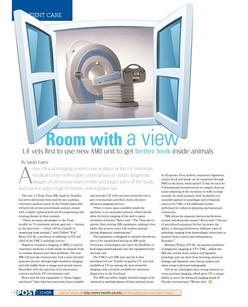
Room with a view<br />
UF vets fi rst to use <strong>new</strong> MRI unit to get better look k inside animals<br />
By Sarah Carey<br />
A<br />
<strong>new</strong> clinical imaging system now in place at the UF Veterinary<br />
Medical Center will enable veterinarians to obtain diagnostic<br />
images <strong>of</strong> previously inaccessible and larger parts <strong>of</strong> the body,<br />
such as the upper legs <strong>of</strong> horses, veterinarians say.<br />
The <strong>new</strong> 1.5 Tesla Titan MR, made by Toshiba,<br />
has never previously been used by any academic<br />
veterinary medical center in the United States and<br />
will provide private practitioners and pet owners<br />
with a highly sophisticated tool for pinpointing and<br />
treating disease in their animals.<br />
“There are many advantages to the Titan,<br />
notably its 71-centimeter patient aperture — known<br />
as the open bore — which will be a benefi t in<br />
examining large animals,” said Clifford “Kip”<br />
Berry, D.V.M., a pr<strong>of</strong>essor <strong>of</strong> radiology at UF and<br />
chief <strong>of</strong> the VMC’s radiology service.<br />
Magnetic resonance imaging, or MRI, is used in<br />
veterinary medicine to look inside an animal’s body<br />
to evaluate diseases and other problems. The <strong>new</strong><br />
MR will provide veterinarians with a more detailed<br />
anatomic picture through high-resolution imaging<br />
and will enable them to image arterial and venous<br />
blood fl ow with the injection <strong>of</strong> an intravenous<br />
contrast medium, UF veterinarians said.<br />
Berry said the <strong>new</strong> equipment is “faster, bigger<br />
and better” than what has previously been available<br />
8 POST<br />
11 09<br />
and provides UF with one more powerful tool to<br />
give veterinarians and their clients the most<br />
advanced imaging services.<br />
“There is more space available inside the<br />
machine to accommodate patients, which should<br />
allow for better imaging <strong>of</strong> the mid to upper<br />
extremity <strong>of</strong> horses,” Berry said. “The Titan also is<br />
quieter than existing MR equipment, making it less<br />
likely that acoustic noise will awaken patients<br />
during diagnostic examinations.”<br />
The equipment is designed so animals should not<br />
have to be repositioned during an MR study.<br />
Veterinary technologists also have the fl exibility to<br />
load large animal patients into the equipment from<br />
the back end.<br />
The VMC’s <strong>new</strong> MR unit and the 8-slice<br />
multidetector row Toshiba Acquilion CT unit now<br />
available at UF are among the most powerful<br />
imaging tools currently available for veterinary<br />
diagnostics in the Southeast.<br />
The MR unit allows highly detailed images to be<br />
obtained in multiple planes <strong>of</strong> bone and s<strong>of</strong>t tissue<br />
Visit us online @ http://<strong>new</strong>s.health.ufl .edu for the latest <strong>new</strong>s and HSC events.<br />
in all species. Foot, fetlock, suspensory ligaments,<br />
carpus, hock and heads can be examined through<br />
MRI in the horse, while spiral CT may be used for<br />
3-dimensional reconstruction in complex fracture<br />
repair planning <strong>of</strong> the extremity or stifl e in large<br />
animals. In small animals, both modalities are<br />
routinely applied to neurologic and orthopedic<br />
cases at the VMC, with additional studies<br />
performed for radiation planning and metastasis<br />
evaluations.<br />
“MR allows for exquisite distinction between<br />
normal and abnormal tissues,” Berry said. “The use<br />
<strong>of</strong> specialized sequences further increases the<br />
ability to distinguish between different types <strong>of</strong><br />
pathology ranging from hemorrhagic infarctions to<br />
primary brain tumors and infl ammatory<br />
disorders.”<br />
Matthew Winter, D.V.M., an assistant pr<strong>of</strong>essor<br />
<strong>of</strong> diagnostic imaging at UF’s VMC, added that<br />
MR also reveals bone, tendon and ligament<br />
pathology and can show bone bruising, meniscal<br />
damage and ligament tears that go undetected<br />
when using traditional radiography.<br />
“All <strong>of</strong> our radiologists have strong interests in<br />
cross-sectional imaging, which gives UF a unique<br />
ability to serve the advanced imaging needs <strong>of</strong><br />
<strong>Florida</strong> veterinarians,” Winter said. P