examination of the eye: methods of diagnosis and instrumentation
examination of the eye: methods of diagnosis and instrumentation
examination of the eye: methods of diagnosis and instrumentation

You also want an ePaper? Increase the reach of your titles
YUMPU automatically turns print PDFs into web optimized ePapers that Google loves.
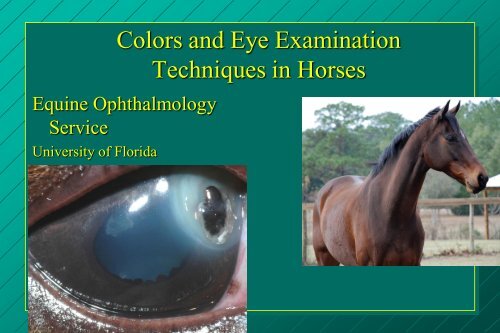
Colors <strong>and</strong> Eye ExaminationEquine OphthalmologyServiceUniversity <strong>of</strong> FloridaTechniques in Horses
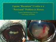
• There are really only 3ophthalmic diseases!!1. Corneal ulcers2. Uveitis3. Everything else!!
Heine C-002-14-400
Heine C-002.14.602
Obvious Things• Just st<strong>and</strong> back <strong>and</strong> lookat <strong>the</strong> symmetry– Lashes– Discomfort <strong>and</strong> squinting– Tearing– Colors– Pupil– Clarity <strong>of</strong> cornea <strong>and</strong> lens– The normal <strong>eye</strong> is “shiny”– Anatomy: anterior to posterior95677Champagne RMH
Lashes pointing down can be earlysign <strong>of</strong> <strong>eye</strong> painLashes(ponies can look through <strong>the</strong>ir lashes)
Nuclear Sclerosis
Ocular Discomfort Level107656Dusty UF Res
Corneal “Colors”• White cornea: abscess ornecrosis• Blue cornea: edema• Red cornea: vessels– Superficial (tree-like) <strong>and</strong>deep vessels (brush).– Note intensity <strong>of</strong> <strong>the</strong> red
Dark is thinShiny is thin
Vascular Patterns• Redness– Very Red– Pale• Symmetry– Asymmetry189711154841194013
Asymmetrical vascularizationCallie
• Edema– Endo<strong>the</strong>lial– Uveitis• Subepi<strong>the</strong>lial scar• Cornealabrasion/ulcer• Subepi<strong>the</strong>lialinflitrate– Immune mediated– FungalCorneal HazeFlyMountOakely IMMKShooter
Epi<strong>the</strong>lial edema Sunshine
Chief BarberIMMK
Jupiter: DSA
Chronic Recurrent DeepImmune Mediated Keratitis• Green fluidfilled lacunaeform in <strong>the</strong>stroma“Alexiej”
May SEK: fungi
C<strong>and</strong>leabraSEK
Pupil Size• Dilated– Glaucoma– Retinal Disease– Optic NerveDisease• Miotic– Uveitis
• Deep corneal scrapingsat <strong>the</strong> edge <strong>of</strong> <strong>the</strong> ulcerto detect bacteria <strong>and</strong>fungal hyphae• Superficial swabbingcannot be expected toyield microbes in a highpercentage <strong>of</strong> cases.• Scrape with h<strong>and</strong>le end<strong>of</strong> scalpel blade.
• Fluorescein: Every <strong>eye</strong> exhibiting signs <strong>of</strong>pain should be stained!!– Detects a corneal epi<strong>the</strong>lial defect or “ulcer”.– Cobalt blue filter aids detection <strong>of</strong> abrasions.
Use fluorescein full strength <strong>and</strong> dilute rose bengal.
Ulcers are best defined with fluorescein.
Silvers<strong>and</strong>s<strong>of</strong>timePredescemetocele
Cobalt Blue Filters• www.slitlamp.com: bluminator
Corneal abrasion: partial epi<strong>the</strong>lial cell layer loss
• Erosion/abrasions are“ulcers” with partialepi<strong>the</strong>lial cell loss• Weak fluoresceinstaining.• Diluting <strong>the</strong> fluoresceinwill make it difficult toidentifyerosions/abrasions
Rose bengal• Tear film integrity• Mucin layer blocks RB
Stain Use/Order• Fluorescein stain first. Identifies ulcers if +.• Rose bengal stain second.– Rose bengal retention indicates tear film instability.• Mucin tear layer blocks RB staining• At risk <strong>of</strong> fungal colonization/invasion• KCS• Viral keratitis• Edema• Scar tissue– Rose bengal stains exposed epi<strong>the</strong>lial cells, mucous <strong>and</strong> stroma(slow absorption)• Not a good prognosis if FL+ <strong>and</strong> RB+• Tear Film Breakup Time in RB+ <strong>eye</strong>s
• Tear film breakup time(TFBUT)In KCS <strong>the</strong> tear film is unstable,<strong>and</strong> “breaks” up faster.• A strip <strong>of</strong> fluorescein is applied in<strong>the</strong> lower <strong>eye</strong>lid fornix <strong>and</strong> <strong>the</strong>nremoved.• The lid is blinked <strong>and</strong> held openuntil <strong>the</strong> appearance <strong>of</strong> a break ordark spot• Normal is 21.8 ± 10.0 seconds• TFBUT <strong>of</strong> 10 seconds or less isconsistent with KCS.
Schirmer Tear Test: 14-34 mm wetting /minute
Flare“Aqueous flare" is a sign <strong>of</strong>uveitis.It is protein from leaky irisblood vessels.Fibrin
H<strong>and</strong>held SlitlampsKowa SL 15 Heine HSL 150Slitlamps <strong>and</strong> flare
Gray 238816
Endo<strong>the</strong>liumEpi<strong>the</strong>lium3 mm diameter stromal abscess
• Very active region– cell division– metabolism– not just structuraladhesion– anterior cell free zone• Opacifies followingulceration <strong>and</strong> PKs• Can mimic appearance<strong>of</strong> trace flare in AC
Oh Great! Ano<strong>the</strong>r Problem!!• Viral• Fungal• Immune Mediated Keratitis• Post Ulceration
Subepi<strong>the</strong>lial Keratomycosis:“Early Keratomycosis”Cornea looks hazy:Focal or generalizedC<strong>and</strong>leabra
Herpes Keratitis:EHV-1, -2 <strong>and</strong> -5 in Germany, USA, AustraliaSubepi<strong>the</strong>lial abscesses
Chronic Superficial ImmuneMediated Keratitis• Insidious onset• Usually unilateral• Slight oculardiscomfort
Chronic Superficial Immune• Prominentsuperficialvascularisation• Epi<strong>the</strong>lial oedema• Lesions underupper lid• Rarely under third<strong>eye</strong>lidMediated Keratitis
IOPTonopen• 23.3 ± 6.9 mmHg (range in <strong>the</strong> horse is up to 37 mmHg!!)• Manometry determined IOP = (1.38 X Tonopen IOP) + 2.3mmHg
TonoVet
Up: ~ 17.5 mmHgDown: ~25.7 mmHg• Head should be up when measuring IOP (87%increased when down)
The AngleOpenClosedStretched Angle
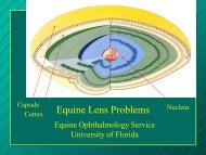
The lens: used for up close vision– Cataracts are lens opacities. Most horses have <strong>the</strong>m.– nuclear sclerosis: aging change seen in older horses– Lens position changes are a big problem in <strong>the</strong> horseFocal cataractWhat about <strong>the</strong> pupil <strong>and</strong> cataracts?
OphthalmoscopyDennis E Brooks DVM PhDUniversity <strong>of</strong> Florida• The direct ophthalmoscope:– lateral magnification: 7.9X– axial magnification: 84X• Indirect ophthalmoscopy:– 5.5 D lens: 3.86X lateral <strong>and</strong> 20.1Xaxial– 14 D lens: 1.18X lateral <strong>and</strong> 1.86Xaxial– 20 D lens: 0.79X <strong>and</strong> 0.84X lateral<strong>and</strong> axial respectively.
• Examine <strong>the</strong> fundus with a bright light,indirect lens, <strong>and</strong> directophthalmoscope.– indirect technique first (low magnification)– direct technique last (high magnification)
• Mydriasis lasts 4-6 hours• Do not use atropine fordiagnostics!• Prepurchase dilatation??
Dan Scott Assoc,retinal diagnostic light
IndirectDirectDifferent ophthalmoscopes providedifferent degrees <strong>of</strong> magnification.Panoptic
Ultrasonography
CorneaIrisLensVitreousDave WilkieRetina
Repay