SBRT&StereoscopicIGRT_2012 [Compatibility Mode]
SBRT&StereoscopicIGRT_2012 [Compatibility Mode]
SBRT&StereoscopicIGRT_2012 [Compatibility Mode]
Create successful ePaper yourself
Turn your PDF publications into a flip-book with our unique Google optimized e-Paper software.
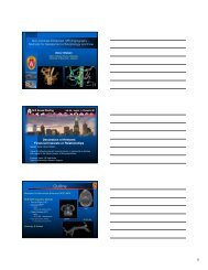
Patient Immobilization<br />
With arms over the head.<br />
Flat lung board with T-bar holder for the arms.<br />
Vacuum Bag<br />
Maximum Intensity Projection (MIP)<br />
CT data is acquired using a Bellows or RPM<br />
device that records a breathing wave.<br />
Maximum intensity values are assigned to pixels<br />
at locations where a tumor moves to over time.<br />
Are a derived dataset of images that show a<br />
“composite” of the tumor volume over the time<br />
period that the CT data was acquired.<br />
4D CT Simulation for LUNG SBRT<br />
CT Scanning procedure for moving target<br />
One free breathing scan – 3 mm slices, entire chest.<br />
One shorter, time-correlated scan – 3 mm slices, to<br />
include the tumor area.<br />
Breathing Waveform is acquired along with CT data<br />
using a Bellows or RPM device.<br />
The CT data acquired in one breath can be binned into<br />
10 equally spaced intervals along the waveform and<br />
reconstructed.<br />
The 10 intervals are called Phases.<br />
Moving Target virtual simulation - Fusion<br />
GTV<br />
MIP dataset<br />
Volume propagation from MIP dataset<br />
to<br />
free breathing dataset<br />
ITV<br />
Free Breathing<br />
dataset<br />
6


![SBRT&StereoscopicIGRT_2012 [Compatibility Mode]](https://img.yumpu.com/16889220/6/500x640/sbrtampstereoscopicigrt-2012-compatibility-mode.jpg)


