Formulation Development and Evaluation of Aceclofenac ... - japer
Formulation Development and Evaluation of Aceclofenac ... - japer
Formulation Development and Evaluation of Aceclofenac ... - japer

You also want an ePaper? Increase the reach of your titles
YUMPU automatically turns print PDFs into web optimized ePapers that Google loves.
Original Article<br />
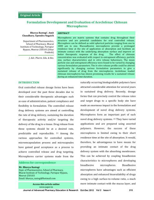
<strong>Formulation</strong> <strong>Development</strong> <strong>and</strong> <strong>Evaluation</strong> <strong>of</strong> Acecl<strong>of</strong>enac Chitosan<br />
Microspheres<br />
Bhavya Rastogi *, Amit<br />
Chaudhary, Upendra Nagaich<br />
Department <strong>of</strong> Pharmaceutics,<br />
School <strong>of</strong> Pharmacy, Bharat<br />
Institute <strong>of</strong> Technology, Partapur<br />
Bypass, Meerut-250103 (Uttar<br />
Pradesh)<br />
INTRODUCTION<br />
J. Adv. Pharm. Edu. & Res.<br />
Oral controlled release dosage forms have been<br />
developed over the past three decades due to<br />
their considerable therapeutic advantages such<br />
as ease <strong>of</strong> administration, patient compliance <strong>and</strong><br />
flexibility in formulation. The controlled release<br />
drug delivery systems are aimed at controlling<br />
the rate <strong>of</strong> drug delivery, sustaining the duration<br />
<strong>of</strong> therapeutic activity <strong>and</strong>/or targeting the<br />
delivery <strong>of</strong> the drug to a tissue. Drug release from<br />
these systems should be at a desired rate,<br />
predictable <strong>and</strong> reproducible. [1] Among the<br />
various approaches for controlled systems,<br />
microencapsulation process <strong>and</strong> microcapsules<br />
have gained good acceptance as a process to<br />
achieve controlled release <strong>and</strong> drug targeting.<br />
Microspheres carrier systems made from the<br />
Address for correspondence<br />
Mr. Bhavya Rastogi<br />
Dept. <strong>of</strong> Pharmaceutics, School <strong>of</strong> Pharmacy,<br />
Bharat Institute <strong>of</strong> Technology, Partapur Bypass,<br />
Meerut-250103<br />
Email: bhavya_rastogi@hotmail.com<br />
Access this article online<br />
www.<strong>japer</strong>.in<br />
ABSTRACT<br />
Microspheres are matrix systems that contains drug throughout their<br />
structure <strong>and</strong> are potential c<strong>and</strong>idates for oral controlled release.<br />
Microsphere can be defined as solid spherical particles ranging from one to<br />
1000 μm in size. Mucoadhesive microspheres provide a prolonged<br />
residence time at the site <strong>of</strong> application or absorption <strong>and</strong> facilitate an<br />
intimate contact with the underlying absorption surface <strong>and</strong> improve or<br />
better therapeutic response <strong>of</strong> the drug. The effect <strong>of</strong> chitosan<br />
concentration was evaluated with respect to entrapment efficiency, particle<br />
size, surface characteristics <strong>and</strong> in vitro release behaviours. The mean<br />
particle size <strong>and</strong> entrapment efficiency were found to be varied by changing<br />
various formulation parameters. The in vitro release pr<strong>of</strong>ile could be altered<br />
significantly by changing various formulation parameters to give a<br />
sustained release <strong>of</strong> drug from the microspheres. Acel<strong>of</strong>enac loaded<br />
chitosan microspheres has shown promising results for a sustained release<br />
during an enhanced time duration.<br />
naturally occurring biodegradable polymers have<br />
attracted considerable attention for several years<br />
in sustained drug delivery. Recently, dosage<br />
forms that can precisely control the release rates<br />
<strong>and</strong> target drugs to a specific body site have<br />
made an enormous impact in the formulation <strong>and</strong><br />
development <strong>of</strong> novel drug delivery systems.<br />
Microspheres form an important part <strong>of</strong> such<br />
novel drug delivery systems. [2] They have varied<br />
applications <strong>and</strong> are prepared using assorted<br />
polymers. However, the success <strong>of</strong> these<br />
microspheres is limited owing to their short<br />
residence time at the site <strong>of</strong> absorption. It would,<br />
therefore, be advantageous to have means for<br />
providing an intimate contact <strong>of</strong> the drug<br />
delivery system with the absorbing membranes.<br />
This can be achieved by coupling bioadhesion<br />
characteristics to microspheres <strong>and</strong> developing<br />
bioadhesive microspheres. Bioadhesive<br />
microspheres have advantages such as efficient<br />
absorption <strong>and</strong> enhanced bioavailability <strong>of</strong> drugs<br />
owing to a high surface-to-volume ratio, a much<br />
more intimate contact with the mucus layer, <strong>and</strong><br />
Journal <strong>of</strong> Advanced Pharmacy Education & Research Oct-Dec 2012 Vol 2 Issue 4<br />
215
Bhavya Rastogi, et al.: <strong>Formulation</strong> <strong>Development</strong> <strong>and</strong> <strong>Evaluation</strong> <strong>of</strong> Acecl<strong>of</strong>enac Chitosan Microspheres<br />
specific targeting <strong>of</strong> drugs to the absorption site.<br />
[3] Chitosan (obtained by deacetylation <strong>of</strong> chitin)<br />
is a cationic polymer that has been proposed for<br />
use in microsphere systems by a number <strong>of</strong><br />
authors. Chitosan was selected as a polymer in<br />
the preparation <strong>of</strong> mucoadhesive microspheres<br />
because <strong>of</strong> its good mucoadhesive <strong>and</strong><br />
biodegradable properties. [4] Hence, there is a<br />
need to develop an oral drug delivery system that<br />
is convenient for patients. Various synthetic <strong>and</strong><br />
natural polymers like alginate, chitosan <strong>and</strong><br />
polyesters have been used to develop drug<br />
delivery systems for entrapping <strong>and</strong> delivering<br />
drugs orally. Chitosan is used in cosmetics <strong>and</strong> is<br />
under investigation for use in a number <strong>of</strong><br />
pharmaceutical formulations. The suitability <strong>and</strong><br />
performance <strong>of</strong> Chitosan as a component <strong>of</strong><br />
pharmaceutical formulations for drug delivery<br />
applications has been investigated in numerous<br />
studies. These include controlled drug delivery<br />
applications, use as a component <strong>of</strong><br />
mucoadhesive dosage forms, rapid release<br />
dosage forms, improved peptide delivery, colonic<br />
drug delivery systems, <strong>and</strong> use for gene delivery.<br />
Chitosan has been processed into several<br />
pharmaceutical forms including gels, films, beads,<br />
microspheres, tablets, <strong>and</strong> coatings for<br />
liposomes. [5]<br />
The objective <strong>of</strong> the present investigation was to<br />
develop an extended <strong>and</strong> controlled release<br />
composition <strong>and</strong> formulation <strong>of</strong> Acecl<strong>of</strong>enac<br />
using chitosan polymer along with sodium tri-<br />
poly phosphate.<br />
MATERIAL AND METHODS<br />
Acecl<strong>of</strong>enac was obtained as a gift sample from<br />
Cotec Health Care Pvt. Ltd, Roorkee, India.<br />
Chitosan was purchased from Sigma Aldrich,<br />
USA. Sodium tripolyphosphate (TPP) <strong>and</strong> all<br />
other reagents were <strong>of</strong> analytical grade.<br />
Preparation <strong>of</strong> Microspheres<br />
Chitosan microspheres were prepared by<br />
ionotropic gelation method. In this method<br />
chitosan stock solution (1% w/v) was prepared<br />
by dissolving chitosan in acetic acid (1% v/v) at<br />
room temperature. The drug was dissolved<br />
directly into the above prepared chitosan<br />
solution. 10 ml <strong>of</strong> this bubble free solution was<br />
dropped through a disposable syringe needle<br />
into a gently agitating 100ml <strong>of</strong> 2% (w/v) sodium<br />
tripolyphosphate solution. The dropping rate <strong>and</strong><br />
falling distance were kept constant. The solution<br />
was magnetically stirred for half an hour<br />
followed by filtration <strong>and</strong> rinsing with distilled<br />
water. Gel like beads were obtained which was<br />
air dried for twenty four hours followed by oven<br />
drying for six hours at 40˚C. [6]<br />
Table 1: <strong>Formulation</strong> table <strong>of</strong> Acecl<strong>of</strong>enac<br />
Chitosan<br />
<strong>Formulation</strong><br />
Code<br />
Acecl<strong>of</strong>enac<br />
(gm)<br />
Chitosan<br />
(gm)<br />
Sod. TPP<br />
(gm)<br />
F1 0.1 0.9 0.2<br />
F2 0.1 0.8 0.2<br />
F3 0.1 0.7 0.2<br />
F4 0.1 0.6 0.2<br />
F5 0.1 0.5 0.2<br />
F6 0.1 0.4 0.2<br />
F7 0.1 0.3 0.2<br />
F8 0.1 0.2 0.2<br />
Characterization <strong>of</strong> Acecl<strong>of</strong>enac Chitosan<br />
Microspheres<br />
Particle size analysis<br />
Particle size analysis plays an important role in<br />
determining the release characteristics <strong>and</strong><br />
chitosan property. Mean particle size for all<br />
formulation was determined by dividing the total<br />
216 Journal <strong>of</strong> Advanced Pharmacy Education & Research Oct-Dec 2012 Vol 2 Issue 4
weight size <strong>of</strong> formulation to % total weight <strong>of</strong><br />
chitosan microspheres. [7]<br />
Drug Entrapment<br />
The various formulations <strong>of</strong> the chitosan<br />
microspheres were subjected for drug content.<br />
50 mg <strong>of</strong> chitosan microspheres from all batches<br />
were accurately weighed <strong>and</strong> crushed. The<br />
powdered <strong>of</strong> microspheres were dissolved with<br />
10ml ethanol in 100ml volumetric flask <strong>and</strong><br />
makeup the volume with 0.1 N HCl. This resulting<br />
solution is than filtered through whatmann filter<br />
paper No. 44. After filtration, from this solution<br />
10 ml was taken out <strong>and</strong> diluted up to 100 ml<br />
with 0.1 N HCl. Again from this solution 2 ml was<br />
taken out <strong>and</strong> diluted up to 10 m1 with 0.1 N HCI<br />
<strong>and</strong> the absorbance was measured at 275 nm<br />
against 0.1 N HCI as a blank. The percentage<br />
drug entrapment was calculated as follows. [8]<br />
Calculated drug concentration<br />
% Drug entrapment = x 100<br />
Theoretical drug concentration<br />
Shape <strong>and</strong> Surface Characterization <strong>of</strong><br />
Chitosan Microspheres by Scanning Electron<br />
Microscopy<br />
Bhavya Rastogi, et al.: <strong>Formulation</strong> <strong>Development</strong> <strong>and</strong> <strong>Evaluation</strong> <strong>of</strong> Acecl<strong>of</strong>enac Chitosan Microspheres<br />
From the formulated batches <strong>of</strong> chitosan<br />
microspheres, formulation (F4) which showed an<br />
appropriate balance between the buoyancy <strong>and</strong><br />
the percentage release were examined for<br />
surface morphology <strong>and</strong> shape using scanning<br />
electron microscope Hitachi, Japan, Trichy.<br />
Sample was fixed on carbon tape <strong>and</strong> fine gold<br />
sputtering was applied in a high vacuum<br />
evaporator. The acceleration voltage was set at<br />
20KV during scanning. Microphotographs were<br />
taken on different magnification <strong>and</strong> higher<br />
magnification (200X) was used for surface<br />
morphology. [9]<br />
In vitro Release Studies:<br />
The drug release rate from chitosan micro<br />
spheres was carried out using the USP<br />
dissolution paddle assembly. A weighed amount<br />
<strong>of</strong> micro spheres equivalent to 100 mg drug were<br />
dispersed in 900 ml <strong>of</strong> phosphate buffer 6.8<br />
maintained at 37 ± 0.5°C <strong>and</strong> stirred at 100 RPM.<br />
At preselected time intervals one ml sample was<br />
withdrawn <strong>and</strong> replaced with equal amount <strong>of</strong><br />
phosphate buffer 6.8. The collected samples<br />
were suitably diluted with phosphate buffer 6.8<br />
<strong>and</strong> analyzed spectrophotometrically at 275 nm<br />
to determine the concentration <strong>of</strong> drug present in<br />
the dissolution medium. The dissolution studies<br />
were repeated using phosphate buffer pH 6.8 as<br />
dissolution medium. [10]<br />
In-Vivo Anti - Inflammatory Study<br />
It has been reported in the literatures that<br />
acecl<strong>of</strong>enac may have therapeutic potential as<br />
anti-inflammatory agent either alone or in<br />
combination with nonsteroidal anti-<br />
inflammatory drug. Therefore the in-vivo<br />
500µg/kg <strong>of</strong> formulation release behaviour <strong>of</strong> the<br />
best formulation F4 was studied by measuring<br />
anti-inflammatory activity in adult male wistar<br />
rats using cotton pellet granuloma method. The<br />
male wistar rats were divided in to three groups,<br />
each group consisting <strong>of</strong> 6 animals. One group<br />
served as control, second group served as<br />
st<strong>and</strong>ard, received 500µg/kg <strong>of</strong> acecl<strong>of</strong>enac as<br />
solution in water in two divided dose orally,<br />
while third group received chitosan<br />
microspheres containing acecl<strong>of</strong>enac orally<br />
require to release about 500µg/kg <strong>of</strong><br />
acecl<strong>of</strong>enac) once daily during the experiment.<br />
The rats with an average weight <strong>of</strong> 150g were<br />
anaesthetized with ether. The cotton pellets each<br />
Journal <strong>of</strong> Advanced Pharmacy Education & Research Oct-Dec 2012 Vol 2 Issue 4<br />
217
Bhavya Rastogi, et al.: <strong>Formulation</strong> <strong>Development</strong> <strong>and</strong> <strong>Evaluation</strong> <strong>of</strong> Acecl<strong>of</strong>enac Chitosan Microspheres<br />
weighing 10 ± 1 mg were prepared <strong>and</strong> sterilized<br />
in hot air oven at 120°C for 3 hours. The<br />
abdomen was shaved cleanly, swabbed with 70%<br />
(v/v) ethanol <strong>and</strong> small incision was made in the<br />
lower abdomen <strong>of</strong> the rat. Using a blunt forceps,<br />
one sterile cotton pellet <strong>of</strong> known weight was<br />
placed in each aexilla <strong>and</strong> groin region <strong>and</strong> then<br />
incision was closed with sutures. On 8 th day<br />
albino rat were sacrificed <strong>and</strong> four pellets were<br />
removed. The pellets were dried overnight at<br />
55°C <strong>and</strong> weighed. The difference between the<br />
final weight <strong>of</strong> the pellet after drying <strong>and</strong> its<br />
initial weight was taken as the granuloma tissue<br />
weight. Results were expressed as percentage<br />
inhibition <strong>of</strong> granuloma in drug treated groups<br />
compared with the control group. [11]<br />
Stability Study<br />
Stability study was carried out for the F4<br />
formulation by exposing it to different<br />
temperature 5-8°C, 27°C <strong>and</strong> 42°C for 45 days.<br />
The sample was analyzed for drug content at the<br />
regular intervals. It was found that no<br />
remarkable change in the drug content <strong>of</strong> F4<br />
formulation. This indicates that F4 was stable for<br />
following temperature. [12]<br />
RESULT AND DISCUSSION<br />
Controlled release microspheres were prepared<br />
using chitosan. The mean particle size <strong>of</strong> the<br />
microspheres significantly increased with<br />
increasing the concentration <strong>of</strong> chitosan <strong>and</strong> was<br />
in the range 613 µm to 869 µm as shown in Table<br />
size decreased. The SEM photographs showed<br />
that the fabricated microspheres were spherical<br />
with a smooth surface <strong>and</strong> exhibited a range <strong>of</strong><br />
sizes within each batch as shown in Fig. 1.<br />
Table 2: <strong>Evaluation</strong> characteristics <strong>of</strong> Acecl<strong>of</strong>enac<br />
Chitosan Microsphere<br />
<strong>Formulation</strong><br />
Code<br />
Mean Particle Size<br />
(µm)<br />
Entrapent<br />
Efficiency<br />
%<br />
Yield<br />
F1 869 74.12 84.67<br />
F2 826 71.56 81.53<br />
F3 800 67.23 76.89<br />
F4 790 64.76 72.56<br />
F5 762 60.01 70.34<br />
F6 758 55.38 68.03<br />
F7 664 49.47 59.44<br />
F8 613 43.14 55.10<br />
The incorporation efficiency <strong>of</strong> Acecl<strong>of</strong>enac was good<br />
in all loadings <strong>and</strong> incorporation efficiency was<br />
increased with the polymer concentration. The high<br />
entrapment efficiency <strong>of</strong> the drug was believed to be<br />
due to its poor solubility in liquid paraffin. As the<br />
polymer concentration increased, the viscosity <strong>of</strong><br />
polymer solution was increased <strong>and</strong> responsible for<br />
formation <strong>of</strong> larger microspheres. In-vitro acecl<strong>of</strong>enac<br />
release studies were performed in 0.1N HCl for 2hrs<br />
<strong>and</strong> 7.4pH for 10hrs. The cumulative release <strong>of</strong><br />
acecl<strong>of</strong>enac significantly decreased with increasing<br />
acrycoat S 100 concentration. The increased density <strong>of</strong><br />
the polymer matrix at higher concentrations results in<br />
an increased diffusional path length. This may<br />
decrease the over-all drug release from the polymer<br />
matrix. Furthermore, smaller microspheres were<br />
formed at a lower polymer concentration <strong>and</strong> had a<br />
larger surface area exposed to dissolution medium,<br />
giving rise to faster drug release. Acecl<strong>of</strong>enac release<br />
was higher in the case <strong>of</strong> microspheres prepared at a<br />
higher agitation speed but the difference in drug<br />
2. The viscosity <strong>of</strong> the medium increases at a release was not statistically significant, but at low<br />
higher polymer concentration resulting in<br />
agitation speed the release rate was slow (Fig. 2).<br />
enhanced interfacial tension with diminished<br />
Table 3 contains the in vivo study data. Present<br />
shearing efficiency <strong>and</strong> increased particle size. As<br />
the stirring rate was increased, the mean particle<br />
investigation promised the therapeutic efficacy <strong>of</strong> the<br />
drug. Stability studies also shown satisfactory results<br />
(Table 4).<br />
218 Journal <strong>of</strong> Advanced Pharmacy Education & Research Oct-Dec 2012 Vol 2 Issue 4
S.<br />
No<br />
Treatment<br />
Dose<br />
(µg/kg)<br />
Table 3: Result Of Anti Inflammatory Activity Measurement<br />
Weight <strong>of</strong> dry cotton pellet<br />
granuloma<br />
(mg)<br />
Before After*<br />
Weight <strong>of</strong> dry granuloma<br />
(mg)<br />
Percentage Decreases in granuloma<br />
(%)<br />
1. Control - 10.1 84.72 74.62 -<br />
2. St<strong>and</strong>ard 500 10 .2 67.98 57.73 22.63<br />
3.<br />
Cumulative % drug release<br />
Bhavya Rastogi, et al.: <strong>Formulation</strong> <strong>Development</strong> <strong>and</strong> <strong>Evaluation</strong> <strong>of</strong> Acecl<strong>of</strong>enac Chitosan Microspheres<br />
F4<br />
formulation<br />
500 10 .1 51.17 40.07 45.49<br />
Table 4: Stability study data <strong>of</strong> the optimized acecl<strong>of</strong>enac chitosan microsphere<br />
S. No Days % Drug Remaining 5-8°C % Drug Remaining 27 ± 2°C % Drug Remaining 42 ± 2°C<br />
1. 0 100 ± 00 100 ± 00 100 ± 00<br />
2. 14 99.6 ± 0.015 99.9 ± 0.003 99.4 ± 0.041<br />
3. 28 99.5 ± 0.013 99.8 ± 0.027 99.2 ± 0.036<br />
4. 45 99.4 ± 0.15 99.6 ± 0.012 99.1 ± 0.02<br />
Fig. 1: SEM <strong>of</strong> Acecl<strong>of</strong>enac Chitosan<br />
Microsphere<br />
100<br />
90<br />
80<br />
70<br />
60<br />
50<br />
40<br />
30<br />
20<br />
10<br />
0<br />
0 2 4 6 8 10 12<br />
Time (hrs)<br />
F1 F2 F3 F4<br />
F5 F6 F7 F8<br />
Fig. 2: Release rate for Acecl<strong>of</strong>enac Chitosan<br />
Microsphere<br />
REFERENCE<br />
1. J.K. Patel, R.P. Patel, A.F. Amin, M.M. Patel (2005).<br />
<strong>Formulation</strong> <strong>and</strong> evaluation <strong>of</strong> mucoadhesive<br />
glipizide microspheres. AAPS Pharm Sci Tech. 6,<br />
E49-55.<br />
2. K.V. Ranga Rao, K.P. Devi (1988). Swelling<br />
controlled release systems: recent developments<br />
<strong>and</strong> application. Int J Pharm. 48, 1-16.<br />
3. Jayakrishnan A, Latha MS. Biodegradable<br />
polymeric microspheres as drug carriers. In: Jain<br />
NK, Editor. Controlled <strong>and</strong> Novel drug delivery.<br />
New Delhi: CBS publishers. 1997. pp 236-255.<br />
4. Kreuter J., <strong>Evaluation</strong> <strong>of</strong> nanoparticles as drug-<br />
delivery systems, Pharm. Acta. Helv, 1983, 58,<br />
196-208.<br />
5. Rasool D, Elham R, Efat F. Gelatin microspheres<br />
for controlled release <strong>of</strong> All trans Retinoic acid<br />
topical formulation <strong>and</strong> Drug Delivery<br />
<strong>Evaluation</strong>. Iranian Journal <strong>of</strong> Pharmaceutical<br />
Research 2003; 47-50.<br />
6. Rossler B, Kreuter J, Scherer D. Collagen<br />
microparticles: Preparation <strong>and</strong> properties. J.<br />
Microencapsul.1995; 49-57.<br />
7. Veerareddy PR, Tedla S, B<strong>and</strong>a SR, B<strong>and</strong>ari S,<br />
Jukanti R. Preparation <strong>and</strong> evaluation <strong>of</strong><br />
mucoadhesive cefdinir microcapsules. J Adv<br />
Pharm Tech Res 2011;2:115-20<br />
8. Yoshifumi M, Youko K, Takashi I, Kyoko K,<br />
Susumu K, Drug Release Pr<strong>of</strong>ile from Calcium-<br />
Induced Alginate-Phosphate Composite Gel<br />
Beads, International Journal <strong>of</strong> Polymer Science<br />
2009; 1-4.<br />
Journal <strong>of</strong> Advanced Pharmacy Education & Research Oct-Dec 2012 Vol 2 Issue 4<br />
219
Bhavya Rastogi, et al.: <strong>Formulation</strong> <strong>Development</strong> <strong>and</strong> <strong>Evaluation</strong> <strong>of</strong> Acecl<strong>of</strong>enac Chitosan Microspheres<br />
9. Mankala SK, Korla AC, Gade S. <strong>Development</strong> <strong>and</strong><br />
evaluation <strong>of</strong> acecl<strong>of</strong>enac-loaded mucoadhesive<br />
microcapsules. J Adv Pharm Tech Res<br />
2011;2:245-54<br />
10. Shilpa A, Agrawal SS Ray AR, Controlled Delivery<br />
<strong>of</strong> Drugs from Alginate Matrix, Journal <strong>of</strong><br />
Macromolecular science Part C-Polymer reviews,<br />
43, 2003, 187.<br />
11. Arora S, Budhiraja RD. Chitosan-alginate<br />
microcapsules <strong>of</strong> amoxicillin for gastric stability<br />
<strong>and</strong> mucoadhesion. J Adv Pharm Tech Res<br />
2012;3:68-74.<br />
12. Grellier M, Granja P L, Fricain J C, Bidarra S J,<br />
Renard M, Bareille R, et al. The effect <strong>of</strong> the co-<br />
immobilization <strong>of</strong> human osteoprogenitors <strong>and</strong><br />
endothelial cells within alginate microspheres on<br />
mineralization in a bone defect. Biomaterials<br />
2009; 30: 3271e8.<br />
Bhavya Rastogi, Amit Chaudhary, Upendra Nagaich,;<br />
<strong>Formulation</strong> <strong>Development</strong> <strong>and</strong> <strong>Evaluation</strong> <strong>of</strong><br />
Acecl<strong>of</strong>enac Chitosan Microspheres; J. Adv. Pharm.<br />
Edu. & Res. 2012: 4: 215-220<br />
Source <strong>of</strong> Support: Nil, Conflict <strong>of</strong> Interest: Nil<br />
220 Journal <strong>of</strong> Advanced Pharmacy Education & Research Oct-Dec 2012 Vol 2 Issue 4