Science of PEMF - PEMF NOW
Science of PEMF - PEMF NOW
Science of PEMF - PEMF NOW

You also want an ePaper? Increase the reach of your titles
YUMPU automatically turns print PDFs into web optimized ePapers that Google loves.
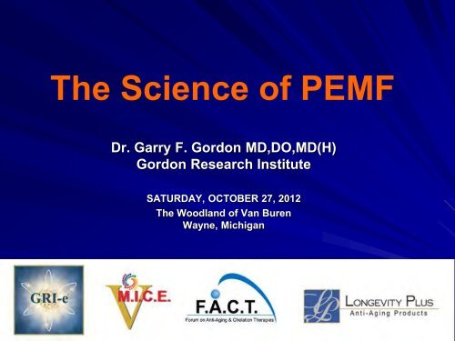
The <strong>Science</strong> <strong>of</strong> <strong>PEMF</strong><br />
Dr. Garry F. Gordon MD,DO,MD(H)<br />
Gordon Research Institute<br />
SATURDAY, OCTOBER 27, 2012<br />
The Woodland <strong>of</strong> Van Buren<br />
Wayne, Michigan
FUNCTIONAL MEDICINE<br />
Predictive, Preventive, Personalized, and Participatory<br />
Functional medicine is the future <strong>of</strong> conventional medicine–available now. It<br />
seeks to identify and address the root causes <strong>of</strong> disease, and views the body<br />
as one integrated system, not a collection <strong>of</strong> independent organs divided up<br />
by medical specialties. It treats the whole system, not just the symptoms.<br />
DNA/Telomerase<br />
Testing<br />
Diet & Lifestyle<br />
Modification<br />
Personalized<br />
Therapies<br />
Community<br />
Group Support<br />
Intensive lifestyle therapy—not just wellness counseling or prevention,<br />
but lifestyle treatment <strong>of</strong> existing chronic disease—focusing on<br />
pre-diabetes, diabetes, and heart disease has been proven<br />
to work better than medication or surgery.
Based on interviews with over three hundred <strong>of</strong> the<br />
world’s top scientists, who are already inventing the<br />
future in their labs, Kaku—in a lucid and engaging<br />
fashion—presents the revolutionary developments in<br />
medicine, computers, quantum physics, and space<br />
travel that will forever change our way <strong>of</strong> life and alter<br />
the course <strong>of</strong> civilization itself.<br />
Dr. Kaku’s astonishing revelations include:<br />
Sensors in your clothing, bathroom, and appliances will<br />
monitor your vitals, and nanobots will scan your DNA<br />
and cells for signs <strong>of</strong> danger, allowing life expectancy to<br />
increase dramatically.<br />
You will control computers and appliances via tiny<br />
sensors that pick up your brain scans.<br />
http://mkaku.org/home/?p=988
THE MAN FROM THE 11TH DIMENSION By Elizabeth Finkel<br />
His mind wanders incredibly complex worlds <strong>of</strong> eleven dimensions and he is trying<br />
to complete Einstein's unfinished masterpiece: a 'theory <strong>of</strong> everything'. Meet one<br />
<strong>of</strong> the world's leading theoretical physicists, Michio Kaku, a founder <strong>of</strong> string field<br />
theory and a man as charming as he is imposing.<br />
Dr. Michio Kaku is the co-creator <strong>of</strong> string field<br />
theory, a branch <strong>of</strong> string theory. He received a<br />
B.S. (summa cum laude) from Harvard University<br />
in 1968 where he came first in his physics class.<br />
He went on to the Berkeley Radiation Laboratory<br />
at the University <strong>of</strong> California, Berkeley and<br />
received a Ph.D. in 1972. In 1973, he held a<br />
lectureship at Princeton University.<br />
Michio continues Einstein’s search for a<br />
“Theory <strong>of</strong> Everything,” seeking to unify the<br />
four fundamental forces <strong>of</strong> the universe—<br />
the strong force, the weak force, gravity and<br />
electromagnetism.<br />
http://www.cosmosmagazine.com/issues/2005/2/ http://www.cosmosmagazine.com/node/99
The Synchronized Universe - A new scientific revolution!<br />
Physicist Dr. Claude Swanson, educated at MIT and Princeton<br />
University, describes the latest discoveries in Energy Medicine.<br />
“We are learning the ‘Secret <strong>of</strong> Life’, how the body’s<br />
trillons <strong>of</strong> cells grow, repair and heal…<br />
Electromagnetism and Earth energies hold<br />
part <strong>of</strong> the answer… we are witnessing the<br />
integration <strong>of</strong> CONSCIOUSNESS with physics“<br />
It is called CHI, PRANA, MANA, ORENDO, WAKEN, BARAKA,<br />
and LIFE FORCE.<br />
It is the energy which enables adepts, Yogis and Shamen to<br />
achieve the miraculous feats they do. It enables QiGong masters<br />
from China to project their energy over thousands <strong>of</strong> miles to heal<br />
injured cells and to cure cancer in laboratory experiments.<br />
Today we have documented pro<strong>of</strong> <strong>of</strong> how this energy changes<br />
the laws <strong>of</strong> physics, bringing together the Theory <strong>of</strong> Relativity<br />
and Quantum Mechanics, and is the explanation for many<br />
strange phenomena which we in the West call "paranormal.“<br />
http://synchronizeduniverse.com/
The Synchronized Universe Model (S.U.M.)<br />
Assumes that all the particles in the universe interact with one another.<br />
Local electrons are tied to distant matter via photons. The “virtual photons” in<br />
space are assumed to be created by the motions <strong>of</strong> other electrons. Most <strong>of</strong> them<br />
are created by the “distant matter” which contains almost all the matter <strong>of</strong> the<br />
universe.<br />
The seemingly random “zig zag” dance they do is not random – it is really the<br />
communication between it and the distant matter – a purposeful, intimate and<br />
conscious dance with one another.<br />
SO ALL THE EXISTING ELECTRONS AND PROTONS AND<br />
OTHER PARTICLES ARE ACTUALLY CONNECTED<br />
TO ONE ANOTHER!<br />
Momentum and energy that is created here (locally) is absorbed there<br />
(universally) and vice versa, virtually instantaneously, able to travel backward in<br />
time as well as forward.<br />
Photons which travel backwards in time are called “advanced waves”, and are a<br />
perfectly valid solution <strong>of</strong> Maxwell’s Equations which govern electromagnetism.<br />
(from pgs 241 – 242 <strong>of</strong> The Synchronized Universe – Claude Swanson, PhD)
Conscious Creation<br />
The law <strong>of</strong> gravity + the law <strong>of</strong> energy + The law <strong>of</strong> observation<br />
The Law <strong>of</strong> Attraction is one <strong>of</strong> the most powerful laws governing the energy <strong>of</strong><br />
the universe. Whether or not we understand it, like gravity, it affects our lives<br />
without fail and without discrimination.<br />
The law <strong>of</strong> attraction operates upon the vibration <strong>of</strong> our thoughts, with “like<br />
attracting like”. Wherever we place our attention, energy and focus, with the<br />
corresponding emotional state or “feelings” about it… those frequencies will<br />
vibrationally attract the same <strong>of</strong> its kind.<br />
(positivity = high vibration = health) (negativity = low vibration = disease)<br />
Applying the Law deliberately to attract what we want in our lives is a practice <strong>of</strong><br />
Conscious Creation through our thought process. It teaches the use <strong>of</strong> ‘attention’<br />
and ‘intention’ to deliberately attract higher vibrations to enhance the quality <strong>of</strong><br />
our lives…<br />
Everything in the universe is made up <strong>of</strong> energy (atoms containing electrons,<br />
protons, neutrons, and quarks), and the law <strong>of</strong> observation states that<br />
ENERGY FOLLOWS THOUGHT.
Cellular communication – electrons and photons<br />
as messengers<br />
Researchers have found that cells are in communication<br />
all the time. The DNA molecule, for example, radiates and<br />
absorbs in the millimeter wave band.<br />
Can this be the source <strong>of</strong> the “Backster Effect”, <strong>of</strong> cell-to-cellcommunication?<br />
Backster Effect – experiment postulating that plants can communicate with other<br />
lifeforms. By measuring the rate at which water rises from a philodendron's root into<br />
its leaves, using a polygraph to record altered electrical resistance signals<br />
from the plant taking up the water – surprisingly the graph tracing began to<br />
show a pattern typical <strong>of</strong> the response you get when you subject a human<br />
to emotional stimulation <strong>of</strong> short duration".<br />
Now It has been proven that a “sick” cell radiates something, and when a<br />
healthy cell receives this radiation, it too becomes sick (Kaznachayev, 1967, 1981,<br />
1982). The opposite also occurs, sick cells can be brought back to health<br />
with radiation from healthy cells.<br />
Can this explain “energy healing”? Source, strength and intention <strong>of</strong> the<br />
energy being radiated?<br />
(from p. 234 <strong>of</strong> The Synchronized Universe – Claude Swanson, PhD)
Dr. Bruce Lipton is an internationally recognized leader<br />
in bridging science and spirit.<br />
The new sciences quantum physics and epigenetics<br />
are revolutionizing our understanding <strong>of</strong> the link<br />
between mind and matter.<br />
By retraining our minds to create healthy beliefs, we<br />
can change the physiology <strong>of</strong> our trillion-celled bodies.<br />
http://www.brucelipton.com/
We’re living the end <strong>of</strong> time. Not the end <strong>of</strong> the world, but the end <strong>of</strong> a world<br />
age – a 5,125 year cycle <strong>of</strong> time – and the way we’ve known the world through<br />
out that time. The present world began in 3,113 B.C. and will end in A.D. 2012.<br />
Because the end <strong>of</strong> anything also marks the beginning <strong>of</strong> what comes next,<br />
we’re also living the start <strong>of</strong> what follows the end <strong>of</strong> time: the next world age,<br />
which ancient traditions called the great cycle.<br />
http://www.greggbraden.com/home/fractal-time-calculator/
Solar Storms – increasing activity and intensity thru 2012<br />
Image provided by NASA, taken<br />
Sunday night, Jan. 22, 2012,<br />
shows a solar flare erupting on<br />
the Sun's northeastern<br />
hemisphere. Space weather<br />
<strong>of</strong>ficials say the strongest solar<br />
storm in more than six years is<br />
already bombarding Earth with<br />
radiation with more to come.<br />
An aurora appears when a<br />
magnetic solar wind slams<br />
into the Earth's magnetic field,<br />
exciting electrons <strong>of</strong> oxygen<br />
and nitrogen.<br />
According to the National Oceanic and Atmospheric Administration, problems can<br />
include current surges in power lines, and interference in the broadcast <strong>of</strong> radio, TV<br />
and telephone signals.<br />
Scientists have been expecting solar eruptions to become more intense as the sun<br />
enters a more active phase <strong>of</strong> its 11-year cycle, with an expected peak in 2013.
The Earth’s Magnetic Field<br />
is Weakening<br />
Over the last 165 years, scientists have<br />
measured the Earth's magnetic field and<br />
have recorded a decline <strong>of</strong> its' strength.<br />
Today the magnetic field <strong>of</strong> the Earth is measured at 0.5 gauss. It is<br />
estimated that the field <strong>of</strong> the Earth 4,000 years ago was 5.0 gauss.<br />
That is a decrease <strong>of</strong> 90%!<br />
In addition, the Earth’s natural magnetic<br />
signal is <strong>of</strong>ten distorted by our modern way<br />
<strong>of</strong> living. The power grid, electrical<br />
appliances, mobile phone's, mobile phone<br />
towers, Satellite signals, TV broadcast<br />
stations, tall buildings, asphalt, draining<br />
pipes and more are responsible for us not<br />
getting the signals we have evolved to. The<br />
immune system suffers because <strong>of</strong> this.
Depression and Earth's weakening<br />
Magnetic Field<br />
Earth’s declining magnetic field may be one <strong>of</strong> the<br />
factors leading to the alarming rise in cases <strong>of</strong> clinical<br />
depression and suicide.<br />
In 2008, Russian scientists found a correlation between Earth’s<br />
declining magnetic field and suicide. Oleg Shumilov <strong>of</strong> the<br />
Institute <strong>of</strong> North Industrial Ecology Problems in Russia, told<br />
the New Scientist the Earth's magnetic field peaked in three<br />
cycles during the year; March to May, another in July with the last in October.<br />
Shumilov argues that many animals can sense the magnetic field, so why should this<br />
not be the case with humans?<br />
Michael Rycr<strong>of</strong>t, formerly head <strong>of</strong> the European Geosciences Society, quoted by the<br />
New Scientist, claims that around 10 to 15% <strong>of</strong> the population are affected by<br />
geomagnetic health problems.<br />
Dementia, depression and mental disorders are on the rise worldwide. If it<br />
turns out Earth is entering a new phase <strong>of</strong> accelerated field declination,<br />
which I believe it is, and artificially induced electro-magnetic field<br />
disturbances continue on Earth; depression and rates <strong>of</strong> suicide on the<br />
planet could start spiking.<br />
http://www.prometeus.nsc.ru/science/scidig/08/apr2.ssi
Electromagn Biol Med. 2010 Aug;29(3):105-12.<br />
A role for the geomagnetic field in<br />
cell regulation.<br />
Lib<strong>of</strong>f AR.<br />
Center for Molecular Biology and Biotechnology, Florida Atlantic University<br />
Abstract<br />
We advance the hypothesis that biological systems utilize the geomagnetic field<br />
(GMF) for functional purposes by means <strong>of</strong> ion cyclotron resonance-like (ICR)<br />
mechanisms.<br />
Numerous ICR-designed experiments have demonstrated that living things<br />
are sensitive, in varying degrees, to magnetic fields that are equivalent to<br />
both changes in the general magnetostatic intensity <strong>of</strong> the GMF, as well as<br />
its temporal perturbations. We propose the existence <strong>of</strong> ICR-like cell<br />
regulation processes, homologous to the way that biochemical messengers<br />
alter the net biological state through competing processes <strong>of</strong> enhancement<br />
and inhibition. In like manner, combinations <strong>of</strong> different resonance<br />
frequencies all coupled to the same local magnetic field provide a unique<br />
means for cell regulation.<br />
PMID:20707644 [PubMed - indexed for MEDLINE]
Bioelectromagnetics. 2009 Jan;30(1):21-8.<br />
Prolonged weakening <strong>of</strong> the geomagnetic<br />
field (GMF) affects the immune system<br />
<strong>of</strong> rats.<br />
Roman A, Tombarkiewicz B.<br />
Department <strong>of</strong> Brain Biochemistry, Institute <strong>of</strong> Pharmacology, Polish Academy <strong>of</strong><br />
<strong>Science</strong>s, Kraków, Poland. roman@if-pan.krakow.pl<br />
We found that the long-term shielding <strong>of</strong> the GMF could influence the<br />
functioning <strong>of</strong> the immune system in a sex-dependent manner.<br />
The deprivation <strong>of</strong> the GMF delayed physiological thymus involution, that<br />
effect being more strongly expressed in females. The weakening <strong>of</strong> the GMF<br />
resulted in an increased number <strong>of</strong> peritoneal macrophages, especially in<br />
males.<br />
The shielding <strong>of</strong> the GMF diminished the ability <strong>of</strong> macrophages to release NO<br />
and to synthesize O2(-), those effects being more powerfully expressed in<br />
males and females, respectively.<br />
It is proposed that the observed changes in the immune system occur as a<br />
consequence <strong>of</strong> the protective effect <strong>of</strong> GMF shielding on the circadian<br />
rhythm-dependent level <strong>of</strong> melatonin.
<strong>PEMF</strong> – The Fifth Element<br />
Most people know we need food (earth),<br />
water (water) and oxygen (air) to survive.<br />
And many people also know they need full<br />
spectrum sunlight (fire) or you get what is<br />
referred to as SAD (seasonal affective disorder).<br />
That makes FOUR critical elements:<br />
EARTH/FOOD WATER FIRE/SUNLIGHT AIR/OXYGEN<br />
However, every organism on earth (that includes people) has evolved to the<br />
natural magnetic signals <strong>of</strong> the earth and that part <strong>of</strong> the solar radiation that<br />
is able to penetrate our atmosphere.<br />
We have learned that these <strong>PEMF</strong> signals are <strong>of</strong> great importance to internal<br />
regulation <strong>of</strong> every organism.<br />
http://www.pemft.net/the-5th-element.html
<strong>PEMF</strong>'s are like a spark plug or catalyst<br />
for energy production in the cell.<br />
Just like a car needs oxygen, fuel and an ignition or spark<br />
plug, so does the human cell need fuel (glucose), oxygen<br />
and a "spark plug" or ignition. This ignition is <strong>PEMF</strong> or<br />
pulsed magnetic energy from both the earth and<br />
movement/exercise on the earth.<br />
We can also think <strong>of</strong> <strong>PEMF</strong> as a battery recharger for the human cell. We now<br />
know that the voltage <strong>of</strong> a healthy cell is about 70-110 millivolts and when we get<br />
sick that voltage drops below 50 millivolts or less and cancer cells are 30 millivolts<br />
or less. Pulsed electromagnetic fields (<strong>PEMF</strong>) act like a catalyst and battery<br />
recharger for the human cells and these <strong>PEMF</strong>'s are critical for human metabolism.<br />
<strong>PEMF</strong>'s also improve microcirculation, oxygenation (up to a 200% increase), help<br />
in nerve regeneration, pain management and many other health promoting<br />
benefits. There are over 1000 clinical studies and over 7000 research papers<br />
validating the therapeutic benefits <strong>of</strong> <strong>PEMF</strong>s.<br />
http://www.pemft.net/the-5th-element.html
Dr. Oz Recommends <strong>PEMF</strong><br />
Pulsating Electromagnetic Therapy<br />
is shifting the paradigm <strong>of</strong><br />
pain management<br />
Pulsed electromagnetic field (<strong>PEMF</strong>)<br />
therapy is FDA-approved to fuse bones<br />
and has been cleared in certain devices<br />
to reduce swelling and joint pain.<br />
Transcranial Magnetic Stimulation (TMS) and Magnetic Resonance Imaging (MRI) work<br />
on the same physics.<br />
All energy is electromagnetic in nature. All atoms, chemicals and cells produce<br />
electromagnetic fields (EMFs).<br />
Every organ in the body produces it own signature bioelectromagnetic field.<br />
<strong>Science</strong> has proven that our bodies actually project their own magnetic fields and that<br />
all 70 trillion cells in the body communicate via electromagnetic frequencies.<br />
Nothing happens in the body without an electromagnetic exchange. When the<br />
electromagnetic activity <strong>of</strong> the body ceases, life ceases.<br />
Watch this amazing 10 minute video to learn more…<br />
http://www.youtube.com/watch?v=cZSOKT-IdFE
Dr. William Pawluk, MD, MSc,<br />
appeared as <strong>PEMF</strong> Specialist on the<br />
Doctor Oz TV Show in November<br />
2011, where they discussed the most<br />
effective types <strong>of</strong> pulsed<br />
electromagnetic field therapy. http://www.drpawluk.com/doctor-oz-article-on-pemfs/<br />
http://www.doctoroz.com/videos/ask-your-doctor-about-pulsed-electromagnetic-field-therapy
http://www.drpawluk.com/<br />
The <strong>PEMF</strong>-100 device is an<br />
innovative, high intensity, very low<br />
frequency pulsed electromagnetic<br />
field generator. It is one <strong>of</strong> the most<br />
intense clinically useful<br />
electromagnetic devices available.<br />
The maximum field intensity is<br />
around 1920 Gauss (192 mT). The<br />
lowest field intensity is still likely<br />
close to 1000 Gauss.
Magnetic Therapy in Eastern Europe: A Review <strong>of</strong> 30 Years <strong>of</strong> Research<br />
By Jiri Jerabek, MD, PhD and William Pawluk, MD, MSc<br />
The book presents information summarizing conditions studied, magnetic field strength and type <strong>of</strong><br />
field used, frequency and duration <strong>of</strong> application and summary <strong>of</strong> actual results. There are detailed<br />
descriptions <strong>of</strong> many studies on both static (permanent) and frequency (pulsed) fields.<br />
Controlled human studies described include:<br />
· Atherosclerosis<br />
· Brain neurosecretion<br />
· Breast fissures<br />
· Burns<br />
· Carpal tunnel syndrome<br />
· Cervicitis<br />
· Chronic bronchitis<br />
· Controlled Studies Animals<br />
· Corneal trauma<br />
· Edema<br />
· Endometriosis<br />
· Femoral artery surgery<br />
· Fractures<br />
· Increased circulation<br />
· Infected skin wounds<br />
· Ischemic heart disease<br />
· Limb grafts<br />
· Liver function<br />
And more…
We are only as healthy as our cells.<br />
“By regenerating the cells in our bodies we can help our cells<br />
become and stay healthy with pulsed electromagnetic fields.<br />
The earth creates magnetic fields, without which life would not be<br />
possible. <strong>Science</strong> teaches that everything is energy. All energy is<br />
electromagnetic in nature. All atoms, chemicals, and cells produce<br />
electromagnetic fields. <strong>Science</strong> has proven that our bodies<br />
actually project their own magnetic fields and our seventy trillion<br />
cells in the body communicate via electromagnetic frequencies.<br />
Disruption <strong>of</strong> electromagnetic energy in cells causes<br />
impaired cell metabolism. This is the final common pathway<br />
<strong>of</strong> disease. If cells are not healthy, the body is not healthy.”<br />
William Pawluk, MD, MSc, and Donna Ganza, ND<br />
Excerpt from 101 Great Ways to Improve Health
Volume 25 | Issue 5 | Page 30<br />
Date: 2011-05-01<br />
Power Failure<br />
Does mitochondrial dysfunction lie at the heart <strong>of</strong><br />
common, complex diseases like cancer and autism?<br />
By Megan Scudellari<br />
Over the last five years, a growing number <strong>of</strong> papers by<br />
researchers around the world have implicated dysfunctional<br />
mitochondria in many elusive diseases, including Parkinson’s, autism, and aging.<br />
Leading the charge is a respected and renowned member <strong>of</strong> the National Academy<br />
<strong>of</strong> <strong>Science</strong>s, Dr. Douglas Wallace, founder <strong>of</strong> the field <strong>of</strong> human mitochondrial<br />
genetics.<br />
“Every one <strong>of</strong> the diseases we can’t solve is absolutely logical if we put<br />
energy at the center,” Dr. Wallace says.<br />
Medicine fails to solve many <strong>of</strong> today’s common, complex diseases, Wallace<br />
asserts, because the fundamental paradigm is wrong: the medical establishment<br />
has spent far too long focusing on anatomy and ignoring energy—specifically,<br />
mitochondria.
The cells <strong>of</strong> living tissue are electrical<br />
direct current (DC) systems<br />
All life generates an electrical DC charge<br />
This natural DC charge is created by the movement <strong>of</strong> ions in and out<br />
<strong>of</strong> cell membranes which are responsible for a healthy cell<br />
membrane’s electrical charge <strong>of</strong> approximately – 70 mV.<br />
Any challenge to the cell, such as oxygen/nutrient deficiency, toxicity,<br />
tissue changes or inflammation, alters ion movement and the charge<br />
on the cell membrane changes.<br />
This altered charge pr<strong>of</strong>oundly affects the homeostasis <strong>of</strong> the cell<br />
and normal metabolic processes, including the movement <strong>of</strong><br />
nutrients into, and waste products <strong>of</strong> metabolism out <strong>of</strong> the cell.<br />
~ Martin Milner, ND
Damaged cells are energy deficient…<br />
They have low oxygen levels, are high in sodium levels, and have a<br />
faltered electrochemical gradient. By inducing a mild electrical current<br />
into damaged cells, <strong>PEMF</strong> therapy slows or stops the release <strong>of</strong> pain and<br />
inflammatory mediators, increases blood flow, and re-establishes normal<br />
cell interaction.<br />
<strong>PEMF</strong> stimulates and restores the electrochemical gradient, the cell starts<br />
pumping sodium out, potassium enters the cell, the swelling resolves,<br />
oxygen starts flowing back in, and pain improves. Due to the density <strong>of</strong><br />
the cell tissue, change requires stronger pulsed magnetic fields to be<br />
able to restore the healthy TMP to its optimal -70 mV.<br />
Several factors influence tissue inflammation and the processes by<br />
which <strong>PEMF</strong> therapy operates to reduce inflammation include complex<br />
mechanical, chemical, electrical and magnetic processes along with<br />
increased circulation, oxygenation and cellular activity.<br />
With reduced inflammation, pain decreases and faster tissue<br />
healing occurs.
PMT-100<br />
The Solution<br />
With more than 40 years <strong>of</strong> clinical studies, researchers believe<br />
that the pulsed signal nudges the body's chemistry so the<br />
healing process may proceed more rapidly.<br />
http://www.pemf.us
Reported <strong>PEMF</strong> Benefits:<br />
Reduced pain<br />
Reduced inflammation<br />
Increased range <strong>of</strong> motion<br />
Faster functional recovery<br />
Reduced muscle loss after surgery<br />
Increased tensile strength in ligaments<br />
Faster healing <strong>of</strong> skin wounds<br />
Enhanced capillary formation<br />
Accelerated nerve regeneration<br />
Reduced tissue necrosis
In the “Beneficial effects <strong>of</strong> electromagnetic fields”,<br />
Bassett C. (Bioelectric Research Center, Columbia University,<br />
NY, 1993)<br />
Study applied time-varying pulsed magnetic fields designed to induce<br />
voltages similar to those produced normally during the dynamic<br />
mechanical deformation <strong>of</strong> connective tissues in an effort to control<br />
cellular function and understand the mechanisms by which <strong>PEMF</strong><br />
treatment operates and concluded:<br />
“As a result, a wide variety <strong>of</strong> challenging musculoskeletal disorders<br />
has been treated successfully over the past two decades... Many <strong>of</strong> the<br />
athermal bioresponses, at the cellular and subcellular levels, have<br />
been identified and found appropriate to correct or modify the<br />
pathologic processes for which <strong>PEMF</strong>s have been used… As<br />
understanding <strong>of</strong> mechanisms expands, specific requirements for field<br />
energetics are being defined and the range <strong>of</strong> treatable ills broadened.<br />
These include nerve regeneration, wound healing, graft behavior,<br />
diabetes, and myocardial and cerebral ischemia (heart attack and<br />
stroke), among other conditions. Preliminary data even suggest<br />
possible benefits in controlling malignancy”.
Attributes <strong>of</strong> <strong>PEMF</strong><br />
How Does <strong>PEMF</strong> Work?<br />
1. Atomic excitement/electron spin to increase and store energy.<br />
2. Molecules tend to align slightly with each magnetic pulse,<br />
making them easier to combine, especially when excited.<br />
3. The pH goes a hundred times more alkaline, which allows<br />
better oxygen uptake, and suppresses some harmful entities.<br />
4. The viscosity shifts on the order <strong>of</strong> 16 fold, allowing liquids to<br />
flow into cell gates, or lymph to thin and flow.<br />
5. Red blood cells separate (probably all take a positive charge<br />
and repel each other) in minutes, allowing more surface area to<br />
transport oxygen.
6. Relaxing <strong>of</strong> the vascular system within minutes <strong>of</strong> completing a<br />
session, which drops blood pressure by up to twenty percent 30<br />
minutes after.<br />
7. Wound healing increases by 30%. There is systemic response to<br />
the sessions as though the body’s functions have been fine tuned,<br />
or turbo charged. Many different problems get better, <strong>of</strong>ten not the<br />
targeted problems only, but things not expected to get better.<br />
8. Bone mending, the quality <strong>of</strong> calcium, is one-third normal time,<br />
and the skin <strong>of</strong> the bone seems to develop cells more like the DNA<br />
dictates.<br />
9. Electroporation is the phenomena wherein the cells gates open<br />
to allow more passage <strong>of</strong> solvent (H20) to dissolve toxins, or allow<br />
better delivery <strong>of</strong> a medicine or herbs.<br />
10. Sodium potassium exchange, which is documented in a US<br />
Army study to reduce pain, <strong>of</strong>ten within minutes <strong>of</strong> treatment.
The Electrical Properties <strong>of</strong> Cancer Cells<br />
By Steve Haltiwanger M.D., C.C.N.<br />
Dr. Aleksandr Samuilovich Presman in his 1970 book Electromagnetic Fields and Life<br />
identified several significant effects <strong>of</strong> the interaction <strong>of</strong> electromagnetic fields with<br />
living organisms.<br />
Electromagnetic fields:<br />
1) have information and communication roles in that they are employed by living<br />
organisms as information conveyors from the environment to the organism, within<br />
the organism and among organisms, and,<br />
2) Are involved in life’s vital processes in that they facilitate pattern formation,<br />
organization and growth control within the organism (Presman, 1970).<br />
If living organisms possess the ability to utilize electromagnetic fields and electricity<br />
there must exist physical structures within the cells that facilitate the sensing,<br />
transducing, storing and transmitting <strong>of</strong> this form <strong>of</strong> energy.<br />
Normal cells possess the ability to communicate information inside themselves and<br />
between other cells. The coordination <strong>of</strong> information by the cells <strong>of</strong> the body is<br />
involved in the regulation and integration <strong>of</strong> cellular functions and cell growth.<br />
When cancer arises cancer cells are no longer regulated by the normal control<br />
mechanisms. (Pg 3)
<strong>PEMF</strong> Therapy Stimulates Cellular Communication<br />
and Replication<br />
DNA synthesis is linked to pulsed, low intensity magnetic fields (Lib<strong>of</strong>f et<br />
al., 1984; Rosch et al., 2004). Proteins are conductors <strong>of</strong> electricity. When<br />
exposed to strong fields, proteins are subject to electrophoresis.<br />
The Ribonucleic Acid (“RNA”) messengers that are synthesized from a<br />
Deoxyribonucleic Acid (“DNA”) template during transcription mediate the<br />
transfer <strong>of</strong> genetic information from the cell nucleus to ribosomes in the<br />
cytoplasm and serve as a template for protein synthesis.<br />
Since RNA mechanically influences the DNA and encoded proteins<br />
influence RNA, the flow <strong>of</strong> information to and from genes may be linked<br />
to changing magnetic fields (Einstein, 1977; Goodman et al., 1983).<br />
Since magnetic fields interact with changing electrical charges and recent<br />
studies (Dandliker et al., 1997) show that DNA conducts electrons along<br />
the stacked bases within the DNA double helix, electro-magnetic fields<br />
may initiate transcription <strong>of</strong> the precursor mRNA by accelerating<br />
electrons moving within the DNA helix (McLean et al., 2003).
British Journal <strong>of</strong> Cancer - 17 January 2012<br />
Treating cancer with amplitude-modulated electromagnetic fields: a potential<br />
paradigm shift, again?<br />
C F Blackman - Integrated Systems Toxicology Division (B-105-03), US Environmental Protection Agency,<br />
Research Triangle Park, NC 27711, USA<br />
The Zimmerman et al (2012) study published here, coupled with the group's two<br />
preceding papers (Barbault et al, 2009; Costa et al, 2011), identify a potential modality<br />
for treating tumours at a dramatic reduction in trauma and cost. This set <strong>of</strong> clinical and<br />
explanatory laboratory results should be understood in the context <strong>of</strong> the history <strong>of</strong><br />
research into the biological effects <strong>of</strong> electromagnetic fields (EMFs).<br />
Costa et al (2011) reported surprising clinical benefits from using the specific AM-EMF<br />
signals to treat advanced hepatocellular carcinoma, stabilising the disease and even<br />
producing partial responses up to 58 months in a subset <strong>of</strong> the patients.<br />
Now Zimmerman et al have examined the growth rate <strong>of</strong> human tumour cell lines from<br />
liver and breast cancers along with normal cells from those tissues exposed to AM-EMF.<br />
Reduced growth rate was observed for tumour cells exposed to tissue-specific AM-EMF,<br />
but no change in growth rate in normal cells derived from the same tissue type, or in<br />
tumour or normal cells from the other tissue type.<br />
(2012) 106, 241–242. doi:10.1038/bjc.2011.576 www.bjcancer.com
Electromagnetic Fields Shrink Tumors<br />
New research shows that low-intensity fields<br />
can inhibit cancer cell proliferation.<br />
By Bob Grant | The Scientist | January 11, 2012<br />
Researchers have demonstrated that small doses <strong>of</strong><br />
electromagnetism can shrink liver and breast<br />
cancer cells without harming surrounding<br />
tissues, according to a report published recently<br />
in the British Journal <strong>of</strong> Cancer.<br />
Very high magnification micrograph <strong>of</strong><br />
fibrolamellar hepatocellular carcinoma<br />
Wikimedia Commons, Nephron<br />
An international team, led by University <strong>of</strong> Alabama at Birmingham oncologist Boris<br />
Pasche, has shown that low-intensity electromagnetic fields can slow the proliferation<br />
<strong>of</strong> and hepatocellular carcinoma (HCC) cells, which are involved with a deadly form <strong>of</strong><br />
liver cancer, and breast cancer cells. “This is a truly novel technique,” Pasche told<br />
The Guardian. “It is innocuous, can be tolerated for long periods <strong>of</strong> time, and could be<br />
used in combination with other therapies.”<br />
In August, Pasche and his colleagues published a British Journal <strong>of</strong> Cancer paper<br />
showing that they could slow tumor growth in some HCC patients by treating them<br />
with low-level electromagnetic fields on a regular basis. In total, 41 patients received<br />
the treatments… after 6 months <strong>of</strong> treatment, tumor growth in 14 <strong>of</strong> those patients had<br />
stabilized, and none experienced negative side effects.<br />
http://the-scientist.com/2012/01/11/electromagnetic-fields-shrink-tumors/
Cancer as a Metabolic Disease<br />
[excerpts from pg(s) 5-6 and 17]<br />
Radiation therapy is given to many cancer<br />
patients. Radiation will kill both cancer cells<br />
and normal cells.<br />
Some normal cells that are not killed outright<br />
can be metabolically transformed into tumor<br />
cells.<br />
Moreover, those tumor cells that survive the<br />
radiation treatment will sometimes grow back<br />
as more aggressive and less manageable<br />
cancers in the future.<br />
Emerging evidence suggests that cancer is a<br />
metabolic rather than genetic disease.<br />
Cancer is a disease <strong>of</strong> defective cellular energy<br />
metabolism, and most <strong>of</strong> the genomic defects<br />
found in cancer arise as secondary<br />
downstream effects <strong>of</strong> defective energy<br />
metabolism.
Advanced cancer patients overoptimistic<br />
about chemotherapy's ability to cure,<br />
study finds…<br />
October 24, 2012<br />
BOSTON––Findings from a nationwide study led by researchers at Dana-Farber Cancer<br />
Institute suggest that patients with advanced lung or colorectal cancer are frequently<br />
mistaken in their beliefs that chemotherapy can cure their disease.<br />
The study, published in the Oct. 25 issue <strong>of</strong> the New England Journal <strong>of</strong> Medicine, found<br />
that 69 percent <strong>of</strong> patients with advanced lung cancer and 81 percent <strong>of</strong> patients with<br />
advanced colorectal cancer did not understand that the chemotherapy they were<br />
receiving was not at all likely to cure their disease.<br />
Their expectations run counter to the fact that although chemotherapy can alleviate pain<br />
and extend life in such patients by weeks or months, it is not a cure for these types <strong>of</strong><br />
advanced cancer except in the rarest <strong>of</strong> circumstances.<br />
"There is a lot <strong>of</strong> harm in not having patients understand the finality <strong>of</strong> the<br />
disease," said Borghaei. Chemo drugs "are very powerful, they have a lot <strong>of</strong><br />
side effects, the chemotherapy is going to harm you more than it helps you,<br />
and it can actually shorten your life. All <strong>of</strong> this should be taken into account."<br />
http://mobile.reuters.com/article/idUSBRE89N1M220121024?irpc=932
40% <strong>of</strong> cancers stem from<br />
factors that we CAN control<br />
Key causes <strong>of</strong><br />
“preventable” cancer<br />
include unhealthy diets,<br />
lack <strong>of</strong> exercise, being<br />
overweight, alcohol and<br />
tobacco…<br />
“we can do more to protect<br />
ourselves against cancer<br />
than our doctors can do for<br />
us” ~ Dr. Anthony Komar<strong>of</strong>f<br />
editor-in-chief, Harvard<br />
Health Letter<br />
http://www.health.harvard.edu/blog/data-show-that-a-healthy-lifestyle-can-lower-cancer-risk<br />
-201208305223
The Prime Cause and Prevention <strong>of</strong> Cancer<br />
Dr.Otto Warburg – 1931 Nobel Laureate<br />
Dr. Warburg stated “Cancerous tissues are acidic, whereas<br />
healthy tissues are alkaline. Water splits into H+ and OH-<br />
ions, if there is an access <strong>of</strong> H+, it is acidic; if there is an<br />
excess <strong>of</strong> OH- ions, then it is alkaline.”<br />
…tumors live in the body anaerobically.<br />
…cell respiration is impaired if the active groups <strong>of</strong> the respiratory enzymes<br />
are removed from the food; and that cell respiration is repaired at once, if<br />
these groups are added again to the food. No way can be imagined that is<br />
scientifically better founded to prevent and cure a disease, the prime cause<br />
<strong>of</strong> which is an impaired respiration.<br />
…the prevention <strong>of</strong> cancer requires no government help, and no extra money.<br />
Healthy tissues are alkaline whereas cancerous tissues<br />
are acidic. Cancer does not survive in an alkaline state.
Mol Aspects Med. 2010 Feb;31(1):60-74. Epub 2009 Dec 6.<br />
The Warburg effect and mitochondrial stability<br />
in cancer cells.<br />
Gogvadze V, Zhivotovsky B, Orrenius S.<br />
Institute <strong>of</strong> Environmental Medicine, Division <strong>of</strong> Toxicology, Karolinska Institutet,<br />
Box 210, Stockholm SE-17177, Sweden.<br />
Abstract<br />
The last decade has witnessed a renaissance <strong>of</strong> Otto Warburg's fundamental<br />
hypothesis, which he put forward more than 80 years ago, that mitochondrial<br />
malfunction and subsequent stimulation <strong>of</strong> cellular glucose utilization lead to the<br />
development <strong>of</strong> cancer.<br />
Since most tumor cells demonstrate a remarkable resistance to drugs that kill nonmalignant<br />
cells, the question has arisen whether such resistance might be a<br />
consequence <strong>of</strong> the abnormalities in tumor mitochondria predicted by Warburg.<br />
The present review discusses potential mechanisms underlying the upregulation <strong>of</strong><br />
glycolysis and silencing <strong>of</strong> mitochondrial activity in cancer cells, and how<br />
pharmaceutical intervention in cellular energy metabolism might make tumor cells<br />
more susceptible to anti-cancer treatment.<br />
2009 Elsevier Ltd. All rights reserved.
CAUSE OF CANCER & pH<br />
by Herman Aihara, author <strong>of</strong> “Acid & Alkaline”<br />
If the condition <strong>of</strong> our extra cellular fluids, especially the blood, becomes<br />
acidic, our physical condition will first manifest tiredness, proneness to<br />
catching colds, etc. When these fluids become more acidic, our condition<br />
then manifests pains and suffering such as headaches, chest pains,<br />
stomach aches, etc.<br />
According to Keiichi Morishita in his Hidden Truth <strong>of</strong> Cancer, If the Blood<br />
develops a more acidic condition, then our body inevitably deposits these<br />
excess acidic substances in some area <strong>of</strong> the body such so that the blood<br />
will not be able to maintain an alkaline condition which causes these areas<br />
such as the cells to become acidic and lowers in oxygen.<br />
Some cells, instead <strong>of</strong> dying - as normal cells do in an acid environment -<br />
survive by becoming abnormal cells. Abnormal, or malignant cells THRIVE<br />
in an acidic and anaerobic (low oxygen) environment.<br />
They do not correspond with brain function, nor with our own DNS memory<br />
code. This is cancer.
pH (Hydrogen potential) and Electrons<br />
An Overlooked Key Nutrient<br />
All physical things are comprised <strong>of</strong> atoms. An atom consists <strong>of</strong> a central<br />
nucleus which is positively charged, and electrons which are negatively<br />
charged in shells or orbits around that central nucleus.<br />
Atoms combine with one another because <strong>of</strong> their desire to lose, gain, or<br />
share electrons.<br />
The phenomenon <strong>of</strong> electrons from one atom being shared with another<br />
atom is essential for construction <strong>of</strong> the complex biochemical compounds,<br />
organelles, cells, tissues, and organs comprising life.<br />
The release <strong>of</strong> energy as electrons move from one energy level to another is<br />
responsible for the energy required in all body processes.<br />
Modern living has created an electron-deficient environment that is<br />
creating electron-deficient bodies. Electron Deficiency is another<br />
way <strong>of</strong> saying something is Acidic.
Electrons – An Overlooked Key Nutrient<br />
All physical things are comprised <strong>of</strong> atoms. An atom consists <strong>of</strong> a central<br />
nucleus which is positively charged, and electrons which are negatively<br />
charged in shells or orbits around that central nucleus.<br />
Atoms combine with one another because <strong>of</strong> their desire to lose, gain, or<br />
share electrons.<br />
The phenomenon <strong>of</strong> electrons from one atom being shared with another<br />
atom is essential for construction <strong>of</strong> the complex biochemical compounds,<br />
organelles, cells, tissues, and organs comprising life.<br />
The release <strong>of</strong> energy as electrons move from one energy level to another is<br />
responsible for the energy required in all body processes.<br />
Modern living has created an electron-deficient environment that is<br />
creating electron-deficient bodies. Electron Deficiency is another<br />
way <strong>of</strong> saying something is Acidic.
Signs and Symptoms <strong>of</strong> Electron Deficiency<br />
Periodontal disease<br />
Dental caries<br />
Bleeding gums<br />
Calculus (calcium scale) on teeth<br />
Halitosis, bad breath<br />
Osteoarthritis<br />
Pseudo-gout<br />
High blood pressure / hypertension<br />
Cancer<br />
Obesity<br />
Osteoporosis<br />
Urinary stones<br />
Premature aging<br />
Muscle atrophy<br />
Allergies<br />
Autoimmune diseases<br />
Repeated infection<br />
Digestive problems<br />
Chronic nasal sinus congestion<br />
Headaches<br />
Poor sleep patterns<br />
Erratic mood swings<br />
Loss <strong>of</strong> good vision and hearing<br />
Depression and psychological<br />
maladies<br />
Loss <strong>of</strong> mental acuity<br />
Low energy<br />
Loss <strong>of</strong> vitality
Clinical Markers <strong>of</strong> Electron Deficiency<br />
1. Urine pH below 5.5 and Salivary pH<br />
below 6.0<br />
2. White Blood Cells or Bacteria in urine<br />
3. Positive Urine Nitrate<br />
4. Free Calcium Risk Index above 0.8<br />
(calculated by multiplying phosphorus by 2.5<br />
and subtracting that from measured calcium).<br />
5. LDH (Lactic Dehydrogenase) < 200<br />
mg./dl.<br />
6. Oxygen Saturation Low<br />
7. Phosphorus level below 3.6 mg./dl<br />
8. Albumin level below 4.0 mg./ dl<br />
9. Calcium Oxalate crystals in the Urine<br />
10. Elevated Monocyte count<br />
11. Elevated Globulin<br />
12. Albumin - Globulin ratio <strong>of</strong> 1.7 or less<br />
13. Elevated Fibrinogen<br />
14. T-cell activation<br />
15. Alteration in the Porphyrin pr<strong>of</strong>ile<br />
16. Elevated malondialdehyde<br />
17. Elevated total conjugated dienes<br />
18. Elevated Pentane, Ethane or<br />
Hydrocarbon levels<br />
19. Increased loss <strong>of</strong> integrity <strong>of</strong> red<br />
blood cells as indicated by a Low-<br />
Normal G-6 PD / High Normal<br />
Total Bilirubin (0.9-1.3).
Oxidation and Reduction<br />
Electron excess and deficiency can also<br />
be understood in terms <strong>of</strong> oxidation and<br />
reduction.<br />
An oxidant is a chemical that is deficient<br />
in electrons and tends to take them from<br />
others. If a compound has its electrons<br />
stolen by an oxidant, it is said to be<br />
oxidized.<br />
A reducing agent is a chemical that<br />
donates electrons to another chemical.<br />
The chemical that receives the electrons<br />
is said to be reduced.<br />
An oxidation-reduction chemical reaction<br />
is one in which some chemicals are<br />
receiving electrons and others are losing<br />
them.<br />
Oxidation-reduction reactions occur<br />
continuously in the body.
Mitochondria<br />
combine hydrogen<br />
derived from dietary<br />
carbs and fats with<br />
oxygen to generate<br />
heat and ATP.<br />
Electrons flowing<br />
through the electron<br />
transport chain,<br />
made up <strong>of</strong> OXPHOS<br />
complexes I through<br />
V, are used to pump<br />
protons out <strong>of</strong> the<br />
mitochondrial<br />
membrane.<br />
This creates an<br />
ELECTRICAL<br />
CHARGE<br />
used to generate<br />
ATP, which powers<br />
most <strong>of</strong> the cell’s<br />
biochemical<br />
reactions.<br />
Mitochondria – The Body’s Powerhouse<br />
http://images.the-scientist.com/content/images/articles/58132/mitochondria_at_work.jpg
Pappas’ equation <strong>of</strong> nuclear fusion<br />
on the level <strong>of</strong> the living cell, indicating its relation to the involved<br />
vital energies as an exothermic reaction:<br />
11Na 23 + 8O 16 + Electrical Excitation + ATP Energy = 19K 39 + Bio Energy<br />
The Sodium-Potassium pump is assumed a molecular exchange, but actually it is a<br />
nuclear process <strong>of</strong> fusion under electrical excitation <strong>of</strong> Na nucleus, firstly by the<br />
charged cell membrane, and secondly via an endothermic catalytic action <strong>of</strong> ATP.<br />
The electrical excitation <strong>of</strong> the Na nucleus may be assisted externally<br />
by appropriate strong electrical nanopulses.<br />
The nuclear fusion <strong>of</strong> Na to K by Oxygen seems to be the most important function <strong>of</strong><br />
the cell and the key to its life and metabolism. A great number <strong>of</strong> other biological and<br />
medical functions and malfunctions are better understood by standard osmosis<br />
related mechanisms alone, and via the above nuclear fusion as well the equivalent to<br />
its reverse for example:<br />
19K 39 = 11Na 23 + 8O 16 - Electrical Current Energy<br />
http://www.papimi.com/PAPIMI%20STUDIES/Pappas%20Pysiology%20<strong>of</strong>%20the%20cell%20manual.pdf
<strong>PEMF</strong> Therapy Increases Energy Storage and Cellular Activity<br />
At the sub-atomic level, as the pulsed fields expand and collapse through a tissue, the<br />
protein molecules, such as the cytochromes in the cells’ mitochondria, gain electrons<br />
and, in doing so, store energy.<br />
The average total energy transmitted to the tissues does not create heat within the<br />
cells, nor cause the cells’ atoms to vibrate much causing a thermal increase, nor cause<br />
an electron to jump to a higher orbit and emit heat as it returns to its orbit <strong>of</strong> origin.<br />
ADP ATP<br />
There is only sufficient average energy for the electron-spin to be increased,<br />
thus, energy gets stored in the cells’ mitochondria by converting ADP<br />
(Adenosine Di-Phosphate) to ATP molecules more rapidly by the addition<br />
<strong>of</strong> the phosphate radical to the ADP.
The ATP molecules store and transport the energy that is then used in the many<br />
chemical processes within the cell that participate in all the metabolic functions<br />
<strong>of</strong> living cells.<br />
This phenomenon is referred to as the electron transport chain and is described in<br />
the diagrams below.
The crisis <strong>of</strong> low energy is reflected in the following<br />
general chain reactions and results :<br />
low transmembrane potential<br />
increased accumulation <strong>of</strong> sodium ions inside the cell : Hypernatremia<br />
increased water molecules attached to sodium molecules inside the cell<br />
associated to hypernatremia<br />
inflammation<br />
increased volume <strong>of</strong> the cell and osmotic pressure inside the cell, damaging<br />
the cell membrane<br />
swelling <strong>of</strong> cell, followed by thinning <strong>of</strong> the cell membrane<br />
cell division<br />
The above conditions further obstruct cell metabolism. When transmembrane<br />
potential drops below 15 mvolts, it leads to cell division and eventually causes<br />
cancerous cells to over populate.<br />
So, we see naturally why the tumor grows or diffuses to adjacent areas and<br />
tissues, a phenomenon known as “cancer diffusion”, i.e., cancers ability to<br />
diffuse to adjacent healthy cells and tissues.<br />
http://www.papimi.com/PAPIMI%20STUDIES/Pappas%20Pysiology%20<strong>of</strong>%20the%20cell%20manual.pdf
Fundamental principle for cancer in relation to the<br />
physical energy condition <strong>of</strong> a cell.<br />
Cancer , is a critically low state <strong>of</strong> energy within a cell and with a<br />
critically low metabolism , in which the cell is being “trapped” for<br />
various reasons.<br />
This critically low energy and metabolism state is manifested by a<br />
low transmembrane potential (TMP) <strong>of</strong> 15 mvolts, which causes a<br />
“chain ” <strong>of</strong> specific malfunctions for the cell, and a general state <strong>of</strong><br />
ischemia (low energy) for the organism .<br />
When a cell is in this particular low energy/metabolism state and has<br />
below TMP <strong>of</strong> 15 mvolts that is responsible for cell metabolism<br />
(Nobel Laureate Albert Szent-Gyorgyi, Cone and others). The<br />
extremely weak TMP <strong>of</strong> 15 mvolts cell divides in two identical parts in<br />
an attempt to survive in larger numbers as a species.<br />
http://www.papimi.com/PAPIMI%20STUDIES/Pappas%20Pysiology%20<strong>of</strong>%20the%20cell%20manual.pdf
<strong>PEMF</strong> induces Electro-poration – Increasing Cellular (TMP)<br />
Transmembrane Potential<br />
Applied <strong>PEMF</strong><br />
stimulates<br />
electroporation <strong>of</strong> the<br />
cell membrane, where<br />
tiny pores or “ion<br />
channels” are opened<br />
during pulses.<br />
This effect increases<br />
trans-membrane<br />
potential, electron<br />
transport, and free<br />
radical scavenging,<br />
which is significantly<br />
important for anti-agine<br />
and treating chronic<br />
diseases including<br />
cancer.
TMP - transmembrane potential is the difference in voltage (or electrical<br />
potential difference) between the interior and exterior <strong>of</strong> a cell (Vinterior − Vexterior).<br />
The membrane potential has two<br />
basic functions. First, it allows a<br />
cell to function as a battery,<br />
providing power to operate a<br />
variety <strong>of</strong> "molecular devices"<br />
embedded in the membrane.<br />
Second, in electrically excitable<br />
cells such as neurons, it is used<br />
for transmitting signals between<br />
different parts <strong>of</strong> a cell. Opening<br />
or closing <strong>of</strong> ion channels at one<br />
point in the membrane produces<br />
a local change in the membrane<br />
potential, which causes electric<br />
current to flow rapidly to other<br />
points in the membrane.<br />
Differences in concentration <strong>of</strong> ions on opposite sides <strong>of</strong> a cellular membrane produce a<br />
voltage difference called the membrane potential. The largest contributions usually come<br />
from sodium (Na+) and chloride (Cl–) ions which have high concentrations in the extracellular<br />
region, and potassium (K+) ions, which along with large protein anions have high<br />
concentrations in the intracellular region. http://en.wikipedia.org/wiki/Membrane_potential
In a study on Chronic Fatigue Syndrome and Electro-medicine, Thomas Valone, Ph.D,<br />
showed that damaged or diseased cells present an abnormally low TMP,<br />
about 80% lower than healthy cells. This signifies a greatly reduced metabolism<br />
and, in particular, impairment <strong>of</strong> the electrogenic Na+/ K+ pump activity associated<br />
with reduced ATP (Adenosine Tri-Phosphate) production.<br />
The Na+/ K+ pump within<br />
the membrane forces a<br />
ratio <strong>of</strong> 3Na+ ions out <strong>of</strong><br />
the cell for every 2K+<br />
ions pumped in for<br />
proper metabolism.<br />
The sodium-potassium<br />
pump uses energy<br />
derived from ATP to<br />
exchange sodium for<br />
potassium ions across<br />
the membrane.<br />
An impaired Na+/ K+ pump results in edema (cellular water accumulation)<br />
and a tendency toward fermentation, a condition known to be favorable<br />
toward cancerous activity.
In electrically excitable cells such as neurons, the TMP is used for<br />
transmitting signals from one part <strong>of</strong> a cell to another.<br />
In non-excitable cells, and in excitable cells in their baseline states, the<br />
TMP is held at a relatively stable value, called the resting potential.<br />
For neurons, typical values <strong>of</strong> the resting potential range from -70 to -80<br />
mV (mill Volts); that is, the interior <strong>of</strong> a cell has a negative baseline voltage.<br />
Each axon has its characteristic resting potential voltage and in each case<br />
the inside is negative relative to the outside.<br />
Opening and closing <strong>of</strong> ion<br />
channels can induce a departure<br />
from the resting potential, called a<br />
depolarization if the interior voltage<br />
rises (say from -70 mV to -65 mV),<br />
or a hyper polarization if the<br />
interior voltage becomes more<br />
negative (for example, changing<br />
from -70 mV to -80 mV).<br />
Special types <strong>of</strong> voltage-dependent<br />
ion channels that generate action<br />
potentials but remain closed at the<br />
resting TMP can be induced to<br />
open by a small depolarization.
<strong>PEMF</strong> Therapy Increases Cellular Membrane Permeability<br />
and Cellular Metabolism<br />
As early as 1940, it was suggested that magnetic fields affect the TMP and the flow <strong>of</strong><br />
ions in and out <strong>of</strong> the cells and might therefore influence cellular membrane<br />
permeability.<br />
It has since been established that magnetic fields can influence ATP (Adenosine Triphosphate)<br />
production; increase the supply <strong>of</strong> oxygen and nutrients via the vascular<br />
and lymphatic systems; improve the removal <strong>of</strong> waste via the lymphatic system; and<br />
help re-balance the distribution <strong>of</strong> ions across the cell membrane.<br />
Healthy cells in tissue have a voltage difference between the inner and outer<br />
membrane referred to as the membrane resting potential that ranges from -70 to -80<br />
mV. This causes a steady flow <strong>of</strong> ions through its voltage-dependant ion channels.<br />
As the magnetic field created fluctuates, it induces an electron flow<br />
or a current in one direction through the living tissue. As electrons<br />
always flow from a negative (cathode) to a positive (anode)<br />
potential, when the magnetic field vanishes, the direction <strong>of</strong> the<br />
electron flow is reversed. Therefore such induced polarized currents<br />
stimulate the exchange <strong>of</strong> ions across the cell membrane.
Physiol Meas. 2004 Aug;25(4):1077-93.<br />
Nanosecond pulsed electric fields modulate cell function<br />
through intracellular signal transduction mechanisms.<br />
Beebe SJ, Blackmore PF, White J, Joshi RP, Schoenbach KH.<br />
Center for Pediatric Research, Eastern Virginia Medical School, Children's Hospital for The King's Daughters, Norfolk, VA, USA.<br />
These studies describe the effects <strong>of</strong> nanosecond (10-300 ns) pulsed electric fields<br />
(nsPEF) on mammalian cell structure and function. As the pulse durations decrease,<br />
effects on the plasma membrane (PM) decrease and effects on intracellular signal<br />
transduction mechanisms increase.<br />
When nsPEF-induced PM electroporation effects occur, they are distinct from classical PM<br />
electroporation effects, suggesting unique, nsPEF-induced PM modulations. In HL-60 cells, nsPEF<br />
that are well below the threshold for PM electroporation and apoptosis induction induce effects that<br />
are similar to purinergic agonistmediated calcium release from intracellular stores, which secondarily<br />
initiate activation capacitive <strong>of</strong> calcium caspase influx through proteases.<br />
store-operated calcium channels in the PM.<br />
Treatment <strong>of</strong> mouse fibrosarcoma tumors with nsPEF also results in apoptosis induction. When<br />
Jurkat cells were transfected by electroporation and then treated with nsPEF, green fluorescent<br />
protein expression was enhanced compared to electroporation alone.<br />
NsPEF with durations and electric field intensities that do or do not cause PM<br />
electroporation, induce apoptosis in mammalian cells with a well-characterized<br />
phenotype typified by externalization <strong>of</strong> phosphatidylserine on the outer PM an<br />
The results indicate that nsPEF activate intracellular mechanisms that can<br />
determine cell function and fate, providing an important new tool for probing<br />
signal transduction mechanisms that modulate cell structure and function and<br />
for potential therapeutic applications for cancer and gene therapy.<br />
PMID:15382843 [PubMed - indexed for MEDLINE]
Alternating electric fields arrest cell proliferation in<br />
animal tumor model and human brain tumors<br />
Yale University School <strong>of</strong> Medicine, New Haven, CT, April 5, 2007<br />
We have recently shown that low intensity, intermediate frequency, electric fields inhibit by an<br />
anti-microtubule mechanism <strong>of</strong> action, cancerous cell growth in vitro. Using implanted<br />
electrodes, these fields were also shown to inhibit the growth <strong>of</strong> dermal tumors in mice. The<br />
present study extends these findings to additional cell lines [human breast carcinoma; MDA-<br />
MB-231, and human non-small-cell lung carcinoma (H1299)] and to animal tumor models<br />
(intradermal B16F1 melanoma and intracranial F-98 glioma) using external insulated<br />
electrodes. These findings led to the initiation <strong>of</strong> a pilot clinical trial <strong>of</strong> the effects <strong>of</strong> TTFields<br />
in 10 patients with recurrent glioblastoma (GBM). Median time to disease progression in<br />
these patients was 26.1 weeks and median overall survival was 62.2 weeks. These time to<br />
disease progression and OS values are more than double the reported medians <strong>of</strong> historical<br />
control patients. No device-related serious adverse events were seen after >70 months <strong>of</strong><br />
cumulative treatment in all <strong>of</strong> the patients. The only device-related side effect seen was a<br />
mild to moderate contact dermatitis beneath the field delivering electrodes.<br />
We conclude that TTFields are a safe and effective new treatment modality<br />
which effectively slows down tumor growth in vitro, in vivo and, as<br />
demonstrated here, in human cancer patients.
Technol Cancer Res Treat. 2011 Jun;10(3):281-6.<br />
Differential sensitivities <strong>of</strong> malignant and normal<br />
skin cells to nanosecond pulsed electric fields.<br />
Yang W, Wu YH, Yin D, Koeffler HP, Sawcer DE, Vernier PT, Gundersen MA.<br />
Ming Hsieh Department <strong>of</strong> Electrical Engineering, Viterbi School <strong>of</strong> Engineering (VSoE), University <strong>of</strong> Southern<br />
California (USC), Los Angeles, CA 90089, USA.<br />
Abstract<br />
Pulsed electric fields with nanosecond duration and high amplitude have effects on biological subjects<br />
and bring new venue in disease diagnosis and therapy. To address this respect, we investigated the<br />
responses <strong>of</strong> paired tumor and normal human skin cells - a basal cell carcinoma (BCC) cell line, and its<br />
sister normal cell line (TE) - to nanosecond, megavolt-per-meter pulses. When BCC (TE 354.T) and TE<br />
(TE 353.SK) cells, cultured under standard conditions, were exposed to 30 ns, 3 MV/m, 50 Hz pulses<br />
and tested for membrane permeabilization, viability, morphology, and caspase activation, we found<br />
that nanoelectropulse exposure: 1) increased cell membrane permeability in both cell<br />
lines but to a greater extent in BCC cells than in normal cells; 2) decreased cell viabilities<br />
with BCC cells affected more than normal cells; 3) induced morphological changes in<br />
both cell lines including condensed and fragmented chromatin with enlarged nuclei; 4)<br />
induced about twice as much caspase activation in BCC cells compared to normal cells.<br />
We concluded that in paired tumor and normal skin cell lines, the response <strong>of</strong><br />
the tumor cells to nanoelectropulse exposure is stronger than the response <strong>of</strong><br />
normal cells, indicating the potential for selectivity in therapeutic applications.<br />
PMID:21517135 [PubMed - in process]
Exercise Alters Epigenetics<br />
Exercise causes short-term changes in DNA methylation<br />
and gene expression in muscle tissue that may have implications for type 2 diabetes.<br />
By Hannah Waters | March 6, 2012<br />
Exercise can delay the onset <strong>of</strong> diabetes by boosting the expression <strong>of</strong> genes<br />
involved in muscle oxidation and glucose regulation. A new study, published on<br />
March 6th in Cell Metabolism, suggests that DNA methylation drives some <strong>of</strong> these<br />
changes, and that they can occur within just a few hours <strong>of</strong> exercise, providing a<br />
potential mechanism for how exercise protects the body from metabolic disease.<br />
People with type 2 diabetes are less responsive to insulin than healthy individuals,<br />
and thus have difficulties maintaining normal blood sugar levels. Certain metabolic<br />
genes, such as those involved in glucose transport and mitochondrial regulation,<br />
have been shown to be expressed at lower levels in diabetics, possibly explaining<br />
their decreased insulin responsiveness.<br />
“Exercise is one therapeutic to maintain sensitivity <strong>of</strong> the organs to insulin and<br />
prevent diabetes,” said molecular physiologist Juleen Zierath <strong>of</strong> the Karolinska<br />
Institute, who in 2009 showed that diabetics have different DNA methylation patterns<br />
in muscle. This suggested “there might be some dynamic changes in methylation”<br />
after exercise, said Zierath, who teamed up with Romain Barres <strong>of</strong> Copenhagen<br />
University and others to further investigate a possible epigenetic mechanism <strong>of</strong><br />
exercise-induced diabetes prevention.<br />
http://the-scientist.com/2012/03/06/exercise-alters-epigenetics/
Natural “exercise” hormone transforms fat cells<br />
Exercise makes cells burn extra energy—that’s one way it helps control weight. It<br />
also generates a newly discovered hormone, called irisin, that transforms energystoring<br />
white fat cells into energy-burning brown fat cells. Irisin also appears to<br />
help prevent or overcome cellular changes that lead to type 2 diabetes.<br />
“Irisin travels throughout the body in the blood and alters fat cells,” explains Dr.<br />
Anthony Komar<strong>of</strong>f, editor in chief <strong>of</strong> the Harvard Health Letter, in the June 2012<br />
issue. “If your goal is to lose weight, you want to increase the number <strong>of</strong> brown fat<br />
cells and decrease white fat cells.”<br />
Fat “color” makes a difference<br />
White adipose tissue, more commonly known as body fat, is the tissue that<br />
dimples thighs, enlarges waists and derrieres, and pads internal organs. Each<br />
white fat cell stores a large droplet <strong>of</strong> fat. Brown fat, in comparison, is chock full <strong>of</strong><br />
energy-burning mitochondria. Its main function is to generate body heat by burning<br />
fat.<br />
http://www.health.harvard.edu/newsletters/Harvard_Health_Letter/2012/June/<br />
major-fat-burning-discovery
Plant Physiol. 2007 January; 143(1): 291–299.<br />
Degradation <strong>of</strong> Oxidized Proteins by Autophagy<br />
during Oxidative Stress in Arabidopsis<br />
Yan Xiong, Anthony L. Contento, Phan Quang Nguyen, and Diane C. Bassham*<br />
Upon encountering oxidative stress, proteins are oxidized extensively by highly<br />
reactive and toxic reactive oxidative species, and these damaged, oxidized proteins<br />
need to be degraded rapidly and effectively. There are two major proteolytic systems<br />
for bulk degradation in eukaryotes, the proteasome and vacuolar autophagy. In<br />
mammalian cells, the 20S proteasome and a specific type <strong>of</strong> vacuolar autophagy,<br />
chaperone-mediated autophagy, are involved in the degradation <strong>of</strong> oxidized proteins in<br />
mild oxidative stress.<br />
Using two macroautophagy markers, monodansylcadaverine and green fluorescent<br />
protein-AtATG8e, we here show that application <strong>of</strong> hydrogen peroxide or the reactive<br />
oxidative species inducer methyl viologen can induce macroautophagy in Arabidopsis<br />
(Arabidopsis thaliana) plants. Macroautophagy-defective RNAi-AtATG18a transgenic<br />
plants are more sensitive to methyl viologen treatment than wild-type plants and<br />
accumulate a higher level <strong>of</strong> oxidized proteins due to a lower degradation rate. In the<br />
presence <strong>of</strong> a vacuolar H+-ATPase inhibitor, concanamycin A, oxidized proteins were<br />
detected in the vacuole <strong>of</strong> wild-type root cells but not RNAi-AtATG18a root cells.<br />
Together, our results indicate that autophagy is involved in degrading oxidized proteins<br />
under oxidative stress conditions in Arabidopsis.<br />
http://www.ncbi.nlm.nih.gov/pmc/articles/PMC1761971/
Exercise as Housecleaning<br />
for the Body<br />
By GRETCHEN REYNOLDS, Columnist<br />
New York Times<br />
February 1, 2012<br />
When ticking <strong>of</strong>f the benefits <strong>of</strong> physical activity, few <strong>of</strong><br />
us would include intracellular housecleaning. But a new<br />
study suggests that the ability <strong>of</strong> exercise to speed the<br />
removal <strong>of</strong> garbage from inside our body’s cells may be one <strong>of</strong> its most valuable, if least<br />
visible, effects.<br />
It’s long been known that cells accumulate flotsam from the wear and tear <strong>of</strong> everyday living.<br />
Broken or misshapen proteins, shreds <strong>of</strong> cellular membranes, invasive viruses or bacteria,<br />
and worn-out, broken-down cellular components, like aged mitochondria, the tiny organelles<br />
within cells that produce energy, form a kind <strong>of</strong> trash heap inside the cell.<br />
Through a process with the expressive name <strong>of</strong> autophagy, or “self-eating,” cells create<br />
specialized membranes that engulf junk in the cell’s cytoplasm and carry it to a part <strong>of</strong> the<br />
cell known as the lysosome, where the trash is broken apart and then burned by the cell for<br />
energy.<br />
Without this efficient system, cells could become choked with trash and malfunction or die.<br />
In recent years, some scientists have begun to suspect that faulty autophagy mechanisms<br />
contribute to the development <strong>of</strong> a range <strong>of</strong> diseases, including diabetes, muscular<br />
dystrophy, Alzheimer’s and cancer. The slowing <strong>of</strong> autophagy as we reach middle age is also<br />
believed to play a role in aging.
The Enigmatic Membrane<br />
By Muriel Mari, Sharon A. Tooze, and Fulvio Reggiori<br />
February 1, 2012<br />
http://the-scientist.com/2012/02/01/the-enigmatic-membrane/<br />
Cells live longer than their internal components. To keep<br />
their cytoplasm clear <strong>of</strong> excess or damaged organelles, as<br />
well as invading pathogens, or to feed themselves in time<br />
<strong>of</strong> nutrient deprivation, cells degrade these unwanted or<br />
potentially harmful structures, and produce needed food<br />
and fuel, using a process they have honed over millions <strong>of</strong><br />
years known as autophagy.<br />
This catabolic process involves the selection and the<br />
sequestration <strong>of</strong> the targeted structures into unique<br />
transport vesicles called autophagosomes, which then<br />
deliver the contents to lysosomes where they are<br />
degraded by lytic enzymes. This conserved eukaryotic<br />
pathway plays a central role in a multitude <strong>of</strong><br />
physiological processes, including programmed cell<br />
death, development, and differentiation.<br />
Autophagy plays a protective role against aging, tumorigenesis, neurodegeneration, and<br />
infection. Given all this, it is not surprising that an impairment <strong>of</strong> autophagy is<br />
correlated with various severe pathologies, including cardiovascular and autoimmune<br />
diseases, neuro- and myodegenerative disorders, and malignancies.
Recent developments reveal a<br />
crucial role for the autophagy<br />
pathway and proteins in<br />
immunity and inflammation.<br />
They balance the beneficial<br />
and detrimental effects <strong>of</strong><br />
immunity and inflammation,<br />
and thereby may protect<br />
against infectious,<br />
autoimmune and<br />
inflammatory diseases.<br />
Autophagy helps the cell fight<br />
infection by some kinds <strong>of</strong><br />
invading bacteria and viruses,<br />
by cleaning them out <strong>of</strong> the<br />
cell's interior without having<br />
to discard the entire cell.<br />
Sustained autophagy may<br />
also increase longevity by<br />
protecting cells against free<br />
radical damage and mutations<br />
in DNA.<br />
http://www.mesaschumacher.com/science-art-and-illustration/medical-illustration/?album=6&gallery=24
"Autophagy is the only way to get rid <strong>of</strong> damaged parts <strong>of</strong> the cell without trashing the<br />
whole thing. So in a nerve cell, for example, you'd want autophagy to correct problems<br />
without destroying the cell." ~ Daniel Klionsky, research pr<strong>of</strong>essor at University <strong>of</strong><br />
Michigan Life <strong>Science</strong>s Institute<br />
Image:Design by D.J. Klionsky and B.A. Rafferty, 3D Modeling and Rendering<br />
by B.A. Rafferty<br />
Autophagy is the<br />
process by which cells<br />
recycle cytoplasm,<br />
proteins, and dispose <strong>of</strong><br />
excess or defective<br />
organelles.<br />
Cells form a doublemembrane<br />
vesicle (blue)<br />
that sequesters cytosol<br />
and organelles (red<br />
oblong).<br />
The resulting<br />
autophagosome fuses<br />
with the lysosome<br />
(green sphere), allowing<br />
the cargo to be broken<br />
down and reused, or<br />
disposed <strong>of</strong>.
Review Article<br />
Journal <strong>of</strong> Molecular and Cellular Cardiology<br />
Volume 51, Issue 4, October 2011, Pages 584–593<br />
Autophagy as a therapeutic target in cardiovascular<br />
disease<br />
Andriy Nemchenko, Mario Chiong, Aslan Turer, Sergio Lavandero, Joseph A. Hill<br />
Abstract - The epidemic <strong>of</strong> heart failure continues apace, and<br />
development <strong>of</strong> novel therapies with clinical efficacy has lagged.<br />
Now, important insights into the molecular circuitry <strong>of</strong> cardiovascular autophagy have<br />
raised the prospect that this cellular pathway <strong>of</strong> protein quality control may be a target<br />
<strong>of</strong> clinical relevance.<br />
Whereas basal levels <strong>of</strong> autophagy are required for cell survival, excessive levels – or<br />
perhaps distinct forms <strong>of</strong> autophagic flux – contribute to disease pathogenesis. Our<br />
challenge will be to distinguish mechanisms that drive adaptive versus maladaptive<br />
autophagy and to manipulate those pathways for therapeutic gain. Recent evidence<br />
suggests this may be possible.<br />
Here, we review the fundamental biology <strong>of</strong> autophagy and its role in a variety <strong>of</strong> forms<br />
<strong>of</strong> cardiovascular disease. We discuss ways in which this evolutionarily conserved<br />
catabolic mechanism can be manipulated, discuss studies presently underway in heart<br />
disease, and provide our perspective on where this exciting field may lead in the<br />
future. This article is part <strong>of</strong> a special issue entitled ‘‘Key Signaling Molecules in<br />
Hypertrophy and Heart Failure.’’<br />
http://www.sciencedirect.com/science/article/pii/S0022282811002483
Article by Martin<br />
Milner, ND<br />
featured in the<br />
May 2010 issue<br />
<strong>of</strong> Townsend<br />
Newsletter,<br />
along with<br />
article by Dr.<br />
Garry Gordon<br />
“Chelation and<br />
Cardiovascular<br />
Disease”
Cellular Workout: Autophagy<br />
The cell’s recycling system, may be responsible<br />
for the health benefits <strong>of</strong> exercise.<br />
By Megan Scudellari | January 18, 2012<br />
It’s indisputable—exercise is good for you. But on<br />
a molecular level, scientists aren’t really sure why.<br />
Published online today in Nature, researchers<br />
show that a cellular housekeeping mechanism,<br />
called autophagy, could be the source <strong>of</strong> the<br />
beneficial effects <strong>of</strong> exercise, including protection<br />
against diabetes.<br />
Targeting the pathway could mimic the health effects <strong>of</strong> exercise—all the<br />
perks with none <strong>of</strong> the sweat—and help treat type II diabetes, the authors<br />
suggest.<br />
Autophagy is an internal recycling system that degrades damaged or unwanted<br />
organelles and proteins in a cell and produces energy. In animal models, this<br />
process has been shown to protect against cancer, neurodegenerative<br />
disorders, infections, diabetes, and more. “Exercise is known to protect against<br />
all these same diseases,” said Beth Levine, a biologist at the University <strong>of</strong> Texas<br />
Southwestern Medical Center, “so it made sense to us that exercise might<br />
induce autophagy.”<br />
The GRIT Group ® Gordon Research Institute Technologies
Exercise Protects the Heart Via Nitric Oxide<br />
Research , School <strong>of</strong> Medicine<br />
May 4, 2011<br />
Exercise both reduces the risk <strong>of</strong> a heart attack and<br />
protects the heart from injury if a heart attack does occur.<br />
For years, doctors have been trying to dissect how this<br />
second benefit <strong>of</strong> exercise works, with the aim <strong>of</strong> finding<br />
ways to protect the heart after a heart attack.<br />
Researchers at Emory University School <strong>of</strong> Medicine<br />
have identified the ability <strong>of</strong> the heart to produce and<br />
store nitric oxide as an important way in which exercise<br />
protects the heart from injury.<br />
Nitric oxide, a short-lived gas generated within the body, turns on chemical<br />
pathways that relax blood vessels to increase blood flow and activate survival<br />
pathways. Both the chemical nitrite and nitrosothiols, where nitric oxide is attached to<br />
proteins via sulfur, appear to act as convertible reservoirs for nitric oxide in situations<br />
where the body needs it, such as a lack <strong>of</strong> blood flow or oxygen.<br />
In experiments with mice, the researchers showed that four weeks <strong>of</strong> being able to run<br />
on a wheel protected the mice from having a blocked coronary artery; the amount <strong>of</strong><br />
heart muscle damaged by the blockage was less after the exercise period. Importantly,<br />
the mice were still protected a week after the wheel was taken away.<br />
http://shared.web.emory.edu/whsc/news/releases/2011/05/exercise-protects-the-heart-via-nitric-oxide.html
<strong>PEMF</strong> Therapy and Nitric Oxide Production<br />
Many cells in the body produce nitric oxide; however, its production by<br />
the vascular endothelium is particularly important in the regulation<br />
<strong>of</strong> blood flow. Abnormal production <strong>of</strong> nitric oxide, as occurs in different<br />
disease states, can adversely affect blood flow and other vascular<br />
functions. Nitric oxide is one <strong>of</strong> the few gaseous signaling molecules<br />
known and is additionally exceptional due to the fact that it is a radical<br />
gas. It is a key vertebrate biological messenger, playing a role in<br />
biological processes.<br />
The March/April 2009 Aesthetic Surgery Journal published a study:<br />
“Evidence-Based Use <strong>of</strong> Pulsed Electromagnetic Field Therapy in<br />
Clinical Plastic Surgery” that summarizes the evolution in the<br />
understanding <strong>of</strong> the physiological effects <strong>of</strong> <strong>PEMF</strong> therapy on cells and<br />
tissues.<br />
Studies emerged suggesting that <strong>PEMF</strong> could modulate the production<br />
<strong>of</strong> growth factors and began to focus on enzyme systems with wellcharacterized<br />
calcium (Ca2+) dependence.
'If Physical Exercise Were a Drug, It Would Be Hitting the Headlines' ;<br />
Exercise Can Help Cancer Survivors, Says New Report<br />
Macmillan Cancer Support - http://www.macmillan.org.uk<br />
08-09-11<br />
EXERCISE is a "wonder drug" for cancer survivors and may even prevent the disease<br />
coming back, according to a report published today. Macmillan Cancer Support said<br />
physical activity should be "prescribed" by doctors after "hard evidence" showed it<br />
can significantly help recovery and prevent other long-term illnesses.<br />
The research also showed exercise had an impact on preventing recurrence <strong>of</strong> a few<br />
specific cancers.<br />
Women with breast cancer who exercise for 150 minutes a week at moderate<br />
intensity have a more than 40% lower risk <strong>of</strong> dying and recurrence <strong>of</strong> disease<br />
compared to women who are active for less than one hour a week.<br />
Results <strong>of</strong> two studies on bowel cancer also show the risk <strong>of</strong> dying or the disease<br />
coming back is cut by about 50% in patients taking six hours a week <strong>of</strong> moderate<br />
intensity exercise.<br />
Prostate cancer patients have around a 30% lower risk <strong>of</strong> dying from the disease<br />
and a 57% lower rate <strong>of</strong> disease progression if they do three hours <strong>of</strong> moderate<br />
intensity exercise a week.
Exercise Associated With Longer Survival<br />
After Brain Cancer Diagnosis<br />
<strong>Science</strong>Daily (June 21, 2011)<br />
Brain cancer patients who are able to exercise live significantly longer than<br />
sedentary patients, scientists at the Duke Cancer Institute report.<br />
The finding, published online June 20 in the Journal <strong>of</strong> Clinical Oncology, adds to<br />
recent research that exercise improves how cancer patients feel during and after<br />
treatments, and may also extend their lives. The study enrolled 243 patients at the<br />
Preston Robert Tisch Brain Tumor Center at Duke with advanced recurrent gliomas,<br />
lethal brain malignancies that typically result in a median life expectancy <strong>of</strong> less than<br />
six months.<br />
The patients who reported participating in<br />
regular, brisk exercise - the equivalent <strong>of</strong> an<br />
energetic walk five days a week for 30 minutes,<br />
had significantly prolonged survival, living a<br />
median 21.84 months vs. 13.03 months for<br />
the most sedentary patients.<br />
24<br />
18<br />
12<br />
6<br />
0<br />
sedentary<br />
exercisers<br />
http://www.sciencedaily.com/releases/2011/06/110621114153.htm
As shown in the following diagram, this mechanism has been<br />
proposed as a working model for <strong>PEMF</strong> therapeutics.
Nitric oxide, known as the 'endothelium-derived relaxing factor', or 'EDRF', is<br />
biosynthesized endogenously from L-arginine, oxygen and NADPH by various nitric<br />
oxide synthase (NOS) enzymes. The endothelium (inner lining) <strong>of</strong> blood vessels<br />
uses nitric oxide to signal the surrounding smooth muscle to relax, thus resulting in<br />
vasodilation and increasing blood flow. Under normal conditions, nitric oxide is<br />
continually being produced by cNOS in the blood vessels.<br />
The activity <strong>of</strong> iNOS is stimulated during inflammation by bacterial endotoxins or<br />
cytokines such as tumor necrosis factor (TNF) and interleukins. During<br />
inflammation, the amount <strong>of</strong> nitric oxide produced by iNOS may be a 1,000-fold<br />
greater than that produced by cNOS.
Intracellular Mechanisms<br />
When nitric oxide forms, it is highly reactive (having a lifetime <strong>of</strong> a few seconds), yet<br />
diffuses freely across membranes, primarely because superoxide anion has a high<br />
affinity for nitric oxide. Superoxide and its products can have vasoactive activities in<br />
addition to their tissue damaging effects.<br />
Nitric oxide also avidly binds to hemoglobin (in red blood cells) and the enzyme<br />
guanylyl cyclase, which is found in vascular smooth muscle cells and most other cells<br />
<strong>of</strong> the body. It also diffuses into the vascular smooth muscle cells adjacent to the<br />
endothelium where it binds to and activates guanylyl cyclase. This enzyme catalyzes<br />
the dephosphorylation <strong>of</strong> GTP to cGMP, which serves as a second messenger for many<br />
important cellular functions, particularly for signaling smooth muscle relaxation.<br />
Because <strong>of</strong> the central role <strong>of</strong> cGMP in nitric<br />
oxide mediated vasodilation, drugs (e.g.,<br />
Viagra®) that inhibit the breakdown <strong>of</strong> cGMP<br />
(cGMP-dependent phosphodiesterase inhibitors)<br />
are used to enhance nitric oxide mediated<br />
vasodilation, particularly in penile erectile tissue<br />
in the treatment <strong>of</strong> erectile dysfunction.<br />
Increased cGMP also has an important antiplatelet,<br />
anti-aggregatory effect. (Cardiovascular<br />
Physiology Concepts by Richard E. Klabunde,<br />
PhD, published in 2005, www.cvphysiology.com<br />
updated in 2008)
Nitric oxide is also generated by phagocytes (monocytes, macrophages, and<br />
neutrophils) and, as such, is part <strong>of</strong> the human immune response. Nitric oxide<br />
has been demonstrated to activate NF-κB in peripheral blood mononuclear<br />
cells, an important protein complex that controls the transcription <strong>of</strong> DNA and<br />
a transcription factor in iNOS gene expression in response to inflammation.<br />
NF-ҡB mechanism <strong>of</strong> action
The Dynamics <strong>of</strong> Pain and <strong>PEMF</strong> Therapy<br />
For most individuals, aside from the multiple benefits <strong>of</strong> the therapy, one <strong>of</strong> the<br />
most relevant effects <strong>of</strong> <strong>PEMF</strong> therapy is the improvement <strong>of</strong> painful conditions<br />
regardless <strong>of</strong> their origin. Pain mechanisms are complex and have peripheral<br />
and central nervous system aspects.<br />
During the last 100 years, theories <strong>of</strong> pain mechanism have evolved from<br />
specificity and summation models to the popular Gate Control Theory. The<br />
latter pain theory, proposed by Melzack/Wall/Casey (Wall and Melzack, 1989)<br />
has become the most important development in the field <strong>of</strong> pain management.<br />
Pain perception is no longer a straightforward afferent transmission <strong>of</strong> pain<br />
signal.<br />
In biology, signal transduction is a mechanism that converts a mechanical or<br />
chemical stimulus to a cell into a specific cellular response. Signal transduction<br />
starts with a signal to a receptor, and ends with a change in cell behavior.<br />
Transmembrane receptors move across the cell membrane, with half <strong>of</strong> the<br />
receptor outside the cell and the other half inside the cell. The signal, such as a<br />
chemical signal, binds to the outer half <strong>of</strong> the receptor, which changes its shape<br />
and conveys another signal inside the cell.
Overview <strong>of</strong> signal transduction pathways
Patient Money<br />
Treating Chronic Pain and Managing<br />
the Bills<br />
By MICHELLE ANDREWS<br />
Published: February 4, 2011<br />
“If you tally up everybody who has chronic,<br />
recurring back, headache and musculoskeletal<br />
problems, it includes almost everybody by the<br />
time people get into their 30s,” said Dr. Perry Fine,<br />
a pr<strong>of</strong>essor <strong>of</strong> anesthesiology at the Pain Research<br />
Center and the University <strong>of</strong> Utah and incoming chairman <strong>of</strong> the American Academy<br />
<strong>of</strong> Pain Medicine.<br />
Given the prevalence <strong>of</strong> chronic pain — <strong>of</strong>ten defined as recurrent pain that lasts<br />
more than three to six months — you might expect that by now medical science<br />
would have figured out how to alleviate it and that health insurers would routinely<br />
cover its treatment.<br />
If only it were that simple. Pain is a sneaky opponent. What one person considers<br />
intolerable may be only moderately uncomfortable to another. This makes treatment<br />
challenging. And insurers <strong>of</strong>ten do not make it any easier.<br />
http://www.nytimes.com/2011/02/05/health/05patient.html
Magnetic Therapy and Migraine Headache; a pulsed electromagnetic<br />
field (<strong>PEMF</strong>) therapy and repetitive transcranial magnetic stimulation<br />
(rTMS/TMS)research bibliography.<br />
http://www.earthpulse.net/migraine.htm<br />
Pulsed magnetic fields beneficially affect migraine's abnormal brain electrophysiology,<br />
which is present to one extent or another in headache syndromes <strong>of</strong> all types. Extensive<br />
research suggests there are electrophysiological abnormalities that coexist with many<br />
different types <strong>of</strong> neurological disease states which can be positively affected by pulsed<br />
magnetic therapy including;<br />
Alzheimer's, epilepsy, Parkinson's, cluster and other headache syndromes,<br />
severe PMS and other exaggerated menstrual related symptoms, insomnia<br />
and other sleeping disorders, attention deficit disorder ADD, attention deficit<br />
hyperactivity disorder ADHD, depression, schizophrenia and other<br />
psychological disorders.<br />
Pulsed magnetic field research suggests that pulsed electromagnetic field therapy can<br />
reduce migraine as well as promote sleep and relaxation in insomniacs. Transcranial<br />
magnetic stimulation (TMS/rTMS) research has proven for decades that pulsed<br />
electromagnetic fields safely reduce anxiety and depression using magnetic fields<br />
that are hundreds, even thousands <strong>of</strong> times the magnetic field density used to produce<br />
deep, restful sleep and improved sense <strong>of</strong> wellbeing induced through EarthPulse<br />
magnetic field supplementation.
<strong>PEMF</strong> Therapy Reduces Pain<br />
Many studies have demonstrated the positive effects <strong>of</strong> <strong>PEMF</strong> therapy on patients<br />
with pain, even as opposed to receiving traditional treatment as well as against a<br />
placebo group getting no treatment. Some studies focused on the rapid, short-term<br />
relief while others demonstrate the long-term effects. The effectiveness <strong>of</strong> <strong>PEMF</strong><br />
therapy has been demonstrated in a wide variety <strong>of</strong> painful conditions.<br />
In a March, 2003 publication on Pain Management with <strong>PEMF</strong> Treatment, Dr. William<br />
Pawluk explains:<br />
”Magnetic fields affect pain perception in many different ways. These<br />
actions are both direct and indirect. Direct effects <strong>of</strong> magnetic fields<br />
are: neuron firing, calcium ion movement, membrane potentials,<br />
endorphin levels, nitric oxide, dopamine levels, acupuncture actions<br />
and nerve regeneration. Indirect benefits <strong>of</strong> magnetic fields on<br />
physiologic function are on: circulation, muscle, edema, tissue<br />
oxygen, inflammation, healing, prostaglandins, cellular metabolism<br />
and cell energy levels… Short-term effects are thought due to a<br />
decrease in cortisol and noradrenaline, and an increase in serotonin,<br />
endorphins and enkephalins. Longer term effects may be due to CNS<br />
and/or peripheral nervous system biochemical and neuronal effects in<br />
which correction <strong>of</strong> pain messages occur; and the pain is not just<br />
masked as in the case <strong>of</strong> medication”.
<strong>PEMF</strong> Therapy Blocks Pain<br />
<strong>PEMF</strong> therapy has shown to be effective at reducing pain both in the short-term<br />
and in the long-term. The ways by which <strong>PEMF</strong> therapy relieves pain include pain<br />
blocking, decreased inflammation, increased cellular flexibility, increased blood<br />
and fluids circulation, and increased tissue oxygenation.<br />
The trans-membrane potential, (“TMP”) is the voltage difference (or electrical<br />
potential difference) between the interior and exterior <strong>of</strong> a cell. An electrochemical<br />
gradient results from a spatial variation <strong>of</strong> both an electrical potential and a<br />
chemical concentration across a membrane. Both components are <strong>of</strong>ten due to ion<br />
gradients, particularly proton gradients, and the result is a type <strong>of</strong> potential energy<br />
available for cellular metabolism. This can be calculated as a thermodynamic<br />
measure, an electrochemical potential that combines the concepts <strong>of</strong> energy stored<br />
in the form <strong>of</strong> chemical potential, which accounts for an ion's concentration<br />
gradient across a cellular membrane, and electrostatics, which accounts for an<br />
ion's tendency to move relative to the TMP.<br />
Differences in concentration <strong>of</strong> ions<br />
on opposite sides <strong>of</strong> a cellular membrane<br />
produce the TMP.
<strong>PEMF</strong> Therapy Increases Blood and Lymphatic Circulation<br />
The arterial and venal blood vessels are intimately associated with the lymphatic<br />
system.. As the blood and lymphatic vessels bring oxygen and nutrients to the cells<br />
and remove their waste products, they are nourishing and detoxifying the cells,<br />
tissues and body.<br />
As <strong>PEMF</strong> therapy<br />
mechanically<br />
stimulates blood<br />
vessels and<br />
blood flow,<br />
the blood vessels<br />
pump blood and<br />
oxygen into<br />
the cells.<br />
Simultaneously,<br />
<strong>PEMF</strong> therapy<br />
mechanically<br />
stimulates the<br />
lymphatic<br />
vessels and waste products are hauled away from the cells more efficiently.<br />
<strong>PEMF</strong> therapy supports immune health by mechanically stimulating lymphatic<br />
drainage and blood flow.
Evidence-based use <strong>of</strong> pulsed electromagnetic field therapy in clinical<br />
plastic surgery.<br />
Strauch B, Herman C, Dabb R, Icznarro LJ, Pilla AA.<br />
Albert Einstein College <strong>of</strong> Medicine, Bronx, NY, USA. bstrauch@montefiore.org<br />
Abstract<br />
Our objective was to review the major scientific breakthroughs and current understanding <strong>of</strong><br />
the mechanism <strong>of</strong> action <strong>of</strong> <strong>PEMF</strong> therapy, providing clinicians with a sound basis for optimal<br />
use.<br />
METHODS: A literature review was conducted, including mechanism <strong>of</strong> action and biologic<br />
and clinical studies <strong>of</strong> <strong>PEMF</strong>. Using case illustrations, a holistic exposition on the clinical use<br />
<strong>of</strong> <strong>PEMF</strong> in plastic surgery was performed.<br />
RESULTS: <strong>PEMF</strong> therapy has been used successfully in the management <strong>of</strong> post surgicai pain<br />
and edema, the treatment <strong>of</strong> chronic wounds, and in facilitating vasodiiatation and angiogenesis.<br />
Using scientific support, the authors present the currently accepted mechanism <strong>of</strong><br />
action <strong>of</strong> <strong>PEMF</strong> therapy.<br />
CONCLUSIONS: This review shows that plastic surgeons have at hand a powerful tool with no<br />
known side effects for the adjunctive, noninvasive, nonpharmacologic management <strong>of</strong><br />
postoperative pain and edema.<br />
Given the recent rapid advances in development <strong>of</strong> portable and economical <strong>PEMF</strong> devices,<br />
what has been <strong>of</strong> most significance to the plastic surgeon is the laboratory and clinical<br />
confirmation <strong>of</strong> decreased pain and swelling following injury or surgery.
Effects <strong>of</strong> pulsed electromagnetic fields on postoperative pain:<br />
a double-blind randomized pilot study in breast augmentation patients.<br />
Hedén P, Pilla AA. Department <strong>of</strong> Plastic Surgery, Akademikliniken, Storängsvägen 10, 115 42,<br />
Stockholm, Sweden. per.heden@ak.se<br />
BACKGROUND: Postoperative pain may be experienced after breast augmentation surgery<br />
despite advances in surgical techniques which minimize trauma. The use <strong>of</strong> pharmacologic<br />
analgesics and narcotics may have undesirable side effects that can add to patient morbidity.<br />
This study reports the use <strong>of</strong> a portable and disposable noninvasive pulsed electromagnetic<br />
field (<strong>PEMF</strong>) device in a double-blind, randomized, placebo-controlled pilot study. This study<br />
was undertaken to determine if <strong>PEMF</strong> could provide pain control after breast augmentation.<br />
METHODS: Forty-t\No healthy females undergoing breast augmentation for aesthetic reasons<br />
entered the study. They were separated into three cohorts, one group (n = 14) received bilateral<br />
<strong>PEMF</strong> treatment, the second group (n = 14) received bilateral sham devices, and in the third<br />
group (n = 14) one <strong>of</strong> the breasts had an active device and the other a sham device. A total <strong>of</strong> 80<br />
breasts were available for final analysis.<br />
RESULTS: VAS data showed that pain had decreased in the active cohort by nearly a factor <strong>of</strong><br />
three times that for the sham cohort by POD 3 (p 0.001), and persisted at this ievel to POD 7.<br />
Patient use <strong>of</strong> postoperative pain medication correspondingly also decreased nearly three times<br />
faster in the active versus the sham cohorts by POD 3 (p 0.001).<br />
CONCLUSION: Pulsed electromagnetic field therapy, adjunctive to standard <strong>of</strong> care, can provide<br />
pain control with a noninvasive modality and reduce morbidity due to pain medication after<br />
breast augmentation surgery.
<strong>PEMF</strong> Therapy Reduces Inflammation<br />
Several factors may contribute to inflammation including injury, tissue damage, a<br />
poor localized circulation with the formation <strong>of</strong> edema. Inflammation causes pain.<br />
Swelling and bruising is an inflammation and discoloration <strong>of</strong> s<strong>of</strong>t tissue caused<br />
by an impact injury or trauma. It can also result from surgery.<br />
Tissue cells are inherently like tiny electrically charged machines. When a cell is<br />
traumatized, the cell’s electrical charge is diminished; this causes normal cell<br />
functions and operations to shut down. Cells that are scarred or fibrotic with<br />
adhesions have a TMP charge <strong>of</strong> approximately -15 mV, degenerative or immunecompromised<br />
cells average -30 mV, both low TMPs.<br />
With the raised TMP, the body releases chemical signals that cause inflammation<br />
swelling and bruising resulting in pain and inhibiting the cell communication<br />
pathways necessary for healing to begin. Numerous clinical studies have<br />
demonstrated that <strong>PEMF</strong> therapy has been successful in reducing inflammation.<br />
<strong>PEMF</strong> therapy treats the cellular source <strong>of</strong> swelling by recharging the<br />
cells with a mild electromagnetic current. This stops the release <strong>of</strong> pain<br />
and inflammatory mediators, reduces inflammatory fluids and allows an<br />
increase in blood flow, therefore increased oxygen intake, to help the<br />
cells heal faster with less swelling, pain and bruising.
<strong>PEMF</strong> Therapy Increases Cellular Membrane Flexibility<br />
and Elasticity<br />
A study entitled “Modulation <strong>of</strong> collagen production in cultured fibroblasts<br />
by a low-frequency pulsed magnetic field” by Murray et al. (Biochim<br />
Biophys Acta) shows that the total protein synthesis was increased in<br />
confluent cells treated with a pulsed magnetic field for the last 24 h <strong>of</strong><br />
culture as well as in cells treated for a total <strong>of</strong> 6 days. However, in 6 daytreated<br />
cultures, collagen accumulation was specifically enhanced as<br />
compared to total protein, whereas after short-term exposure, collagen<br />
production was increased only to the same extent as total protein. These<br />
results indicate that a pulsed magnetic field can specifically increase<br />
collagen production, the major differentiated function <strong>of</strong> fibroblasts,<br />
possibly by altering cyclic-AMP metabolism.<br />
<strong>PEMF</strong> therapy successfully increases membrane flexibility by increasing<br />
the synthesis <strong>of</strong> collagen, a crucial protein that supports membrane<br />
elasticity, within the fibroblasts. In doing so, <strong>PEMF</strong> therapy increases tissue<br />
and muscle flexibility and, in doing so, increases range <strong>of</strong> motion.
<strong>PEMF</strong> Therapy Increases Cellular Genesis<br />
(Cellular Growth and Repair)<br />
The many intra and inter cellular processes and activity stimulated by <strong>PEMF</strong><br />
therapy lead to faster cellular and tissue regeneration. This fact is shown by<br />
the results <strong>of</strong> many studies on a variety <strong>of</strong> tissues, including bones, spine,<br />
cartilage, intestines, blood vessels, nerves, brain, and muscles.<br />
In December 2004, the Swiss Medical Tribune stated that <strong>PEMF</strong> therapy<br />
provided:<br />
“improvement <strong>of</strong> blood circulation, relief from pain, improvement <strong>of</strong> bone<br />
healing and the stimulation <strong>of</strong> nerve cells. Not only is the <strong>PEMF</strong> therapy<br />
effective in disease condition: it is an excellent means <strong>of</strong> preventing stress,<br />
assisting regeneration and recovery after sports exertion… Through<br />
metabolic activation and blood circulation more nutrients and oxygen are<br />
available to muscle cells, less damage is experienced, and efficiency is<br />
improved.”
This patient was diagnosed with parotid cancer<br />
and had surgery and radiation therapy in August<br />
<strong>of</strong> 2007. Following this, his face refused to heal.<br />
The side <strong>of</strong> his face stayed, pretty much as seen<br />
here for the next 3 1/2 years.<br />
In late April, 2011, he started to apply <strong>PEMF</strong><br />
to his face. 5 treatments and 2 weeks later, his<br />
face looked like this.<br />
His face continues to improve with regular <strong>PEMF</strong><br />
treatment sessions. The patient is, <strong>of</strong> course, very<br />
happy with the improvement in his face.<br />
He is also very happy that the <strong>PEMF</strong><br />
treatments have reduced the pelvic pain<br />
and frequent nighttime urination caused by an<br />
inflamed prostate!
Lassie Bit by Another Dog – Tissue Regeneration using <strong>PEMF</strong><br />
The bite injury interrupted the<br />
circulation to the leg causing<br />
tissue necrosis and removal <strong>of</strong><br />
most <strong>of</strong> the leg.<br />
<strong>PEMF</strong> treatments were given<br />
every other day - low intensity<br />
with butterfly at the top <strong>of</strong><br />
the hip for 8 minutes, then<br />
midway down the leg...where<br />
the most growth <strong>of</strong> tissue and skin<br />
was needed for 3 minutes on medium<br />
intensity.<br />
The morning after an evening<br />
treatment new skin and tissue<br />
growth appeared around the<br />
perimeter <strong>of</strong> the open wound, about<br />
the diameter <strong>of</strong> a piece<br />
<strong>of</strong> yarn.<br />
Although it took awhile on this last<br />
section to fill in all the connective<br />
tissue and skin coverage, finally<br />
Lassie is back on her feet.
P R Health Sci J. 2008 Jun;27(2):175-9.<br />
Elimination <strong>of</strong> a pressure ulcer with electrical stimulation--a case study.<br />
Sosa IJ, Reves O, Kuffier DP.<br />
Section <strong>of</strong> Neurological Surgery, University <strong>of</strong> Puerto Rico, San Juan, Puerto Rico.<br />
Abstract<br />
Pressure ulcers, also called decubitus ulcers, are a common challenge <strong>of</strong> humanity and<br />
are exceptionally difficult to heal. They are wounds that are initiated by relatively short<br />
periods <strong>of</strong> pressure on the skin that blocks blood circulation causing the skin and<br />
underlying tissues to die, leading to an open Wound. Pressure release can prevent<br />
further tissue degeneration, and some ulcers heal and disappear by themselves.<br />
However, many pressure ulcers never heal and continue to grow in diameter and depth.<br />
By one year, such unhealing ulcers are referred to as chronic ulcers. Chronic ulcers<br />
frequently jeopardize the life <strong>of</strong> the patient due to infections that become increasingly<br />
deep until they invade bones and the circulatory system.<br />
We report on a patient with a chronic pressure ulcer at his coccyx prominence.<br />
Fourteen months after the ulcer had appeared, a surface pulse electromagnetic force<br />
(<strong>PEMF</strong>) stimulator was applied over T7-T8, 45 cm cephalic to the ulcer, as part <strong>of</strong> a<br />
nerve stimulation study. Although the ulcer had continued to grow both in diameter and<br />
depth for 14 months and showed no signs <strong>of</strong> healing, within 6 days <strong>of</strong> applying the<br />
<strong>PEMF</strong> stimulator, the ulcer began to heal and was fully eliminated after 3 months.<br />
We concluded that the electrical stimulation induced the healing <strong>of</strong> the pressure ulcer.<br />
The ulcer elimination is quite surprising due to the exceptionally low electric being<br />
generated by the stimulator at a distance <strong>of</strong> 45 cm.
Many Severe Diabetic Neuropathy Patients Have Their<br />
Scheduled Amputations Cancelled After<br />
Electrotherapy Treatments .<br />
One Study In Mexico Saved 100% <strong>of</strong> the 87 Patients From Amputation…<br />
December January February 4th February 27th
This patient is diabetic. His right calf and<br />
ankle were very swollen and discolored<br />
and he had 2 toes amputated.<br />
In early 2010 he developed a blister on the<br />
sole <strong>of</strong> his foot that quickly progressed into<br />
the hole seen here in this picture.<br />
After 2 <strong>PEMF</strong> sessions beginning in early<br />
June 2011, the swelling and discoloration<br />
in his calf and ankle diminished<br />
dramatically.<br />
After 4 more sessions, and 19 days total,<br />
the healing was much more than we<br />
expected.
<strong>PEMF</strong> and the spine<br />
In a long-term study entitled: “Spine fusion for discogenic low back pain: outcome<br />
in patients treated with or without pulsed electromagnetic field stimulation”, Marks<br />
RA. (Richardson Orthopaedic Surgery, TX, USA) randomly selected 61 patients who<br />
underwent lumbar fusion surgeries for discogenic low back pain between 1987 and<br />
1994 and had failed to respond to preoperative conservative treatments. Average<br />
follow-up time was 15.6 months postoperatively.<br />
Fusion succeeded in 97.6% <strong>of</strong> the 42 patients who received <strong>PEMF</strong> stimulation for<br />
only 52.6% <strong>of</strong> the 19 patients who did not receive electrical stimulation <strong>of</strong> any kind.<br />
A similar study by Richard A. Silver, M.D. (Tucson Orthopaedic & Fracture Surgery<br />
Associates, Ltd., Tucson, AZ, USA) with 85 patients who had undergone surgery <strong>of</strong><br />
posterior lumbar interbody fusion (PLIF) and had risk factors associated with a poor<br />
prognosis for healing, including smoking, prior back surgery, multiple spinal levels<br />
fused, diabetes millitus, and obesity, roentgenographic examination and clinical<br />
evidence indicated that all but two patients achieved successful fusion. Of the 83<br />
patients with successful spinal fusion, 29 (34.9%) were assessed as "excellent," 45<br />
(54.2%) as "good," 3 (3.6%) as "fair", and 6 (7.2%) as "poor".<br />
Adjunctive treatment with <strong>PEMF</strong> appeared effective in promoting spinal<br />
fusion following PLIF procedures across all patient subgroups.
<strong>PEMF</strong>, cartilage and bones<br />
In a study entitled: “Modification <strong>of</strong> biological behavior <strong>of</strong> cells by Pulsing Electromagnetic<br />
fields”, 20 subjects <strong>of</strong> ages between 57 and 75 years with decreased bone<br />
mineral density as defined by a bone densitometer, were treated with <strong>PEMF</strong> therapy<br />
during a period <strong>of</strong> 12 weeks by Ben Philipson, Curatronic Ltd. (University <strong>of</strong> Hawaii<br />
School <strong>of</strong> Medicine, HI, USA). After a period <strong>of</strong> 6 weeks, the bone density rose in those<br />
patients with an average <strong>of</strong> 5.6%.<br />
Properly applied pulsed electromagnetic fields, if scaled for whole body use, have<br />
clear clinical benefits in the treatment <strong>of</strong> bone diseases and related pain, <strong>of</strong>ten caused<br />
by micro-fractures in vertebrae. In addition, joint pain caused by worn out cartilage<br />
layers can be treated successfully, through electromagnetic stimulation.<br />
<strong>PEMF</strong> application promotes bone union by electric current induction, which changes<br />
the permeability <strong>of</strong> cell membrane allowing more ions across, affects the activity <strong>of</strong><br />
intracellular cyclic adenosine monophosphate (cAMP) and cyclic guanosine<br />
monophosphate (cGMP), and accelerates osteoblast differentiation by activation <strong>of</strong><br />
p38 phosphorylation.<br />
<strong>PEMF</strong> stimulation also increases the partial oxygen pressure and calcium<br />
transport. Repair and growth <strong>of</strong> cartilage is thus stimulated, preventing<br />
grinding <strong>of</strong> the bones.
Bone Has Electrical Qualities<br />
Bone has electrical qualities in its healthy<br />
physiological condition. Healthy bone<br />
maintains a dynamic balance between<br />
positive and negative charges.<br />
A bone fracture changes the polarity at the<br />
fracture site to an electronegative environment.<br />
This negative polarity indicates that the body's<br />
natural repair process has begun.<br />
When human bone is bent or broken, it<br />
generates an electrical field. This low-level<br />
electrical field activates the body's internal<br />
repair mechanism, which in turn stimulates<br />
bone healing.<br />
In some patients, this healing process is<br />
impaired or absent. The fracture fragments<br />
may not mend properly, and a<br />
nonunion results.<br />
http://www.bonestimulation.com/physio/how_it_works.html
Electrical currents have been<br />
used to heal broken bones<br />
since the mid 1800s. However,<br />
it wasn't until the 1950s that<br />
scientists made an important<br />
discovery.<br />
<strong>PEMF</strong> enhances the electrical<br />
polarity by inducing an<br />
electrical field at the fracture<br />
site which supports the natural<br />
healing process and stimulates<br />
fracture repair.<br />
<strong>PEMF</strong> bone growth stimulation<br />
generates a time varying<br />
magnetic field within the body.<br />
The electric potential created<br />
by <strong>PEMF</strong> stimulates fracture<br />
healing.<br />
<strong>PEMF</strong> Bone Growth Stimulation<br />
http://www.bonestimulation.com/physio/how_it_works.html
J Biomed Eng. 1988 Jul;10(4):301-4.<br />
Treatment <strong>of</strong> delayed- and non-union <strong>of</strong><br />
fractures using pulsed electromagnetic fields.<br />
Colson DJ, Browett JP, Fiddian NJ, Watson B.<br />
Department <strong>of</strong> Medical Electronics, St Bartholomew's Hospital, London, UK.<br />
Abstract<br />
A prospective series <strong>of</strong> 32 consecutive patients, with 33 long-bone fractures<br />
suffering from delayed- or non-union were treated by pulsed electromagnetic fields<br />
(<strong>PEMF</strong>) or by <strong>PEMF</strong> with surgery. The management regime for the <strong>PEMF</strong> treatment<br />
was simpler and less rigid than that reported by Bassett et al. and our stimulation<br />
waveform was also different.<br />
Nineteen fractures (100%) treated with surgery and <strong>PEMF</strong> united within<br />
nine months <strong>of</strong> the commencement <strong>of</strong> <strong>PEMF</strong> treatment. Fourteen<br />
fractures were treated with <strong>PEMF</strong> alone. Twelve (86%) united within ten<br />
months and two failed to unite.<br />
The results <strong>of</strong> this study suggest that the stimulating waveform is less critical than<br />
is claimed by Bassett et al. and that a simpler and easier management regime for<br />
<strong>PEMF</strong> treatment can be just as effective. Alternatively <strong>PEMF</strong> may have no effect on<br />
fracture healing.<br />
PMID:3266275[PubMed - indexed for MEDLINE]
Orthopedics. 1992 Jun;15(6):711-9.<br />
Treatment <strong>of</strong> ununited tibial fractures: a comparison<br />
<strong>of</strong> surgery and pulsed electromagnetic fields (<strong>PEMF</strong>).<br />
Gossling HR, Bernstein RA, Abbott J.<br />
Department <strong>of</strong> Orthopedic Surgery, University <strong>of</strong> Connecticut Health Ctr<br />
Abstract<br />
The use <strong>of</strong> pulsed electromagnetic fields (<strong>PEMF</strong>) is gaining acceptance for the<br />
treatment <strong>of</strong> ununited fractures. The results <strong>of</strong> 44 articles published in the English<br />
language literature have been compiled to assess the effectiveness <strong>of</strong> <strong>PEMF</strong> vs<br />
surgical therapy.<br />
After multiple failed surgeries, the success rate <strong>of</strong> <strong>PEMF</strong> is reported to be<br />
greater than with surgery; this discrepancy increases with additional numbers <strong>of</strong><br />
prior surgeries. In infected nonunions, the results <strong>of</strong> surgical treatment decreased by<br />
21% and were less than the results utilizing <strong>PEMF</strong> (69% vs 81%). In open fractures,<br />
surgical healing exceeded <strong>PEMF</strong> (89% vs 78%), whereas in closed injuries <strong>PEMF</strong><br />
cases healed more frequently (85% vs 79%).<br />
In general, <strong>PEMF</strong> treatment <strong>of</strong> ununited fractures has proved to be more<br />
successful than noninvasive traditional management and at least as<br />
effective as surgical therapies. Given the costs and potential dangers <strong>of</strong> surgery,<br />
<strong>PEMF</strong> should be considered an effective alternative. Experience supports its role as a<br />
successful method <strong>of</strong> treatment for ununited fractures <strong>of</strong> the tibia.<br />
PMID:1608864[PubMed - indexed for MEDLINE]
Case Report<br />
Management <strong>of</strong> a tibial periprosthetic fracture following revision<br />
knee arthroplasty using a pulsed electromagnetic field stimulation<br />
device: a case report<br />
Ashtin Doorgakant, Mohammed A Bhutta and Hans Marynissen<br />
Trauma and Orthopaedics, North Western Deanery, East Lancashire Hosp UK<br />
Cases Journal 2009, 2:8706doi:10.4076/1757-1626-2-8706<br />
Periprosthetic fractures associated with total knee<br />
arthroplasty are rare but present a challenging problem<br />
particularly when associated with revision arthroplasty.<br />
Fractures around tibial stems are particularly difficult with no<br />
accepted technique in their management.<br />
This case describes a tibial periprosthetic fracture following a<br />
revision knee arthroplasty, which was successfully managed<br />
with a Pulsed Electro-Magnetic Field (<strong>PEMF</strong>) bone stimulation<br />
device. We believe this to be first reported use <strong>of</strong> a bone<br />
stimulation device in this clinical environment.<br />
Eight months from sustaining the periprosthetic fracture<br />
and 7 months from the application <strong>of</strong> the <strong>PEMF</strong>D complete bony union was<br />
achieved clinically and radiologically (Figure 2C &2D). At 21 months from<br />
fracture and 14 month from bony union the patient is mobilising fully weightbearing<br />
and is asymptomatic.<br />
http://www.casesjournal.com/content/2/1/8706
Med Biol Eng Comput. 1992 May;30(3):257-66.<br />
Treatment <strong>of</strong> chronic wounds by means <strong>of</strong> electric<br />
and electromagnetic fields.<br />
Vodovnik L, Karba R.<br />
Faculty <strong>of</strong> Electrical & Computer Engineering, University <strong>of</strong> Ljubljana, Slovenia.<br />
Abstract<br />
The healing <strong>of</strong> a cutaneous wound is accompanied by endogeneous electrical<br />
phenomena. Not knowing whether they represent merely a side-effect <strong>of</strong> the<br />
physiological processes which take course during healing or whether they play a much<br />
more important role as mediators <strong>of</strong> healing, externally applied electricity was examined<br />
as a therapeutic tool for the enhancement <strong>of</strong> natural regenerative processes. In the<br />
present review a historical literature survey dealing with human applications <strong>of</strong> electric<br />
current for wound healing acceleration is given. It presents a complete palette <strong>of</strong><br />
heterogeneous studies, differing in the parameters <strong>of</strong> applied electric current, in delivery<br />
modes as well as in the types <strong>of</strong> wounds being stimulated.<br />
Few studies have assessed the effectiveness <strong>of</strong> electrical stimulation on chronic<br />
wounds. In spite <strong>of</strong> the extensive work performed in the field <strong>of</strong> electrical wound healing<br />
we remain only part way towards explaining the mechanisms by which electricity<br />
reinforces the regenerative capabilities <strong>of</strong> injured tissue as well as only part way<br />
towards the selection <strong>of</strong> the optimal stimulation method from among the published<br />
reports. Nevertheless, the evidence suggests it is a potentially useful,<br />
accessible and cheap therapy, which might play a valuable role in everyday<br />
practice.
Ann Readapt Med Phys. 2008 Jul;51(6):441-51.<br />
Electrical stimulation and muscle strengthening.<br />
Dehail P, Duclos C, Barat M.<br />
EA 4136 handicap et système nerveux, service de médecine physique et réadaptation,<br />
hôpital Pellegrin Bordeaux cedex, France. patrick.dehail@chu-bordeaux.fr<br />
Abstract<br />
Objective to identify the effects <strong>of</strong> application methods and indications <strong>of</strong> direct<br />
muscle electrostimulation on strength gain.<br />
Literature review and analysis <strong>of</strong> articles from Medline database with the following<br />
entries: muscular or neuromuscular, electromyostimulation, electrical stimulation,<br />
strengthening, strength training, immobilization, muscle dystrophy, bed-rest, bedbound,<br />
knee or hip surgery, postoperative phase, cachexia, sarcopenia, and their<br />
French equivalent.<br />
Because <strong>of</strong> its specific muscle recruitment order, different from that <strong>of</strong> voluntary<br />
contraction, direct muscle electrostimulation is theoretically a complementary tool for<br />
muscle strengthening. It can be used in healthy subjects and in several affections<br />
associated with muscle function loss.<br />
Its interest seems well-established for post-traumatic or postsurgery lower-limb<br />
immobilizations but too few controlled studies have clearly shown the overall benefits<br />
<strong>of</strong> its application in other indications. Whatever the indication, superimposed or<br />
combined electrostimulation techniques are generally more efficient than<br />
electrostimulation alone.
Karen suffers from Guillian Barre Syndrome, a debilitating autoimmune<br />
disease that causes severe nerve damage and paralysis. She was wheelchair bound<br />
for 9 years, was in constant pain, had no feeling in her hands and fingers, was unable<br />
to care for herself or her family, and was suffering from chronic depression as well.<br />
After daily treatments using Pulsed Electro<br />
Magnetic Field (<strong>PEMF</strong>), Karen is walking again.<br />
She is no longer in constant pain and has<br />
regained sensation in her hands and fingers.<br />
She can cook and care for herself and her<br />
family, and is happy to be able to read a book<br />
once again… a favorite past-time that she was<br />
unable to do before <strong>PEMF</strong> because she<br />
couldn’t hold a book or feel to turn the pages.<br />
Karen is also free from her chronic<br />
depression, which is also an indicated and<br />
FDA approved therapy that <strong>PEMF</strong> <strong>of</strong>fers.<br />
View Karen’s story at http://www.gentlemethod.com/<strong>PEMF</strong>_Therapy.html or<br />
On You Tube at http://www.youtube.com/watch?v=InUQagZByfM&feature=player_<br />
embedded
<strong>PEMF</strong> and multiple sclerosis<br />
At the Biologic Effects <strong>of</strong> Light 1998 Symposium, Richards et al. explain the effects<br />
<strong>of</strong> pulsing magnetic field on brain electrical activity in multiple sclerosis:<br />
Recently, a histologic study has also shown that widespread axonal damage<br />
occurs in MS along with demyelination. What is the possible connection between<br />
MS and bio-electromagnetic fields? We recently published a review entitled "Bioelectromagnetic<br />
applications for multiple sclerosis," which examined several<br />
scientific studies that demonstrated the effects <strong>of</strong> electromagnetic fields on nerve<br />
regeneration, brain electrical activity (electro-encephalography), neurochemistry,<br />
and immune system components. All <strong>of</strong> these effects are important for disease<br />
pathology and clinical symptoms in MS”.<br />
MS patients were exposed to a magnetic pulsing device that was either active<br />
(<strong>PEMF</strong>) or inactive (placebo) for two months. Each MS patient received a set <strong>of</strong><br />
tests to evaluate MS disease status before and after wearing the device.<br />
There was a significant improvement in the performance scale combined<br />
rating for bladder control, cognitive function, fatigue level, mobility,<br />
spasticity, and vision. There was also a significant change between pretreatment<br />
and post-treatment in alpha EEG magnitude during the<br />
language task.
Static Magnetic Field Exposure Reproduces Cellular Effects<br />
<strong>of</strong> the Parkinson’s Disease Drug Candidate ZM241385<br />
Zhiyun Wang, Pao-Lin Che, Jian Du, Barbara Ha, Kevin J. Yarema*<br />
Department <strong>of</strong> Biomedical Engineering, The Johns Hopkins University,<br />
Baltimore, Maryland, United States <strong>of</strong> America<br />
Abstract<br />
This study was inspired by coalescing evidence that magnetic therapy may be a viable<br />
treatment option for certain diseases. This premise is based on the ability <strong>of</strong> moderate<br />
strength fields (i.e., 0.1 to 1 Tesla) to alter the biophysical properties <strong>of</strong> lipid bilayers and<br />
in turn modulate cellular signaling pathways. In particular, previous results from our<br />
laboratory (Wang et al., BMC Genomics, 10, 356 (2009)) established that moderate<br />
strength static magnetic field (SMF) exposure altered cellular endpoints associated with<br />
neuronal function and differentiation. Building on this background, the current paper<br />
investigated SMF by focusing on the adenosine A2A receptor (A2AR) in the PC12 rat<br />
adrenal pheochromocytoma cell line that displays metabolic features <strong>of</strong> Parkinson’s<br />
disease (PD).<br />
Methodology and Principal Findings: SMF reproduced several responses elicited by<br />
ZM241385, a selective A2AR antagonist, in PC12 cells including altered calcium flux,<br />
increased ATP levels, reduced cAMP levels, reduced nitric oxide production, reduced<br />
p44/42 MAPK phosphorylation, inhibited proliferation, and reduced iron uptake.<br />
SMF also counteracted several PD-relevant endpoints exacerbated by A2AR agonist<br />
CGS21680 in a manner similar to ZM241385; these include reduction <strong>of</strong> increased<br />
expression <strong>of</strong> A2AR, reversal <strong>of</strong> altered calcium efflux, dampening <strong>of</strong> increased<br />
adenosine production, reduction <strong>of</strong> enhanced proliferation and associated p44/42 MAPK<br />
phosphorylation, and inhibition <strong>of</strong> neurite outgrowth.
Neuromuscular electrical stimulation in neurorehabilitation<br />
Lynne R. Sheffler MD*, John Chae MD<br />
Article first published online: 13 FEB 2007<br />
Abstract<br />
This review provides a comprehensive overview <strong>of</strong> the clinical<br />
uses <strong>of</strong> neuromuscular electrical stimulation (NMES) for functional<br />
and therapeutic applications in subjects with spinal cord injury or<br />
stroke.<br />
Functional applications refer to the use <strong>of</strong> NMES to activate<br />
paralyzed muscles in precise sequence and magnitude to directly accomplish<br />
functional tasks. In therapeutic applications, NMES may lead to a specific effect<br />
that enhances function, but does not directly provide function.<br />
The specific neuroprosthetic or “functional” applications reviewed in this article<br />
include upper- and lower-limb motor movement for self-care tasks and mobility,<br />
respectively, bladder function, and respiratory control.<br />
Specific therapeutic applications include motor relearning, reduction <strong>of</strong> hemiplegic<br />
shoulder pain, muscle strengthening, prevention <strong>of</strong> muscle atrophy, prophylaxis <strong>of</strong><br />
deep venous thrombosis, improvement <strong>of</strong> tissue oxygenation and peripheral<br />
hemodynamic functioning, and cardiopulmonary conditioning.<br />
Perspectives on future developments and clinical applications <strong>of</strong> NMES are<br />
presented. Muscle Nerve, 2007
<strong>PEMF</strong> and the Brain<br />
A four-week double-blind, placebo-controlled study conducted by Uni der<br />
Bundeswehr (Munich, Germany) assessed the efficacy <strong>of</strong> <strong>PEMF</strong> Therapy for Insomnia.<br />
One hundred one patients were randomly assigned to either active treatment (n = 50)<br />
or placebo (n = 51) and allocated to one <strong>of</strong> three diagnostic groups: sleep latency;<br />
interrupted sleep; or nightmares. The results showed 70% (n = 34) <strong>of</strong> the patients<br />
given active <strong>PEMF</strong> treatment experienced substantial or even complete relief <strong>of</strong> their<br />
complaints; 24% (n = 12) reported clear improvement; 6% (n = 3) noted a slight<br />
improvement. Only one placebo patient (2%) had very clear relief; 49% (n = 23)<br />
reported slight or clear improvement; and 49% (n = 23) saw no change in their<br />
symptoms. No adverse effects <strong>of</strong> treatment were reported.<br />
Stunning results were obtained in a study entitled “Protection against focal cerebral<br />
ischemia following exposure to a pulsed electro-magnetic field”, Grant G et.al (1994<br />
Department <strong>of</strong> Neurosurgery, Stanford University, CA, USA) stated: “There is evidence<br />
that electro-magnetic stimulation may accelerate the healing <strong>of</strong> tissue damage<br />
following ischemia. Exposure to pulsed electro-magnetic field attenuated cortical<br />
ischemia edema on MRI at the most anterior coronal level by 65%. On histological<br />
examination, <strong>PEMF</strong> exposure reduced ischemic neuronal damage in this same cortical<br />
area by 69% and by 43% in the striatum. Preliminary data suggest that exposure to a<br />
<strong>PEMF</strong> <strong>of</strong> short duration may have implications for the treatment <strong>of</strong> acute stroke”.
EIGHTEEN YR-OLD YOUNG MAN RECOVERING FROM AUTISM<br />
WITH <strong>PEMF</strong> THERAPY!<br />
Christian was diagnosed with<br />
Cerebral Palsy, Autism and<br />
ADHD. He had anxiety and<br />
anger issues stemming from<br />
associated problems<br />
including limited verbal and<br />
communication skills,<br />
stuttering, inability to focus or<br />
look people in the eye. He<br />
also had physical difficulty<br />
walking and maintaining his<br />
balance.<br />
After just a month and a half <strong>of</strong> <strong>PEMF</strong> treatments, Christian no longer stutters or<br />
loses his balance. He is calmer, more peaceful, has energy and focus and feels like<br />
he has awakened from a long sleep. His mom Christine says “he even has a<br />
girlfriend now!”<br />
View Christian’s story here: http://www.pemf.us/autism-video/
Transcranial Magnetic Stimulation (TMS)<br />
A Cure for Depression?<br />
Dr. Oz investigates<br />
‘Transcranial Magnetic<br />
Stimulation’, or TMS, a<br />
groundbreaking therapy<br />
for depression that’s both<br />
noninvasive and FDA<br />
approved.<br />
TMS is thought to help correct<br />
electrical signaling and<br />
chemical imbalances in the<br />
brain that trigger depression.<br />
In transcranial magnetic stimulation (TMS),<br />
magnetic fields are sent through the scalp and<br />
skull to a particular area <strong>of</strong> the brain. When the<br />
magnetic field enters the brain it creates a small<br />
electrical current. This electrical current stimulates<br />
the brain cells (neurons) in a targeted brain area<br />
and causes them to fire (or send an electrical<br />
impulse).<br />
http://www.doctoroz.com/videos/transcranial-magnetic-stimulation-cure-depression-pt-1
Bill Doyle: Treating cancer with<br />
electric fields<br />
The [Tumor Treating Fields] patients can<br />
undergo all the activities <strong>of</strong> their daily life.<br />
There's none <strong>of</strong> the tiredness. There's none<br />
<strong>of</strong> what is called the ‘chemo head.’<br />
Surgery, chemotherapy and<br />
radiation are the best-known<br />
methods for treating cancer.<br />
At TEDMED, Bill Doyle<br />
presents a new approach,<br />
called Tumor Treating Fields,<br />
which uses electric fields to<br />
interrupt cancer cell division.<br />
Still in its infancy -- and<br />
approved for only certain<br />
types <strong>of</strong> cancer -- the<br />
treatment comes with one big<br />
benefit: quality <strong>of</strong> life.<br />
With his company Novocure,<br />
Bill Doyle works to bring<br />
breakthrough medical<br />
technologies to doctors and<br />
patients.<br />
http://www.ted.com/talks/lang/en/bill_doyle_treating_cancer_with_electric_fields.html
Biol Psychiatgy. 2010 Jul 15;68(2):163-9. Epub 2010 Apr 10.<br />
Transcranial low voltage pulsed electromagnetic fields in patients with<br />
treatment-resistant depression.<br />
|\/Iartinv K, Lunde M, Bech P. Psychiatric Research Unit, Mental Health Center North Zealand, Hillerad, Denmark.<br />
Abstract<br />
BACKGROUND: Approximately 30% <strong>of</strong> patients with depression are resistant to antidepressant drugs.<br />
Repetitive transcranial magnetic stimulation has been found effective in combination with<br />
antidepressants in this patient group. The aim <strong>of</strong> this study was to evaluate the antidepressant effect<br />
<strong>of</strong> a new principle using low-intensity transcranially applied pulsed electromagnetic fields (T-<strong>PEMF</strong>) in<br />
combination with antidepressants in patients with treatment-resistant depression.<br />
METHODS: This was a sham-controlled double-blind study comparing 5 Weeks <strong>of</strong> active or sham T-<br />
<strong>PEMF</strong> in patients with treatment-resistant major depression. The antidepressant treatment, to which<br />
patients had been resistant, was unchanged 4 weeks before and during the study period. Weekly<br />
assessments were performed using both clinician-rated and patientrated scales. The equipment was<br />
designed as a helmet containing seven separate coils located over the skull that generated an<br />
electrical field in tissue with orders <strong>of</strong> magnitude weaker than those generated by rTMS equipment.<br />
RESULTS: Patients on active T-<strong>PEMF</strong> showed a clinically and statistically significant better outcome<br />
than patients treated with sham T-<strong>PEMF</strong>, with an onset <strong>of</strong> action Within the first Weeks <strong>of</strong> therapy.<br />
Effect size on the Hamilton 17-item Depression Rating Scale Was .62 (95% conñdence interval<br />
Treatment-emergent side effects were few and mild.<br />
CONCLUSION: The T-<strong>PEMF</strong> treatment was superior to sham treatment in patients with treatmentresistant<br />
depression. Few side effects were observed. Mechanism <strong>of</strong> the antidepressant action, in light<br />
<strong>of</strong> the known effects <strong>of</strong> <strong>PEMF</strong> stimulation to the brain, is discussed.<br />
Copyright 2010 Society <strong>of</strong> Biological Psychiatry. Published by Elsevier Inc. All rights reserved.
http://www.neurostartms.com/Physician/Clinical-Scientific-Publications-Presentations.aspx
Experimental Magnetic Pulses May Help Heal A<br />
Brain After Stroke<br />
by Jon Hamilton – December 15, 2011<br />
A little brain stimulation seems to speed up recovery<br />
from a stroke.<br />
This isn‘t the sort <strong>of</strong> brain stimulation you get from<br />
conversation. It's done using an electromagnetic coil<br />
placed against the scalp.<br />
Researchers think the treatment encourages brain cells<br />
to form new connections, allowing the brain to rewire<br />
itself to compensate for damage caused by a stroke.<br />
The latest evidence that stimulation works comes from Italy, where researchers treated<br />
patients with a condition called hernispatial neglect.<br />
Scientists suspect the problem occurs when a stroke causes damage that upsets the<br />
normal balance between the two sides <strong>of</strong>the brain. A stroke in the right side <strong>of</strong> the<br />
brain, for example, <strong>of</strong>ten seems to cause the healthy left side <strong>of</strong>the brain to become<br />
overactive and overload circuits involved in perception.<br />
Researcher found that electromagnetic brain stimulation helped stroke<br />
patients recover motor skills, muscle strength and the ability to swallow.
Attenuation <strong>of</strong> interleukin1beta by pulsed electromagnetic fields<br />
after traumatic brain injury<br />
Rasuuli J, Lekhrai R, White NM, Flamm ES, Pilla AA, Strauch B, Íaswer D.<br />
Department <strong>of</strong> Neurosurgery, The Albert Einstein College <strong>of</strong> Medicine and Montefiore Medical Center, 111 E. 210th St.,<br />
Bronx, NY 10467,<br />
United States.<br />
Abstract<br />
Traumatic Brain Injury (TBI) is a major cause <strong>of</strong> morbidity and mortality in civilian and military<br />
populations. Inter1eukin1beta is a proinflammatory cytokine with a key role in the<br />
inflammatory response following TBI and studies indicate that attenuation <strong>of</strong> this cytokine<br />
improves behavioral outcomes. Pulsed electromagnetic fields (<strong>PEMF</strong>) can reduce<br />
inflammation after s<strong>of</strong>t tissue injuries in animals and humans.<br />
Therefore, we explored Whether <strong>PEMF</strong> signals could alter the course <strong>of</strong> production in rats<br />
subjected to closedhead contusive Weightdrop injuries (Marmarou method) and penetrating<br />
needle-stick brain injuries. Protein levels, measured by the Biorad assay, were not altered by<br />
injuries or <strong>PEMF</strong> treatment.<br />
In addition, we verified that levels in cerebrospinal fluid (CSF) were proportional to injury<br />
severity in the contusion model. Results demonstrate that <strong>PEMF</strong> treatment attenuated levels<br />
up to 10~fold in CSF Within 6h after contusive injury and also significantly suppressed Within<br />
after penetrating injury. In contrast, no differences in IL-1 ß were seen between <strong>PEMF</strong>-treated<br />
and control groups in brain homogenates. To the authors' knowledge, this is the first report <strong>of</strong><br />
the use <strong>of</strong> <strong>PEMF</strong> to modulate an inflammatory cytokine after TBI. These results warrant further<br />
studies to assess the effects <strong>of</strong> <strong>PEMF</strong> on other inflammatory markers and functional<br />
outcomes.<br />
Copyright@ 2012 Elsevier Ireland Ltd. All rights reserved.
Alzheimer's disease: improvement <strong>of</strong> visual memory and<br />
visuoconstructive performance by treatment with picotesla<br />
range magnetic fields.<br />
Sandyk R.<br />
NeuroCommunication Research Laboratories, Danbury, CT 06811.<br />
Abstract<br />
Impairments in visual memory and visuoconstructive functions commonly occur in<br />
patients with Alzheimer's disease (AD). Recently, I reported that external application <strong>of</strong><br />
electromagnetic fields (EMF) <strong>of</strong> extremely low intensity (in the picotesla range) and <strong>of</strong><br />
low frequency (in the range <strong>of</strong> 5Hz-8Hz) improved visual memory and visuoperceptive<br />
functions in patients with Parkinson's disease. Since a subgroup <strong>of</strong> Parkinsonian<br />
patients, specifically those with dementia, have coexisting pathological and clinical<br />
features <strong>of</strong> AD, I investigated in two AD patients the effects <strong>of</strong> these extremely weak<br />
EMF on visual memory and visuoconstructive performance.<br />
Treatment with EMF resulted in a dramatic improvement in visual memory and<br />
enhancement <strong>of</strong> visuoconstructive performance which was associated<br />
clinically with improvement in other cognitive functions such as short term<br />
memory, calculations, spatial orientation, judgement and reasoning as well as<br />
level <strong>of</strong> energy, social interactions, and mood. The report demonstrates, for the<br />
first time, that specific cognitive symptoms <strong>of</strong> AD are improved by treatment with EMF<br />
<strong>of</strong> a specific intensity<br />
and frequency.<br />
http://www.pemf.us/docs/ScienStudies_Magnetic64.pdf
<strong>PEMF</strong> and Stem Cells - Research<br />
Stimulation <strong>of</strong> osteogenic differentiation in human osteoprogenitor<br />
cells by pulsed electromagnetic fields: an in vitro study.<br />
Jansen JH, van der Jagt OP, Punt BJ, Verhaar JA, van Leeuwen JP, Weinans H, Jahr H.<br />
BMC Musculoskelet Disord. 2010 Aug 23;11:188.PMID: 20731873 [PubMed - in process]<br />
Modulation <strong>of</strong> osteogenesis in human mesenchymal stem cells by specific pulsed<br />
electromagnetic field stimulation.<br />
Tsai MT, Li WJ, Tuan RS, Chang WH.<br />
J Orthop Res. 2009 Sep;27(9):1169-74.PMID: 19274753 [PubMed - indexed for MEDLINE]<br />
Effect <strong>of</strong> pulsed electromagnetic field on the proliferation and differentiation potential<br />
<strong>of</strong> human bone marrow mesenchymal stem cells.<br />
Sun LY, Hsieh DK, Yu TC, Chiu HT, Lu SF, Luo GH, Kuo TK, Lee OK, Chiou TW.<br />
Bioelectromagnetics. 2009 May;30(4):251-60.PMID: 19204973 [PubMed - indexed for MEDLINE]<br />
Osteoprotegerin (OPG) production by cells in the osteoblast lineage is regulated by<br />
pulsed electromagnetic fields in cultures grown on calcium phosphate substrates.<br />
Schwartz Z, Fisher M, Lohmann CH, Simon BJ, Boyan BD.<br />
Ann Biomed Eng. 2009 Mar;37(3):437-44. Epub 2009 Jan 13.PMID: 19139991 [PubMed - indexed for MEDLINE]<br />
Pulsed electromagnetic fields enhance BMP-2 dependent osteoblastic differentiation <strong>of</strong><br />
human mesenchymal stem cells.<br />
Schwartz Z, Simon BJ, Duran MA, Barabino G, Chaudhri R, Boyan BD.<br />
J Orthop Res. 2008 Sep;26(9):1250-5.PMID: 18404656 [PubMed - indexed for MEDLINE]<br />
http://www.ncbi.nlm.nih.gov/sites/entrez?db=pubmed&cmd=DetailsSearch&term=<strong>PEMF</strong>+stem+cells<br />
&log$=activity
Recharge Your Life with <strong>PEMF</strong>!<br />
Advanced cellular exercise providing electro-nutrient therapy<br />
<strong>PEMF</strong> Restores Inner Energy – like a “battery charger”<br />
Boost cellular energy and nutrient uptake<br />
Enhance total body detoxification
Electromagnetic Therapy<br />
for energy production and cellular detoxification<br />
In an article published in Plos One, November 2010, volume 5, issue 11<br />
(Wang), page 4, Johns Hopkins’ researchers found a 38% increase in<br />
ATP production in P12 cells that were placed in a static magnetic field<br />
device that we supplied.<br />
This increase could be much higher in vivo with the brain's pulsed DC<br />
electromagnetic field interacting with an enhanced earth-type field<br />
resulting in increased resonance <strong>of</strong> the mitochondria. All <strong>of</strong> this<br />
leading to enhance electron transfer in the creb cycle resulting in more<br />
ATP production.<br />
↑ ATP equals ↑ Na+ K+ pump function<br />
which leads to ↑ charge <strong>of</strong> the cell<br />
wall and ↑ metal excretion.
M.I.C.E. = Magnetically Induced Cellular<br />
EXERCISE
FIGHT-EM with MICE is an acronym that stands for:<br />
F = Food and Focus - related aspect and leaky gut, and Focus (positive mental<br />
outlook): Acidophilus, Avoid food sensitivities (wheat, dairy) food supplements to<br />
include Vitamin C and D 2<br />
I = Infections - causing cancer, cardiovascular disease, autoimmune diseases:<br />
Ozone/UVB, HBO, Silver, Vitamins A, C and D including IV Vit C<br />
G = Genetics - and epigenetics and methylation issues needed for detoxing<br />
B-12, MSM, TMG, 5’MTHF<br />
H = Heavy Metals and Hormones - Daily detoxification <strong>of</strong> mercury, lead; Hormonal<br />
balance and support for both men and women: Oral Chelation, Zeolite, DHEA,<br />
HRT, Melatonin, GH Support, Thyroid<br />
T = Toxins - BPA, phtalates, and other toxins including household chemicals and<br />
everyday products: Exercise, IR/FIR Sauna, <strong>PEMF</strong>, Magnetics, Electrotherapy,<br />
cold (s<strong>of</strong>t) lasers.<br />
E = Energy and Exercise - <strong>PEMF</strong> or pulsed electromagnetic frequency therapy that<br />
promotes healing through<br />
Magnetically Induced Cellular Exercise, or MICE
http://www.pulse4life.com/
Pulsed Electro-magnetic Frequency (<strong>PEMF</strong>) Therapy<br />
“How It Works”<br />
Visit Gordon Research Institute online to<br />
read more about the applications and<br />
benefits <strong>of</strong> <strong>PEMF</strong> such as:<br />
Reduced pain<br />
Reduced inflammation<br />
Increased range <strong>of</strong> motion<br />
Faster functional recovery<br />
Reduced muscle loss after surgery<br />
Increased tensile strength in ligaments<br />
Faster healing <strong>of</strong> skin wounds<br />
Enhanced capillary formation<br />
Accelerated nerve regeneration<br />
Reduced tissue necrosis<br />
The scientific white paper with full study<br />
references and illustrations is entitled<br />
“How <strong>PEMF</strong> Works”<br />
and can be found under the health topic <strong>PEMF</strong>.<br />
http://gordonresearch.com/articles_<strong>PEMF</strong>/docs/<strong>PEMF</strong>howitworks.pdf
Health Consultations<br />
Get a personalized health consultation! Dr. Garry Gordon <strong>of</strong>fers his 53+<br />
years <strong>of</strong> advanced medical experience to you via telephone, or in- person,<br />
for $300 per hour.<br />
Arrangements may be made with Katie Isham, by calling her at the Gordon<br />
Research Institute at 928-472-4263, ext. 134, Fax to 928-474-3819, or by<br />
emailing Katie at kisham@longevityplus.com, to learn more.<br />
Appointments may include a review <strong>of</strong> all prior medical records and/or any new<br />
tests that can be ordered in preparation for your personalized consultation. Test<br />
panels can be more focused on ANTI-AGING, or cancer, depending on your<br />
concerns.<br />
Since Dr. Gordon does not accept insurance, he has made arrangements for cash<br />
paying patients to obtain substantial discounts <strong>of</strong> 70% or more for any blood tests<br />
that he orders. In Addition, Dr. Gordon now <strong>of</strong>fers the most advanced and<br />
comprehensive 72 gene test panel available anywhere for $425.<br />
<strong>NOW</strong> YOU too can have your OWN PERSONALIZED $10,000 PILL!
FACT (Forum on Anti-Aging and Chelation Therapy)<br />
FACT is the world’s largest FREE<br />
online school <strong>of</strong> Advanced Alternative<br />
Medicine with over 3000 practicing<br />
Healthcare Pr<strong>of</strong>essionals participating<br />
and sharing their wisdom and<br />
experience with one another.<br />
For registering for today’s Electron Deficiency Webinar, you are also<br />
receiving a complimentary FACT subscription!<br />
As a member <strong>of</strong> FACT, you will have access to the most current<br />
protocols for managing any health condition, including, Autism, ADHD,<br />
Alzheimers’s, Cancer, Chronic Infections, Diabetes, Fibromyaligia, Guillian<br />
Barre Syndrome, Heart Disease and Hypertension, Insomnia, Lyme Disease,<br />
Menopause, Metabolic Syndrome, Multiple Sclerosis, Obesity, Parkinson’s<br />
Disease, and more.<br />
When you have a need to know, FACT is your resource! And It’s FREE!<br />
You may opt out <strong>of</strong> membership at any time simply by sending an email request to “unsubscribe”.
Gordon Research<br />
Consulting<br />
Services<br />
Health Coaching<br />
and Health History<br />
Review<br />
Comprehensive<br />
Blood Lipid Panels<br />
Customized Diet<br />
and Nutritional<br />
Supplement<br />
Protocols<br />
Smart DNA Gene<br />
Testing<br />
Telomere Testing
THANK YOU<br />
Garry F. Gordon MD, DO, MD(H)<br />
GORDON RESEARCH INSTITUTE<br />
600 N. Beeline Hwy, Payson, AZ 85541<br />
PH 928-472-4263 Fax 1-928-474-3819<br />
ggordon@gordonresearch.com<br />
www.gordonresearch.com