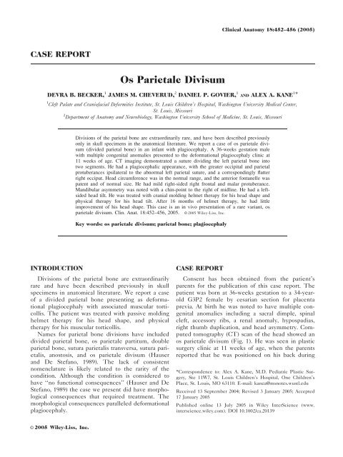
Os parietale divisum - Department of Anatomy & Neurobiology
Os parietale divisum - Department of Anatomy & Neurobiology
Os parietale divisum - Department of Anatomy & Neurobiology
Create successful ePaper yourself
Turn your PDF publications into a flip-book with our unique Google optimized e-Paper software.
CASE REPORT<br />
<strong>Os</strong> Parietale Divisum<br />
DEVRA B. BECKER, 1 JAMES M. CHEVERUD, 2 DANIEL P. GOVIER, 1 AND ALEX A. KANE 1 *<br />
1 Cleft Palate and Crani<strong>of</strong>acial Deformities Institute, St. Louis Children’s Hospital, Washington University Medical Center,<br />
St. Louis, Missouri<br />
2 <strong>Department</strong> <strong>of</strong> <strong>Anatomy</strong> and <strong>Neurobiology</strong>, Washington University School <strong>of</strong> Medicine, St. Louis, Missouri<br />
INTRODUCTION<br />
Divisions <strong>of</strong> the parietal bone are extraordinarily rare, and have been described previously<br />
only in skull specimens in the anatomical literature. We report a case <strong>of</strong> os <strong>parietale</strong> <strong>divisum</strong><br />
(divided parietal bone) in an infant with plagiocephaly. A 36-weeks gestation male<br />
with multiple congenital anomalies presented to the deformational plagiocephaly clinic at<br />
11 weeks <strong>of</strong> age. CT imaging demonstrated a suture dividing the left parietal bone into<br />
two segments. He had a plagiocephalic appearance, with the greater occipital and parietal<br />
protuberances ipsilateral to the abnormal left parietal suture, and a correspondingly flatter<br />
right occiput. Head circumference was in the normal range, and the anterior fontanelle was<br />
patent and <strong>of</strong> normal size. He had mild right-sided right frontal and malar protuberance.<br />
Mandibular asymmetry was noted with a chin-point to the right <strong>of</strong> midline. He had a leftsided<br />
head tilt. He was treated with cranial molding helmet therapy for his head shape and<br />
physical therapy for his head tilt. After 16 months <strong>of</strong> helmet therapy, he had little<br />
improvement <strong>of</strong> his head shape. This case is an in vivo presentation <strong>of</strong> a rare variant, os<br />
<strong>parietale</strong> <strong>divisum</strong>. Clin. Anat. 18:452–456, 2005. VC 2005 Wiley-Liss, Inc.<br />
Key words: os <strong>parietale</strong> <strong>divisum</strong>; parietal bone; plagiocephaly<br />
Divisions <strong>of</strong> the parietal bone are extraordinarily<br />
rare and have been described previously in skull<br />
specimens in anatomical literature. We report a case<br />
<strong>of</strong> a divided parietal bone presenting as deformational<br />
plagiocephaly with associated muscular torticollis.<br />
The patient was treated with passive molding<br />
helmet therapy for his head shape, and physical<br />
therapy for his muscular torticollis.<br />
Names for parietal bone divisions have included<br />
divided parietal bone, os <strong>parietale</strong> partitum, double<br />
parietal bone, sutura parietalis transversa, sutura parietalis,<br />
anostosis, and os <strong>parietale</strong> <strong>divisum</strong> (Hauser<br />
and De Stefano, 1989). The lack <strong>of</strong> consistent<br />
nomenclature is likely related to the rarity <strong>of</strong> the<br />
condition. Although the condition is considered to<br />
have ‘‘no functional consequences’’ (Hauser and De<br />
Stefano, 1989) the case we present did have morphological<br />
consequences that required treatment. The<br />
morphological consequences paralleled deformational<br />
plagiocephaly.<br />
VC 2005 Wiley-Liss, Inc.<br />
CASE REPORT<br />
Clinical <strong>Anatomy</strong> 18:452–456 (2005)<br />
Consent has been obtained from the patient’s<br />
parents for the publication <strong>of</strong> this case report. The<br />
patient was born at 36-weeks gestation to a 34-yearold<br />
G3P2 female by cesarian section for placenta<br />
previa. At birth he was noted to have multiple congenital<br />
anomalies including a sacral dimple, spinal<br />
cleft, accessory ribs, a renal anomaly, hypospadias,<br />
right thumb duplication, and head asymmetry. Computed<br />
tomography (CT) scan <strong>of</strong> the head showed an<br />
os <strong>parietale</strong> <strong>divisum</strong> (Fig. 1). He was seen in plastic<br />
surgery clinic at 11 weeks <strong>of</strong> age, when the parents<br />
reported that he was positioned on his back during<br />
*Correspondence to: Alex A. Kane, M.D. Pediatric Plastic Surgery,<br />
Ste 11W7, St. Louis Children’s Hospital, One Children’s<br />
Place, St. Louis, MO 63110. E-mail: kanea@msnotes.wustl.edu<br />
Received 13 September 2004; Revised 3 January 2005; Accepted<br />
17 January 2005<br />
Published online 13 July 2005 in Wiley InterScience (www.<br />
interscience.wiley.com). DOI 10.1002/ca.20139
Fig. 1. Three-dimensional reconstructions <strong>of</strong> head CT carried out shortly after birth. The accessory<br />
parietal suture is marked by the black arrow.<br />
sleep, and that they noted that his head shape was<br />
worse than at birth. On exam, he was noted to have<br />
a head circumference <strong>of</strong> 39.5 cm (22nd percentile<br />
corrected). He had right-sided occipital flattening<br />
and a right frontal and malar protuberance (Fig. 2).<br />
Mandibular asymmetry was noted with a chin-point<br />
to the right <strong>of</strong> midline. Anterior fontanelle was patent<br />
and non-bulging. Neck range <strong>of</strong> motion was full.<br />
CT imaging <strong>of</strong> the head demonstrated a suture completely<br />
dividing the left parietal bone into two segments<br />
in the antero-posterior dimension, and no<br />
other abnormalities. Parietal formina were not noted<br />
to be enlarged (Fig. 3).<br />
He was seen again in follow-up at 5 months <strong>of</strong> age, at<br />
which time his head circumference was noted to be 41.5<br />
cm (12th percentile) and he was noted to have continued<br />
right occipital flattening with right malar protuberance.<br />
He was begun on a course <strong>of</strong> molding-helmet<br />
therapy per the deformational plagiocephaly protocol.<br />
After 1 month <strong>of</strong> helmet therapy, the patient had<br />
some improvement <strong>of</strong> his right occipital flattening<br />
on physical exam. He was, however, noted to have<br />
developed left-sided head-tilt with constraint noted<br />
in his left sternocleidomastoid muscle. He was<br />
started on physical therapy.<br />
CT imaging <strong>of</strong> the head was repeated at 10<br />
months <strong>of</strong> age, and demonstrated persistence <strong>of</strong> the<br />
accessory parietal suture (Fig. 4). At 1 year <strong>of</strong> age,<br />
the patient continued helmet therapy at approximately<br />
23 hrs/day. His occipital contour was noted<br />
to be improved from baseline, although his frontal<br />
contour was unchanged.<br />
Fig. 2. Appearance <strong>of</strong> the patient in frontal (left) and birds eye<br />
(right) views.<br />
<strong>Os</strong> Parietale Divisum<br />
453
454 Becker et al.<br />
Fig. 3. CT scan 3D reconstructions <strong>of</strong> the subject at 3.5 months <strong>of</strong> age. The accessory parietal suture is marked by the black arrow.<br />
At 16–17 months <strong>of</strong> age, he continued to wear<br />
the helmet for 15 hrs/day, and his family reported<br />
positive changes in the shape <strong>of</strong> his head. Despite<br />
this report, on clinical exam his head circumfer-<br />
ence was 45.5 cm (4th percentile), and his<br />
head shape was noted to have a persistent mild<br />
to moderately severe right occipital flattening<br />
(Fig. 5).<br />
Fig. 4. CT scan 3D reconstructions at 10 months <strong>of</strong> age. There has been some interval closure <strong>of</strong> the accessory parietal suture.
DISCUSSION<br />
Hrdlic˘ka’s encyclopedic 155-page manuscript on<br />
divisions <strong>of</strong> the parietal bone, published in 1903,<br />
belies the extraordinary rarity <strong>of</strong> the variant. Though<br />
he reported to cite every case in the literature, the<br />
number <strong>of</strong> complete antero-posterior divisions <strong>of</strong> the<br />
parietal bone in the very young totaled three. It is<br />
not surprising, then, that 7 years later Berry noted,<br />
in his description <strong>of</strong> an Australian aboriginal skull,<br />
that ‘‘...[os <strong>parietale</strong> partitum] is a condition <strong>of</strong> sufficient<br />
rarity to justify the description <strong>of</strong> any specimen<br />
in which it occurs.’’ Berry examined 3,400 human<br />
crania and found only two with os <strong>parietale</strong> <strong>divisum</strong><br />
(Berry, 1910). Berry’s exhortation did not portend<br />
further descriptions; for years the literature remained<br />
largely silent on this condition. There has been,<br />
however, some relatively recent work. Shapiro (1972)<br />
reviews the literature and reports his own findings.<br />
He confirms the condition is rare, and notes that in<br />
his review <strong>of</strong> 25,000 radiographs he found only three<br />
cases <strong>of</strong> complete parietal divisions. In addition,<br />
Fenton et al. (2000) report two cases <strong>of</strong> divided parietal<br />
bones found at autopsy. In these cases, radiographs<br />
<strong>of</strong> two infants were obtained in the context<br />
<strong>of</strong> workup for bleeding disorders, and the divided<br />
parietal bones were initially interpreted as linear<br />
parietal fractures.<br />
Nomenclature associated with the condition has<br />
not been uniform. There are no fewer than six terms<br />
for the condition: os <strong>parietale</strong> <strong>divisum</strong>, os <strong>parietale</strong>,<br />
partitum, double parietal bone, sutura parietalis<br />
transversa, and sutura parietalis transversa (Hauser<br />
and De Stefano, 1989). The lack <strong>of</strong> consistency in<br />
<strong>Os</strong> Parietale Divisum<br />
Fig. 5. Appearance <strong>of</strong> the patient at 16 months in frontal (left) and birds eye (right) views. Note his torticollis.<br />
455<br />
nomenclature is likely related to the rarity <strong>of</strong> the<br />
condition.<br />
Furthermore, anatomic variation exists. The divisions<br />
can be either complete or incomplete, and<br />
can further be antero-posterior (parallel with the sagittal<br />
suture), supero-inferior, or oblique. All <strong>of</strong> these<br />
subtypes were described by Hrdlic˘ka in his article<br />
(1903). Hauser and De Stefano (1989) also include<br />
the number <strong>of</strong> subdivided parts in their classification.<br />
Some authors have reported a preference for the left<br />
side. The condition is known to occur in other mammals,<br />
specifically apes and monkeys, as well as<br />
humans.<br />
There has been debate over the presumed<br />
embryologic processes that cause the condition, and<br />
the most popular is the two-center theory <strong>of</strong> ossification,<br />
which holds that there are two centers <strong>of</strong> ossification.<br />
The two-center theory <strong>of</strong> ossification is attributed<br />
to Toldt in 1883 by Shapiro (1972). During<br />
development, intramembranous bone forms in mesenchymal<br />
tissue. First, mesenchymal cells differentiate<br />
into osteoblasts. Those osteoblasts form ossification<br />
centers, which generate radial ossification to the<br />
bone edges. Failure <strong>of</strong> the ossification centers to<br />
meet may lead to an accessory suture. The two-center<br />
theory <strong>of</strong> ossification was used as the basis for an<br />
explanation <strong>of</strong> os <strong>parietale</strong> <strong>divisum</strong> in an Australian<br />
skull by Turner in 1890 (Turner, 1890).<br />
The phenotype <strong>of</strong> our case is consistent with<br />
other reported cases <strong>of</strong> os <strong>parietale</strong> partitum (Bessell-<br />
Browne and Thonell, 2004), and with what is<br />
expected based on our knowledge <strong>of</strong> craniosynostosis<br />
and deformational plagiocephaly. ‘‘Virchow’s Law’’<br />
predicts that growth will occur parallel to a synostosed
456 Becker et al.<br />
suture and will be inhibited perpendicular to it. This<br />
explains the asymmetries associated with unicoronal<br />
synostosis, as well as some <strong>of</strong> the key differences<br />
between synostotic plagiocephaly (<strong>of</strong> the unicoronal<br />
and unilambdoid types) and non-synostotic deformational<br />
plagiocephaly. Chin deviation will be contralateral<br />
in synostosis and ipsilateral in deformational<br />
plagiocephaly, and the ipsilateral cheek is forward in<br />
synostosis and backward in deformational plagiocephaly<br />
(resulting in a relative prominence <strong>of</strong> the<br />
contralateral cheek). In this case <strong>of</strong> os <strong>parietale</strong> <strong>divisum</strong>,<br />
the patient had ipsilateral chin deviation, and contralateral<br />
cheek prominence, consistent with deformational<br />
plagiocephaly. It is interesting to note, and<br />
not simple to explain why, the patient’s occipital<br />
flattening was contralateral to the divided parietal<br />
bone.<br />
Although we noted an association <strong>of</strong> os <strong>parietale</strong><br />
<strong>divisum</strong> with deformational plagiocephaly the exact<br />
causal relationship between the two is not known.<br />
Plagiocephaly has not been reported in skull specimens<br />
with os <strong>parietale</strong> <strong>divisum</strong>. It is possible that os<br />
<strong>parietale</strong> <strong>divisum</strong> predisposes a supinely positioned<br />
child to deformational plagiocephaly. An accessory<br />
parietal suture certainly provides an outlet for abnormal<br />
force vectors, and the ultimate phenotype is predictably<br />
consistent with deformational plagiocephaly.<br />
Given that our patient was born with multiple congenital<br />
anomalies, he might have been predisposed<br />
to the development <strong>of</strong> deformational plagiocephaly,<br />
and therefore the presence <strong>of</strong> an accessory parietal<br />
suture was not causally related.<br />
CONCLUSION<br />
<strong>Os</strong> <strong>parietale</strong> <strong>divisum</strong> is a rare variant in which the<br />
parietal bone is divided. Our patient presented with<br />
deformational plagiocephaly.<br />
REFERENCES<br />
Berry RJA. 1910. A case <strong>of</strong> os <strong>parietale</strong> bipartitum in an Australian<br />
aboriginal skull. J Anat Physiol 44:73–82.<br />
Bessell-Browne RJ, Thonell S. 2004. Bipartite parietal bone:<br />
a rare cause <strong>of</strong> plagiocephaly. Australas Radiol 48:248–250.<br />
Fenton LZ, Sirotnak AP, Handler MH. 2000. Parietal pseud<strong>of</strong>racture<br />
and spontaneous intracranial hemorrhage suggesting<br />
nonaccidental trauma: report <strong>of</strong> 2 cases. Pediatr<br />
Neurosurg 33:318–322.<br />
Hauser G, De Stefano GF. 1989. Epigenetic variants <strong>of</strong> the<br />
human skull. Stuttgart: Schweizerbart.<br />
Hrdlic˘ka A. 1903. Divisions <strong>of</strong> the parietal bone in man and<br />
other mammals. Bull Am Mus Nat Hist 29:231–386.<br />
Shapiro R. 1972. Anomalous parietal sutures and the bipartite<br />
parietal bone. Am J Roentgenol Radium Ther Nucl Med<br />
115:569–577.<br />
Turner W. 1890. Double right parietal bone in an Australian<br />
skull. J Anat Physiol 25:473–474.


