Roles of SNARE Proteins in Synaptic Vesicle Fusion - Department of ...
Roles of SNARE Proteins in Synaptic Vesicle Fusion - Department of ...
Roles of SNARE Proteins in Synaptic Vesicle Fusion - Department of ...

You also want an ePaper? Increase the reach of your titles
YUMPU automatically turns print PDFs into web optimized ePapers that Google loves.
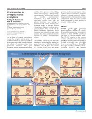
36 M.T. Palfreyman, E.M. Jorgensen<br />
<strong>SNARE</strong> Discovery: A Convergence <strong>of</strong> Genetics<br />
and Biochemistry<br />
To understand the mechanisms <strong>of</strong> synaptic vesicle fusion, it is useful to th<strong>in</strong>k about<br />
the evolution <strong>of</strong> neurotransmission. Eukaryotic cells separate cellular functions <strong>in</strong>to<br />
membrane-bound organelles. The content <strong>of</strong> these organelles are moved between<br />
compartments and the extracellular environment by transport vesicles. Cellular compartments<br />
must be kept dist<strong>in</strong>ct, but membrane-impermeable cargo must be transferred<br />
to the target organelle. To transfer cargo the lipid bilayers <strong>of</strong> the vesicle and<br />
the target must merge so that their lum<strong>in</strong>al contents can <strong>in</strong>term<strong>in</strong>gle. In some cases,<br />
cargo must be secreted <strong>in</strong>to the extracellular space via exocytosis. It was perhaps a<br />
small step for the cell to develop a mechanism for calcium-dependent regulation <strong>of</strong><br />
exocytosis, but it was a giant leap for evolution. The nervous system is arguably the<br />
universe’s greatest <strong>in</strong>vention.<br />
A convergence <strong>of</strong> <strong>in</strong>dependent tracks led to the identification <strong>of</strong> <strong>SNARE</strong>s as the<br />
central players <strong>in</strong> membrane fusion. In the late 1980s <strong>SNARE</strong> prote<strong>in</strong>s were<br />
identified <strong>in</strong> the bra<strong>in</strong> as components <strong>of</strong> the synapse. Specifically, synaptobrev<strong>in</strong><br />
(also called vesicle-associated membrane prote<strong>in</strong> [VAMP]) was purified from synaptic<br />
vesicles (1). Subsequently, two additional <strong>SNARE</strong>s, syntax<strong>in</strong> and SNAP-25<br />
(synaptosome-associated prote<strong>in</strong> <strong>of</strong> 25 kDa), were found localized to the plasma<br />
membrane <strong>of</strong> neurons (2–4). The identification <strong>of</strong> homologues among the yeast sec<br />
genes l<strong>in</strong>ked the mechanisms <strong>of</strong> synaptic function to vesicular traffick<strong>in</strong>g (5,6) and<br />
h<strong>in</strong>ted at the universality <strong>of</strong> membrane fusion. Although the <strong>SNARE</strong> prote<strong>in</strong>s were<br />
well placed to mediate synaptic vesicle fusion and were related to prote<strong>in</strong>s required<br />
for traffick<strong>in</strong>g, there was at this po<strong>in</strong>t no evidence that these prote<strong>in</strong>s functioned <strong>in</strong><br />
calcium-dependent exocytosis <strong>of</strong> synaptic vesicles.<br />
The groups <strong>of</strong> He<strong>in</strong>er Niemann, Re<strong>in</strong>hard Jahn, and Cesare Montecucco were<br />
look<strong>in</strong>g for the targets <strong>of</strong> the clostridial tox<strong>in</strong>s. The clostridial tox<strong>in</strong>s from the<br />
anaerobic bacteria Clostridium botul<strong>in</strong>um and Clostridium tetani can potently<br />
<strong>in</strong>hibit neurotransmission (7). Thus, it was reasoned that their targets would identify<br />
essential prote<strong>in</strong>s <strong>in</strong> synaptic transmission. Botul<strong>in</strong>um and tetanus tox<strong>in</strong>s cleave<br />
the <strong>SNARE</strong> prote<strong>in</strong>s, demonstrat<strong>in</strong>g the central role <strong>of</strong> the <strong>SNARE</strong>s <strong>in</strong> synaptic<br />
vesicle release (8–11). These were the first functional data that the <strong>SNARE</strong>s were<br />
<strong>in</strong>volved <strong>in</strong> neurotransmission (12,13). The central role <strong>of</strong> the <strong>SNARE</strong>s <strong>in</strong> neurotransmission<br />
would later be confirmed from electrophysiologic studies on null<br />
mutants <strong>in</strong> the <strong>SNARE</strong> prote<strong>in</strong>s <strong>in</strong> Drosophila, mice, and Caenorhabditis elegans<br />
(14–19). Thus, the functional data identified the <strong>SNARE</strong>s as perpetrators but their<br />
association had not been described.<br />
The discovery that these prote<strong>in</strong>s formed a complex was demonstrated soon<br />
after. Jim Rothman’s group was tak<strong>in</strong>g a biochemical approach to understand traffick<strong>in</strong>g<br />
<strong>in</strong> the Golgi apparatus. The tox<strong>in</strong> N-ethylmaleimide (NEM) potently <strong>in</strong>hibits<br />
Golgi traffick<strong>in</strong>g (20). Wilson et al (21) found that the target <strong>of</strong> NEM was the<br />
mammalian homologue <strong>of</strong> a previously cloned yeast gene SEC18 (22). Rothman’s<br />
group named this new prote<strong>in</strong> the NEM-sensitive factor (NSF) (23), and NSF was<br />
Wang_Ch03.<strong>in</strong>dd 36 5/15/2008 5:27:11 PM