Roles of SNARE Proteins in Synaptic Vesicle Fusion - Department of ...
Roles of SNARE Proteins in Synaptic Vesicle Fusion - Department of ...
Roles of SNARE Proteins in Synaptic Vesicle Fusion - Department of ...
Create successful ePaper yourself
Turn your PDF publications into a flip-book with our unique Google optimized e-Paper software.
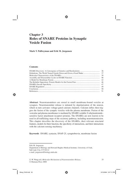
Chapter 3<br />
<strong>Roles</strong> <strong>of</strong> <strong>SNARE</strong> <strong>Prote<strong>in</strong>s</strong> <strong>in</strong> <strong>Synaptic</strong><br />
<strong>Vesicle</strong> <strong>Fusion</strong><br />
Mark T. Palfreyman and Erik M. Jorgensen<br />
Contents<br />
<strong>SNARE</strong> Discovery: A Convergence <strong>of</strong> Genetics and Biochemistry .......................................... 36<br />
Defi nitions: The World Turned Upside Down and Given a Good Shake .................................. 37<br />
Molecular Characteristics <strong>of</strong> the <strong>SNARE</strong>s ................................................................................. 38<br />
Assembly and Disassembly Cycles <strong>in</strong> <strong>SNARE</strong> Function .......................................................... 40<br />
A Model for Membrane <strong>Fusion</strong> ................................................................................................. 42<br />
The Reliable Opposition: Prote<strong>in</strong> Models for the <strong>Fusion</strong> Pore .................................................. 46<br />
<strong>SNARE</strong>s Encode Specifi city ...................................................................................................... 47<br />
<strong>SNARE</strong> Regulation .................................................................................................................... 49<br />
Conclusion ................................................................................................................................. 50<br />
References .................................................................................................................................. 51<br />
Abstract Neurotransmitters are stored <strong>in</strong> small membrane-bound vesicles at<br />
synapses. Neurotransmitter release is <strong>in</strong>itiated by depolarization <strong>of</strong> the neuron,<br />
which <strong>in</strong> turn activates voltage-gated calcium channels. Calcium <strong>in</strong>flux then triggers<br />
the fusion <strong>of</strong> the synaptic vesicles with the plasma membrane. <strong>Fusion</strong> <strong>of</strong> the<br />
vesicular and plasma membranes is mediated by <strong>SNARE</strong> (soluble N-ethylmaleimide–<br />
sensitive factor attachment receptor) prote<strong>in</strong>s. The <strong>SNARE</strong>s are now known to be<br />
used <strong>in</strong> all traffick<strong>in</strong>g steps <strong>of</strong> the secretory pathway, <strong>in</strong>clud<strong>in</strong>g neurotransmission.<br />
This chapter describes the discovery <strong>of</strong> the <strong>SNARE</strong>s, their relevant structural<br />
features, models for their function, the specificity <strong>of</strong> <strong>in</strong>teractions, and their <strong>in</strong>teractions<br />
with the calcium-sens<strong>in</strong>g mach<strong>in</strong>ery.<br />
Keywords <strong>SNARE</strong>, syntax<strong>in</strong>, SNAP-25, synaptobrev<strong>in</strong>, membrane fusion<br />
Erik M. Jorgensen<br />
<strong>Department</strong> <strong>of</strong> Biology and Howard Hughes Medical Institute, University <strong>of</strong> Utah,<br />
Salt Lake City, UT 84112<br />
e-mail: jorgensen@biology.utah.edu<br />
Z.-W. Wang (ed.) Molecular Mechanisms <strong>of</strong> Neurotransmitter Release, 35<br />
© Humana Press 2008<br />
Wang_Ch03.<strong>in</strong>dd 35 5/15/2008 5:27:11 PM
36 M.T. Palfreyman, E.M. Jorgensen<br />
<strong>SNARE</strong> Discovery: A Convergence <strong>of</strong> Genetics<br />
and Biochemistry<br />
To understand the mechanisms <strong>of</strong> synaptic vesicle fusion, it is useful to th<strong>in</strong>k about<br />
the evolution <strong>of</strong> neurotransmission. Eukaryotic cells separate cellular functions <strong>in</strong>to<br />
membrane-bound organelles. The content <strong>of</strong> these organelles are moved between<br />
compartments and the extracellular environment by transport vesicles. Cellular compartments<br />
must be kept dist<strong>in</strong>ct, but membrane-impermeable cargo must be transferred<br />
to the target organelle. To transfer cargo the lipid bilayers <strong>of</strong> the vesicle and<br />
the target must merge so that their lum<strong>in</strong>al contents can <strong>in</strong>term<strong>in</strong>gle. In some cases,<br />
cargo must be secreted <strong>in</strong>to the extracellular space via exocytosis. It was perhaps a<br />
small step for the cell to develop a mechanism for calcium-dependent regulation <strong>of</strong><br />
exocytosis, but it was a giant leap for evolution. The nervous system is arguably the<br />
universe’s greatest <strong>in</strong>vention.<br />
A convergence <strong>of</strong> <strong>in</strong>dependent tracks led to the identification <strong>of</strong> <strong>SNARE</strong>s as the<br />
central players <strong>in</strong> membrane fusion. In the late 1980s <strong>SNARE</strong> prote<strong>in</strong>s were<br />
identified <strong>in</strong> the bra<strong>in</strong> as components <strong>of</strong> the synapse. Specifically, synaptobrev<strong>in</strong><br />
(also called vesicle-associated membrane prote<strong>in</strong> [VAMP]) was purified from synaptic<br />
vesicles (1). Subsequently, two additional <strong>SNARE</strong>s, syntax<strong>in</strong> and SNAP-25<br />
(synaptosome-associated prote<strong>in</strong> <strong>of</strong> 25 kDa), were found localized to the plasma<br />
membrane <strong>of</strong> neurons (2–4). The identification <strong>of</strong> homologues among the yeast sec<br />
genes l<strong>in</strong>ked the mechanisms <strong>of</strong> synaptic function to vesicular traffick<strong>in</strong>g (5,6) and<br />
h<strong>in</strong>ted at the universality <strong>of</strong> membrane fusion. Although the <strong>SNARE</strong> prote<strong>in</strong>s were<br />
well placed to mediate synaptic vesicle fusion and were related to prote<strong>in</strong>s required<br />
for traffick<strong>in</strong>g, there was at this po<strong>in</strong>t no evidence that these prote<strong>in</strong>s functioned <strong>in</strong><br />
calcium-dependent exocytosis <strong>of</strong> synaptic vesicles.<br />
The groups <strong>of</strong> He<strong>in</strong>er Niemann, Re<strong>in</strong>hard Jahn, and Cesare Montecucco were<br />
look<strong>in</strong>g for the targets <strong>of</strong> the clostridial tox<strong>in</strong>s. The clostridial tox<strong>in</strong>s from the<br />
anaerobic bacteria Clostridium botul<strong>in</strong>um and Clostridium tetani can potently<br />
<strong>in</strong>hibit neurotransmission (7). Thus, it was reasoned that their targets would identify<br />
essential prote<strong>in</strong>s <strong>in</strong> synaptic transmission. Botul<strong>in</strong>um and tetanus tox<strong>in</strong>s cleave<br />
the <strong>SNARE</strong> prote<strong>in</strong>s, demonstrat<strong>in</strong>g the central role <strong>of</strong> the <strong>SNARE</strong>s <strong>in</strong> synaptic<br />
vesicle release (8–11). These were the first functional data that the <strong>SNARE</strong>s were<br />
<strong>in</strong>volved <strong>in</strong> neurotransmission (12,13). The central role <strong>of</strong> the <strong>SNARE</strong>s <strong>in</strong> neurotransmission<br />
would later be confirmed from electrophysiologic studies on null<br />
mutants <strong>in</strong> the <strong>SNARE</strong> prote<strong>in</strong>s <strong>in</strong> Drosophila, mice, and Caenorhabditis elegans<br />
(14–19). Thus, the functional data identified the <strong>SNARE</strong>s as perpetrators but their<br />
association had not been described.<br />
The discovery that these prote<strong>in</strong>s formed a complex was demonstrated soon<br />
after. Jim Rothman’s group was tak<strong>in</strong>g a biochemical approach to understand traffick<strong>in</strong>g<br />
<strong>in</strong> the Golgi apparatus. The tox<strong>in</strong> N-ethylmaleimide (NEM) potently <strong>in</strong>hibits<br />
Golgi traffick<strong>in</strong>g (20). Wilson et al (21) found that the target <strong>of</strong> NEM was the<br />
mammalian homologue <strong>of</strong> a previously cloned yeast gene SEC18 (22). Rothman’s<br />
group named this new prote<strong>in</strong> the NEM-sensitive factor (NSF) (23), and NSF was<br />
Wang_Ch03.<strong>in</strong>dd 36 5/15/2008 5:27:11 PM
3 <strong>Roles</strong> <strong>of</strong> <strong>SNARE</strong> <strong>Prote<strong>in</strong>s</strong> <strong>in</strong> <strong>Synaptic</strong> <strong>Vesicle</strong> <strong>Fusion</strong> 37<br />
found to b<strong>in</strong>d, via the action <strong>of</strong> the soluble NSF adaptors (SNAPs) (24), to a set <strong>of</strong><br />
prote<strong>in</strong>s from bra<strong>in</strong> detergent extracts that came to be collectively known as the<br />
soluble N-ethylmaleimide–sensitive factor attachment receptor prote<strong>in</strong>s (<strong>SNARE</strong>s).<br />
The evidence for <strong>SNARE</strong> <strong>in</strong>volvement <strong>in</strong> synaptic vesicle exocytosis was now<br />
overwhelm<strong>in</strong>g, but a list <strong>of</strong> names <strong>in</strong> a complex did not constitute a model.<br />
The first coherent model, called the <strong>SNARE</strong> hypothesis, would arise from the<br />
meld<strong>in</strong>g <strong>of</strong> the genetic and biochemical observations described above. Although<br />
wrong <strong>in</strong> detail, it would catalyze a number <strong>of</strong> hypothesis-driven experiments that<br />
would lead to more accurate models. Based on the f<strong>in</strong>d<strong>in</strong>g that unique <strong>SNARE</strong>s are<br />
found at each <strong>of</strong> the traffick<strong>in</strong>g steps (25,26), Thomas Söllner and Jim Rothman<br />
proposed that <strong>SNARE</strong> <strong>in</strong>teractions provided the specificity for vesicular traffick<strong>in</strong>g<br />
by tether<strong>in</strong>g the vesicle to its target membrane (27,28). The <strong>SNARE</strong>s would then<br />
be acted on by the adenos<strong>in</strong>e triphosphatase (ATPase) NSF which, by disassembl<strong>in</strong>g<br />
the <strong>SNARE</strong>s, would drive fusion (27,29).<br />
Further experiments from Bill Wickner’s lab, us<strong>in</strong>g a purified vacuole fusion<br />
assay, demonstrated that NSF acted not at the f<strong>in</strong>al step <strong>of</strong> fusion, but rather to<br />
recover monomeric <strong>SNARE</strong>s for use <strong>in</strong> further rounds <strong>of</strong> fusion (28,29,30,31). NSF<br />
was act<strong>in</strong>g as a chaperone to separate the embrac<strong>in</strong>g <strong>SNARE</strong>s on the plasma membrane<br />
to reactivate the system for further fusion (32,33). Thus assembly <strong>of</strong> the<br />
<strong>SNARE</strong>s, not disassembly, catalyzes fusion.<br />
F<strong>in</strong>ally, Rothman’s group demonstrated that the <strong>SNARE</strong>s alone could fuse membranes.<br />
The <strong>SNARE</strong>s were <strong>in</strong>corporated <strong>in</strong>to vesicles composed <strong>of</strong> artificial lipid<br />
bilayers. Donor vesicles conta<strong>in</strong><strong>in</strong>g synaptobrev<strong>in</strong> were capable <strong>of</strong> fusion with<br />
acceptor vesicles conta<strong>in</strong><strong>in</strong>g syntax<strong>in</strong> and SNAP-25 (34). This experiment was<br />
extended to native membranes by eng<strong>in</strong>eer<strong>in</strong>g <strong>SNARE</strong>s to face out <strong>of</strong> the cell; <strong>in</strong><br />
this configuration the <strong>SNARE</strong>s could <strong>in</strong>duce fusion <strong>of</strong> whole cells (35). Thus, the<br />
current th<strong>in</strong>k<strong>in</strong>g is that the <strong>SNARE</strong>s function <strong>in</strong> the f<strong>in</strong>al steps <strong>in</strong> fusion and represent<br />
the m<strong>in</strong>imal fusion mach<strong>in</strong>ery.<br />
In the follow<strong>in</strong>g sections we briefly def<strong>in</strong>e the steps lead<strong>in</strong>g to fusion, <strong>in</strong>troduce<br />
the structure <strong>of</strong> the <strong>SNARE</strong> prote<strong>in</strong>s, present a model for fusion, discuss <strong>SNARE</strong><br />
specificity, and f<strong>in</strong>ally touch on the regulation <strong>of</strong> the <strong>SNARE</strong> complex by other<br />
prote<strong>in</strong>s.<br />
Def<strong>in</strong>itions: The World Turned Upside<br />
Down and Given a Good Shake<br />
In the past, synaptic vesicles were thought to dock with the plasma membrane, and<br />
then undergo a maturation step <strong>in</strong> which they became release ready. Depolarization<br />
activated a calcium sensor that then allowed the vesicle to fuse with the plasma<br />
membrane. Only a subset <strong>of</strong> docked vesicles were considered to be <strong>in</strong> the readily<br />
releasable pool (36). Thus, the life <strong>of</strong> a vesicle could be divided <strong>in</strong>to four steps:<br />
dock<strong>in</strong>g, matur<strong>in</strong>g to release-ready, calcium sens<strong>in</strong>g, and fus<strong>in</strong>g. The def<strong>in</strong>ition <strong>of</strong><br />
these stages <strong>in</strong> vesicle fusion relied on morphologic and electrophysiologic criteria.<br />
Wang_Ch03.<strong>in</strong>dd 37 5/15/2008 5:27:11 PM
38 M.T. Palfreyman, E.M. Jorgensen<br />
Current studies have sought to associate these pools with particular molecular <strong>in</strong>teractions<br />
and thereby more precisely def<strong>in</strong>e these states.<br />
Paradoxically, recent studies have tended to confuse rather than clarify the states<br />
<strong>of</strong> a vesicle. Although some have argued that very few docked vesicles are <strong>in</strong> the<br />
readily releasable pool (36,37), others suggest that docked vesicles are equivalent<br />
to the readily releasable pool (38–41). Studies <strong>of</strong> <strong>SNARE</strong> prote<strong>in</strong>s have also muddied<br />
our previously clean def<strong>in</strong>itions <strong>of</strong> these pools. The assembly <strong>of</strong> <strong>SNARE</strong> prote<strong>in</strong>s<br />
between synaptic vesicle and plasma membrane is def<strong>in</strong>ed as vesicle<br />
“prim<strong>in</strong>g.” Initial studies suggested that prim<strong>in</strong>g occurred after dock<strong>in</strong>g (15).<br />
However, recent studies suggest that the primed state may correspond to “docked”<br />
vesicles as observed <strong>in</strong> electron micrographs (14). Thus, the morphologic, electrophysiologic,<br />
and molecular def<strong>in</strong>itions have seem<strong>in</strong>gly converged on a s<strong>in</strong>gle state.<br />
It is hoped that as the actions <strong>of</strong> various prote<strong>in</strong>s are more precisely understood, we<br />
will once aga<strong>in</strong> ref<strong>in</strong>e synaptic vesicle fusion <strong>in</strong>to discrete steps.<br />
There is one last sorry note concern<strong>in</strong>g our attempts to def<strong>in</strong>e steps <strong>in</strong> vesicle<br />
fusion: the term<strong>in</strong>ology used for synaptic vesicle fusion is at odds with the term<strong>in</strong>ology<br />
used <strong>in</strong> yeast. In yeast, prim<strong>in</strong>g” refers to the generation <strong>of</strong> free <strong>SNARE</strong>s rather<br />
than the formation <strong>of</strong> the <strong>SNARE</strong> complex, tether<strong>in</strong>g rather than dock<strong>in</strong>g describes<br />
the <strong>in</strong>itial membrane association, and dock<strong>in</strong>g <strong>in</strong>cludes <strong>SNARE</strong> engagement. Only<br />
the word fusion seems to mean the same th<strong>in</strong>g <strong>in</strong> these different languages.<br />
Molecular Characteristics <strong>of</strong> the <strong>SNARE</strong>s<br />
The <strong>SNARE</strong> prote<strong>in</strong>s are characterized by a conserved 60- to 70-am<strong>in</strong>o-acid <strong>SNARE</strong><br />
motif. Phylogenetic analysis <strong>in</strong>dicates that <strong>SNARE</strong> prote<strong>in</strong>s can be divided <strong>in</strong>to four<br />
families (25,42,43). The <strong>in</strong>dividual <strong>SNARE</strong> motifs are largely unstructured <strong>in</strong> solution,<br />
but when all four family members are mixed, the <strong>SNARE</strong> motifs come together to form<br />
a four-helix parallel bundle known as the core complex (Fig. 3.1A,B) (44). The <strong>SNARE</strong><br />
complex is remarkably stable and can only be separated by boil<strong>in</strong>g <strong>in</strong> the presence <strong>of</strong><br />
sodium dedocyl sulfonate (SDS) (45,46). The hydrophobic residues <strong>of</strong> the alpha-helical<br />
<strong>SNARE</strong> motifs are oriented <strong>in</strong>ward to form layers like those <strong>in</strong> the coiled coil doma<strong>in</strong>s<br />
<strong>of</strong> classical leuc<strong>in</strong>e zippers. However, the layer <strong>in</strong> the middle <strong>of</strong> the complex, called the<br />
“0” layer, is formed by ionic <strong>in</strong>teractions between an arg<strong>in</strong><strong>in</strong>e (R-<strong>SNARE</strong>) and three<br />
glutam<strong>in</strong>es (Qa, Qb, and Qc <strong>SNARE</strong>s) (Fig. 3.1B,C). The role for these conserved residues<br />
buried <strong>in</strong> the hydrophobic core is briefly discussed <strong>in</strong> the next section. At each<br />
fusion site a unique <strong>SNARE</strong> complex consist<strong>in</strong>g <strong>of</strong> all four flavors is formed. While<br />
other complexes have been observed <strong>in</strong> vitro, the only complexes that have been shown<br />
to efficiently support fusion are QabcR complexes (47–52).<br />
The <strong>SNARE</strong>s that are used for synaptic vesicle exocytosis are synaptobrev<strong>in</strong><br />
(R-<strong>SNARE</strong>, also called VAMP2), syntax<strong>in</strong> 1a (Qa <strong>SNARE</strong>), and SNAP-25 (conta<strong>in</strong>s<br />
both the Qb and Qc <strong>SNARE</strong> motifs) (Fig. 3.1) (1–4,53).<br />
In addition to the <strong>SNARE</strong> motifs, all three <strong>SNARE</strong>s conta<strong>in</strong> sequences that<br />
anchor them to the membrane (Fig. 3.1A). Syntax<strong>in</strong> and synaptobrev<strong>in</strong> are anchored<br />
Wang_Ch03.<strong>in</strong>dd 38 5/15/2008 5:27:12 PM
3 <strong>Roles</strong> <strong>of</strong> <strong>SNARE</strong> <strong>Prote<strong>in</strong>s</strong> <strong>in</strong> <strong>Synaptic</strong> <strong>Vesicle</strong> <strong>Fusion</strong> 39<br />
Fig. 3.1 Molecular description <strong>of</strong> the <strong>SNARE</strong>s. By assembl<strong>in</strong>g <strong>in</strong>to a four-helix parallel bundle,<br />
the <strong>SNARE</strong>s bridge the gap between the two membranes dest<strong>in</strong>ed to fuse. (a) In the case <strong>of</strong> the<br />
neuronal <strong>SNARE</strong>s, syntax<strong>in</strong> (red) and SNAP-25 (green) are found on the plasma membrane and<br />
synaptobrev<strong>in</strong> (blue) is associated with the synaptic vesicle. The 60- to 70-am<strong>in</strong>o-acid <strong>SNARE</strong><br />
motifs form a four-helix bundle. Syntax<strong>in</strong> and synaptobrev<strong>in</strong> contribute one <strong>SNARE</strong> motif and<br />
SNAP-25 contributes two. Syntax<strong>in</strong> conta<strong>in</strong>s an additional regulatory doma<strong>in</strong> composed <strong>of</strong> three<br />
alpha-helices called the Habc doma<strong>in</strong>. Syntax<strong>in</strong> and synaptobrev<strong>in</strong> are transmembrane prote<strong>in</strong>s,<br />
while SNAP-25 is attached to the membrane via palmitoylation <strong>of</strong> the l<strong>in</strong>ker region. (b) The wire<br />
frame model shows the backbone <strong>of</strong> the <strong>SNARE</strong> motifs. The N-term<strong>in</strong>i are at the left and the Cterm<strong>in</strong>i<br />
are at the right, match<strong>in</strong>g the illustration <strong>in</strong> (A). The am<strong>in</strong>o acids fac<strong>in</strong>g toward the center<br />
<strong>of</strong> this helix (denoted as layers −7 to +8) are largely hydrophobic <strong>in</strong> nature with the notable exception<br />
<strong>of</strong> the zero layer. (c) In the zero layer charged residues are oriented toward the center <strong>of</strong> the<br />
helix. Syntax<strong>in</strong> contributes one glutam<strong>in</strong>e (Qa), SNAP-25 contributes two glutam<strong>in</strong>es (Qb and<br />
Qc), and synaptobrev<strong>in</strong> contributes one arg<strong>in</strong><strong>in</strong>e (R). (A: Courtesy <strong>of</strong> Enfu Hui and Edw<strong>in</strong> R.<br />
Chapman. B: Adapted from Fasshauer et al [40]. C: Adapted from Bracher et al [193].)<br />
via transmembrane doma<strong>in</strong>s. SNAP-25 is anchored via the palmitoylation <strong>of</strong><br />
cyste<strong>in</strong>es <strong>in</strong> the l<strong>in</strong>ker region connect<strong>in</strong>g the two <strong>SNARE</strong> motifs. In all <strong>SNARE</strong>based<br />
fusion reactions, each <strong>of</strong> the two membranes dest<strong>in</strong>ed to fuse must conta<strong>in</strong> a<br />
<strong>SNARE</strong> with a transmembrane doma<strong>in</strong>; otherwise fusion will not occur (54).<br />
Wang_Ch03.<strong>in</strong>dd 39 5/15/2008 5:27:12 PM
40 M.T. Palfreyman, E.M. Jorgensen<br />
Fig. 3.2 Acceptor complex and zipper model for <strong>SNARE</strong> assembly. The Q <strong>SNARE</strong>s syntax<strong>in</strong> and<br />
SNAP-25 assemble on the plasma membrane. This Qabc acceptor complex then contacts the<br />
distal N-term<strong>in</strong>us <strong>of</strong> synaptobrev<strong>in</strong> on the synaptic vesicle. This conformation is known as a<br />
“loose” <strong>SNARE</strong> complex. The “zipper<strong>in</strong>g” <strong>of</strong> the rest <strong>of</strong> the <strong>SNARE</strong>s <strong>in</strong>to the complex serves two<br />
potential functions. First, full assembly <strong>of</strong> the <strong>SNARE</strong>s leads to close proximity between the<br />
membranes dest<strong>in</strong>ed to fuse. Second, the zipper<strong>in</strong>g might provide torque that is transferred to the<br />
transmembrane doma<strong>in</strong> lead<strong>in</strong>g to full fusion<br />
Synaptobrev<strong>in</strong> is located on synaptic vesicles, while syntax<strong>in</strong> and SNAP-25 are on<br />
the plasma membrane. The assembly <strong>of</strong> synaptobrev<strong>in</strong>, syntax<strong>in</strong>, and SNAP-25 <strong>in</strong>to<br />
the <strong>SNARE</strong> complex would thus bridge the vesicle and plasma membrane, form<strong>in</strong>g<br />
what is known as a trans <strong>SNARE</strong> complex (Fig. 3.1A).<br />
Assembly and Disassembly Cycles <strong>in</strong> <strong>SNARE</strong> Function<br />
The steps <strong>in</strong> the assembly <strong>of</strong> the trans <strong>SNARE</strong> complex are still <strong>in</strong> dispute. Based<br />
on biochemical experiments us<strong>in</strong>g the yeast <strong>SNARE</strong>s, it was proposed that homologues<br />
<strong>of</strong> syntax<strong>in</strong> (Sso1p) and SNAP-25 (Sec9p) might form an “acceptor complex”<br />
(55) (Fig. 3.2). A syntax<strong>in</strong>–SNAP-25 complex was also subsequently proposed<br />
for the neuronal <strong>SNARE</strong>s (56). This acceptor complex greatly speeds up the assembly<br />
<strong>of</strong> the core complex (57). However, it is not known whether this complex exists <strong>in</strong><br />
vivo. It has been shown that SNAP-25 and syntax<strong>in</strong> can stably associate <strong>in</strong> cells (58).<br />
Specifically, a fluorescently tagged SNAP-25 generated an <strong>in</strong>tramolecular fluorescence<br />
resonance energy transfer (FRET) signal upon assembly with syntax<strong>in</strong> <strong>in</strong><br />
PC12 cells.<br />
This acceptor complex compris<strong>in</strong>g one SNAP-25 molecule and one syntax<strong>in</strong> molecule<br />
is highly reactive and will rapidly <strong>in</strong>corporate a second syntax<strong>in</strong> molecule to<br />
form a dead-end Qaabc complex (55–58). This dead-end complex might be prevented<br />
<strong>in</strong> vivo by the action <strong>of</strong> tomosyn, a molecule with an R-<strong>SNARE</strong> doma<strong>in</strong> (59,60). By<br />
occupy<strong>in</strong>g the synaptobrev<strong>in</strong> position <strong>in</strong> the complex, tomosyn might prevent the<br />
accumulation <strong>of</strong> the nonproductive Qaabc complexes and thus promote <strong>SNARE</strong> complex<br />
formation (60,61). However, this model is not consistent with the largely <strong>in</strong>hibitory<br />
role for tomosyn; genetic knockouts yield large <strong>in</strong>creases <strong>in</strong> synaptic vesicle<br />
release (62,63). Tomosyn is thus more likely to b<strong>in</strong>d to the acceptor complexes and,<br />
just like the Qaabc complexes, might form <strong>in</strong>active complexes (62–64).<br />
Wang_Ch03.<strong>in</strong>dd 40 5/15/2008 5:27:12 PM
3 <strong>Roles</strong> <strong>of</strong> <strong>SNARE</strong> <strong>Prote<strong>in</strong>s</strong> <strong>in</strong> <strong>Synaptic</strong> <strong>Vesicle</strong> <strong>Fusion</strong> 41<br />
A second prote<strong>in</strong> family that might serve to stabilize the acceptor complex is<br />
the SM (Sec1/Munc-18) family. At the synapse these prote<strong>in</strong>s are called Unc18<br />
prote<strong>in</strong>s (UNC-18 <strong>in</strong> C. elegans, Munc18 <strong>in</strong> mammals, and ROP <strong>in</strong> Drosophila).<br />
It was orig<strong>in</strong>ally thought that Unc18 exclusively bound to syntax<strong>in</strong> monomers<br />
(65–69). However, more recent experiments have suggested alternative modes <strong>of</strong><br />
b<strong>in</strong>d<strong>in</strong>g (70–73). When reconstituted <strong>in</strong>to lawns <strong>of</strong> plasma membrane, Unc18 was<br />
displaced from syntax<strong>in</strong> by synaptobrev<strong>in</strong> but only when SNAP-25 was also<br />
present (70). Unc18 might therefore stabilize a syntax<strong>in</strong>/SNAP-25 acceptor complex<br />
await<strong>in</strong>g synaptobrev<strong>in</strong> (70). Nonetheless, it is still at present unclear how<br />
acceptor complexes are ma<strong>in</strong>ta<strong>in</strong>ed or even whether they are true <strong>in</strong>termediates <strong>in</strong><br />
core complex assembly. Indeed, synaptobrev<strong>in</strong> and syntax<strong>in</strong> have been shown to<br />
assemble <strong>in</strong> vitro <strong>in</strong> the absence <strong>of</strong> SNAP-25 (74–76), suggest<strong>in</strong>g that SNAP-25<br />
might jo<strong>in</strong> the complex last. It has even been suggested that syntax<strong>in</strong> might be the<br />
last molecule to enter the core complex <strong>in</strong> vivo (77). Until <strong>SNARE</strong> assembly can<br />
be monitored <strong>in</strong> vivo, we are forced to rely on these studies <strong>of</strong> <strong>in</strong> vitro <strong>SNARE</strong><br />
<strong>in</strong>teractions.<br />
Once synaptobrev<strong>in</strong> enters the complex it is proposed to make contact at the<br />
N-term<strong>in</strong>al portion <strong>of</strong> the <strong>SNARE</strong> doma<strong>in</strong> distal from the membrane. This conformation<br />
<strong>of</strong> the <strong>SNARE</strong>s is termed a loose configuration and is then thought to zipper down<br />
to a tight conformation (Fig. 3.2). <strong>Synaptic</strong> vesicles are held <strong>in</strong> a release-ready state<br />
<strong>in</strong> which the trans <strong>SNARE</strong> complex is likely to be arrested <strong>in</strong> a partially zippered<br />
state. Calcium b<strong>in</strong>d<strong>in</strong>g to synaptotagm<strong>in</strong> would release arrest so that the <strong>SNARE</strong><br />
complex could fully zipper to the tight conformation. This transition to the tight<br />
conformation would pull the transmembrane doma<strong>in</strong>s <strong>of</strong> the <strong>SNARE</strong>s, and hence<br />
the membranes, <strong>in</strong>to close proximity and <strong>in</strong>duce fusion (78,79). Models for the<br />
action <strong>of</strong> <strong>SNARE</strong>s <strong>in</strong> membrane fusion are described below.<br />
Once the two membranes have merged, the core complex is now located <strong>in</strong> a<br />
s<strong>in</strong>gle membrane and is referred to as a cis <strong>SNARE</strong> complex. To undergo further<br />
rounds <strong>of</strong> fusion, this cis complex must be disassembled and the <strong>SNARE</strong>s repartitioned<br />
to their appropriate compartments. Disassembly is mediated by the<br />
action <strong>of</strong> NSF and the SNAPs. Together NSF and the SNAPs are able to disassemble<br />
all <strong>SNARE</strong> complexes thus far tested (80). The ATPase NSF itself does<br />
not directly b<strong>in</strong>d <strong>SNARE</strong>s; <strong>in</strong>stead, it b<strong>in</strong>ds <strong>SNARE</strong>s through the action <strong>of</strong> the<br />
SNAPs. The SNAPs b<strong>in</strong>d to the surface <strong>of</strong> the cis <strong>SNARE</strong>s around the central<br />
zero layer, which conta<strong>in</strong>s the conserved Q and R residues (81). The disassembly<br />
<strong>of</strong> the mammalian core complexes <strong>in</strong> PC12 cells is <strong>in</strong>hibited by mutation <strong>in</strong> these<br />
conserved residues (82). However, the disassembly <strong>of</strong> the C. elegans core complex<br />
is not affected by the same mutations (83). An alternative model for the<br />
function <strong>of</strong> these conserved residues is that they have a role before fusion <strong>in</strong> gett<strong>in</strong>g<br />
the four helixes to align <strong>in</strong> register to ensure that their transmembrane<br />
doma<strong>in</strong>s are directly opposed at their C-term<strong>in</strong>al ends (42,78). It has also been<br />
proposed that they might function <strong>in</strong> the prevention <strong>of</strong> full <strong>SNARE</strong> zipper<strong>in</strong>g<br />
(77). The next section explores how the formation <strong>of</strong> these <strong>SNARE</strong> complexes<br />
might catalyze fusion.<br />
Wang_Ch03.<strong>in</strong>dd 41 5/15/2008 5:27:13 PM
42 M.T. Palfreyman, E.M. Jorgensen<br />
A Model for Membrane <strong>Fusion</strong><br />
Membranes do not spontaneously fuse, because <strong>of</strong> the high repulsive forces between<br />
two phospholipid bilayers 1 to 2 nm apart. How might the <strong>SNARE</strong>s fuse membranes?<br />
Three characteristics <strong>of</strong> the <strong>SNARE</strong>s are central to the current models for<br />
their function <strong>in</strong> fus<strong>in</strong>g membranes. First, the assembled <strong>SNARE</strong> complex is<br />
remarkably stable. The formation <strong>of</strong> the <strong>SNARE</strong> complex is therefore an energy<br />
source that can be used to overcome barriers to fusion. Second, the <strong>SNARE</strong> complex<br />
must consist <strong>of</strong> at least two <strong>SNARE</strong> molecules with transmembrane doma<strong>in</strong>s (84).<br />
The transmembrane doma<strong>in</strong>s must be <strong>in</strong>serted <strong>in</strong>to both <strong>of</strong> the membranes dest<strong>in</strong>ed<br />
to fuse (54). Third, the <strong>SNARE</strong>s assemble <strong>in</strong> a parallel orientation (44,78,79,85).<br />
Due to the parallel orientation <strong>of</strong> the <strong>SNARE</strong> motifs, <strong>SNARE</strong> assembly leads to the<br />
close apposition <strong>of</strong> the transmembrane doma<strong>in</strong>s and hence the membranes themselves.<br />
This section describes how the assembly <strong>of</strong> the <strong>SNARE</strong> complexes might<br />
lead the membranes through the sequential <strong>in</strong>termediates <strong>of</strong> a lipid stalk, a hemifusion<br />
diaphragm, an <strong>in</strong>itial fusion pore, and f<strong>in</strong>ally full fusion (Fig. 3.3).<br />
The stability <strong>of</strong> the <strong>SNARE</strong> complexes comb<strong>in</strong>ed with their parallel orientation<br />
led to the idea that their formation might provide the driv<strong>in</strong>g force for fusion. By first<br />
assembl<strong>in</strong>g at their N-term<strong>in</strong>als and subsequently “zipper<strong>in</strong>g” down to their membrane<br />
proximal C-term<strong>in</strong>als, the assembly <strong>of</strong> the <strong>SNARE</strong>s would br<strong>in</strong>g the transmembrane<br />
doma<strong>in</strong>s <strong>of</strong> synaptobrev<strong>in</strong> and syntax<strong>in</strong> <strong>in</strong>to close proximity (77–79, 86–88)<br />
(Fig. 3.2). Evidence for zipper<strong>in</strong>g comes from two complementary experiments.<br />
First, biochemical and structural studies have shown that the membrane proximal<br />
doma<strong>in</strong> <strong>of</strong> syntax<strong>in</strong> becomes sequentially more ordered upon b<strong>in</strong>d<strong>in</strong>g synaptobrev<strong>in</strong><br />
<strong>in</strong> a directed N- to C-term<strong>in</strong>al fashion (55,57,87,89). The temperatures for assembly<br />
and disassembly <strong>of</strong> <strong>SNARE</strong> complex differ by as much as 10°C. Thus, assembly and<br />
dissociation follow different reaction pathways. This hysteresis suggests a k<strong>in</strong>etic<br />
barrier between folded and unfolded states (45). Mutations <strong>in</strong> the N-term<strong>in</strong>al hydrophobic<br />
core <strong>of</strong> the <strong>SNARE</strong> complex selectively slowed <strong>SNARE</strong> assembly, while<br />
those <strong>in</strong> the C-term<strong>in</strong>al did not slow assembly (56,87), suggest<strong>in</strong>g that the N-term<strong>in</strong>al<br />
nucleates <strong>SNARE</strong> assembly. The k<strong>in</strong>etic barrier to assembly also suggests that loose<br />
<strong>SNARE</strong> complexes could be an <strong>in</strong>termediate.<br />
The second l<strong>in</strong>e <strong>of</strong> evidence for zipper<strong>in</strong>g comes from <strong>in</strong> vivo disruption studies<br />
us<strong>in</strong>g clostridial tox<strong>in</strong>s, antibodies directed toward the <strong>SNARE</strong> motifs, and mutations<br />
<strong>in</strong> the hydrophobic core <strong>of</strong> the <strong>SNARE</strong> complex (77,86–88,90). The tox<strong>in</strong> and<br />
antibody disruption studies demonstrated that the N-term<strong>in</strong>i <strong>of</strong> <strong>SNARE</strong>s become<br />
resistant to cleavage or antibody block at early stages, while C-term<strong>in</strong>i are only resistant<br />
to disruptions at late stages. As a specific example, Hua et al <strong>in</strong>jected either botul<strong>in</strong>um<br />
tox<strong>in</strong> D, which cleaves free synaptobrev<strong>in</strong> at the N-term<strong>in</strong>al side <strong>of</strong> the <strong>SNARE</strong><br />
motif, or botul<strong>in</strong>um tox<strong>in</strong> B, which cleaves synaptobrev<strong>in</strong> toward the C-term<strong>in</strong>al<br />
side <strong>of</strong> the <strong>SNARE</strong> motif (88). <strong>SNARE</strong>s cannot be cleaved once they have assembled<br />
<strong>in</strong>to the four helix <strong>SNARE</strong> complex (46). Exocytosis from the crayfish neuromuscular<br />
junction was not sensitive to cleavage at the N-term<strong>in</strong>us <strong>of</strong> the <strong>SNARE</strong> motif,<br />
suggest<strong>in</strong>g that this region was protected, presumably by the <strong>SNARE</strong> complex. By<br />
contrast, neurotransmitter release was blocked by cleavage at the C-term<strong>in</strong>us <strong>of</strong> the<br />
Wang_Ch03.<strong>in</strong>dd 42 5/15/2008 5:27:13 PM
3 <strong>Roles</strong> <strong>of</strong> <strong>SNARE</strong> <strong>Prote<strong>in</strong>s</strong> <strong>in</strong> <strong>Synaptic</strong> <strong>Vesicle</strong> <strong>Fusion</strong> 43<br />
Fig. 3.3 A model for <strong>SNARE</strong>-mediated membrane fusion. The high repulsive forces between<br />
lipid membranes prevent them from fus<strong>in</strong>g. The <strong>SNARE</strong>s are thought to provide the energy that<br />
enables the lipid rearrangements required for fusion. Pair<strong>in</strong>g <strong>of</strong> the <strong>SNARE</strong>s br<strong>in</strong>gs the membranes<br />
<strong>in</strong>to close proximity and leads to the merger <strong>of</strong> the proximal leaflet <strong>of</strong> the membranes to<br />
form a lipid stalk. The lipid stalk can then expand <strong>in</strong>to a hemifusion diaphragm. <strong>Fusion</strong> is likely<br />
to require the transfer <strong>of</strong> energy from the <strong>SNARE</strong> motif to the transmembrane doma<strong>in</strong>s. It is<br />
thought that the weakest po<strong>in</strong>ts lie at the edge <strong>of</strong> the hemifusion diaphragm. A rupture <strong>in</strong> the<br />
membrane at one <strong>of</strong> these po<strong>in</strong>ts leads to fusion <strong>of</strong> the distal leaflet <strong>of</strong> the membranes and completes<br />
the fusion process. Regions <strong>of</strong> negative lipid curvature are <strong>in</strong>dicated by arrowheads <strong>in</strong> the<br />
stalk. (Courtesy <strong>of</strong> Enfu Hui and Edw<strong>in</strong> R. Chapman.)<br />
Wang_Ch03.<strong>in</strong>dd 43 5/15/2008 5:27:13 PM
44 M.T. Palfreyman, E.M. Jorgensen<br />
<strong>SNARE</strong> motif (86). Importantly, once the neuromuscular junction was electrically<br />
stimulated, botul<strong>in</strong>um tox<strong>in</strong> B was able to block exocytosis, demonstrat<strong>in</strong>g that the<br />
crayfish synaptobrev<strong>in</strong> monomers were <strong>in</strong>deed targets for the tox<strong>in</strong>. Thus, these<br />
data suggested that the N-term<strong>in</strong>us, but not the C-term<strong>in</strong>us, <strong>of</strong> synaptobrev<strong>in</strong>, is<br />
zippered <strong>in</strong>to a <strong>SNARE</strong> complex <strong>in</strong> primed vesicles; presumably, calcium <strong>in</strong>flux<br />
stimulates full zipper<strong>in</strong>g and membrane fusion.<br />
As a second example, the mutations <strong>in</strong> the hydrophobic core <strong>of</strong> the <strong>SNARE</strong><br />
complex have been expressed <strong>in</strong> neurosecretory chromaff<strong>in</strong> cells (87,90). Mutations<br />
<strong>in</strong> the C-term<strong>in</strong>al hydrophobic core <strong>in</strong>crementally reduced the k<strong>in</strong>etics <strong>of</strong> the rapid<br />
component <strong>of</strong> secretion, while those <strong>in</strong> the N-term<strong>in</strong>al reduced the susta<strong>in</strong>ed component<br />
<strong>of</strong> release, which is thought to correspond to engagement <strong>of</strong> new <strong>SNARE</strong><br />
complexes (87). Importantly, the N-term<strong>in</strong>al mutants did not change the k<strong>in</strong>etics <strong>of</strong><br />
the fast or slow components <strong>of</strong> release, only the amplitude <strong>of</strong> the response. Thus, it<br />
was <strong>in</strong>terpreted that the C-term<strong>in</strong>al mutations were slow<strong>in</strong>g “zipper<strong>in</strong>g” while those<br />
<strong>in</strong> the N-term<strong>in</strong>al were disrupt<strong>in</strong>g nucleation <strong>of</strong> the <strong>SNARE</strong> complexes (87). By<br />
contrast, when <strong>SNARE</strong>s bear<strong>in</strong>g mutations <strong>in</strong> the hydrophobic core were <strong>in</strong>troduced<br />
<strong>in</strong>to the neurosecretory PC12 cells, there was no gradient <strong>in</strong> the efficacy <strong>of</strong><br />
mutations <strong>in</strong> the k<strong>in</strong>etics <strong>of</strong> exocytosis (90).<br />
The zipper<strong>in</strong>g <strong>of</strong> the membrane proximal portion <strong>of</strong> the <strong>SNARE</strong> complex<br />
likely serves two functions. First, the <strong>SNARE</strong>s are thought to catalyze the formation<br />
<strong>of</strong> a “hemifusion” transition state <strong>in</strong> which the proximal membrane leaflets have<br />
merged. This state can be achieved with comparatively low-energy requirements<br />
(91–94) and might simply need the <strong>SNARE</strong>s to br<strong>in</strong>g the membranes <strong>in</strong>to close<br />
proximity (95). Second, the <strong>SNARE</strong>s have been proposed to open up a fusion<br />
pore. This step requires the transmembrane doma<strong>in</strong>s <strong>of</strong> the <strong>SNARE</strong>s and likely<br />
<strong>in</strong>volves the transfer <strong>of</strong> energy from the zipper<strong>in</strong>g <strong>of</strong> the <strong>SNARE</strong> cytoplasmic<br />
doma<strong>in</strong>s be<strong>in</strong>g passed to the transmembrane doma<strong>in</strong> <strong>in</strong> order to locally disrupt<br />
lipid membranes (96).<br />
Inspired by experiments <strong>in</strong> viral fusion and model<strong>in</strong>g <strong>of</strong> lipid bilayers, it is proposed<br />
that the <strong>in</strong>itial steps <strong>of</strong> membrane merger result <strong>in</strong> a lipid stalk (97,98). The<br />
stalk corresponds to an hourglass-like structure that may conta<strong>in</strong> as few as a dozen<br />
lipid molecules (98–100). The expansion <strong>of</strong> the stalk then results <strong>in</strong> a hemifusion<br />
diaphragm (91,101). These steps are not as highly energetically unfavorable as later<br />
steps and can be experimentally observed by dehydration <strong>of</strong> planar lipid bilayers,<br />
even <strong>in</strong> the absence <strong>of</strong> <strong>SNARE</strong>s (92,93,100,102). Direct evidence for lipid stalks<br />
has come from x-ray–scatter<strong>in</strong>g experiments that have given us a structure <strong>of</strong> this<br />
<strong>in</strong>termediate (100). The hemifusion state has been shown to be a metastable <strong>in</strong>termediate<br />
<strong>in</strong> vivo and can be observed for extensive periods <strong>of</strong> time <strong>in</strong> certa<strong>in</strong> fusion<br />
reactions (103). Importantly, <strong>in</strong> vitro liposome fusion experiments have shown that<br />
hemifusion is an <strong>in</strong>termediate <strong>in</strong> the fusion pathway mediated by the synaptic vesicle<br />
<strong>SNARE</strong>s (104–106). Hemifusion <strong>in</strong>termediates have also been seen at central<br />
synapses us<strong>in</strong>g conical electron tomography; hemifused vesicles corresponded to<br />
those vesicles that were docked at the active zone (107).<br />
Aside from the tomography and x-ray–scatter<strong>in</strong>g experiments, the evidence<br />
for stalk <strong>in</strong>termediates and hemifusion diaphragms comes from two observations:<br />
Wang_Ch03.<strong>in</strong>dd 44 5/15/2008 5:27:14 PM
3 <strong>Roles</strong> <strong>of</strong> <strong>SNARE</strong> <strong>Prote<strong>in</strong>s</strong> <strong>in</strong> <strong>Synaptic</strong> <strong>Vesicle</strong> <strong>Fusion</strong> 45<br />
the sensitivity <strong>of</strong> the fusion reaction to lipids <strong>of</strong> different <strong>in</strong>tr<strong>in</strong>sic curvature<br />
(108), and the exchange <strong>of</strong> lipid membrane without lum<strong>in</strong>al content mix<strong>in</strong>g<br />
(103,109–111). The <strong>in</strong>tr<strong>in</strong>sic curvature <strong>of</strong> lipids is determ<strong>in</strong>ed by the ratio <strong>of</strong> the<br />
size <strong>of</strong> the lipid head group to their acyl cha<strong>in</strong> tails. For example, a lipid with a<br />
s<strong>in</strong>gle acyl cha<strong>in</strong> would promote a positive <strong>in</strong>tr<strong>in</strong>sic curvature (convex). At stalk<br />
structures and hemifusion diaphragms the outer, nonfused monolayer must adopt<br />
a negative curvature (concave) (arrowheads <strong>in</strong> Fig. 3.3) compared to the fused<br />
proximal monolayer, which adopts a net positive curvature (Fig. 3.3). This model<br />
predicts that, when added at the f<strong>in</strong>al steps <strong>of</strong> fusion, lipids with negative curvature<br />
would stimulate fusion while those with positive curvature would h<strong>in</strong>der<br />
fusion. Indeed, for all fusion reactions thus far tested, this prediction has been<br />
borne out (112). The application <strong>of</strong> lipids with altered curvature has been particularly<br />
useful <strong>in</strong> determ<strong>in</strong><strong>in</strong>g at which step fusion is arrested <strong>in</strong> various experimental<br />
manipulations (91,95,112).<br />
When the <strong>SNARE</strong> transmembrane doma<strong>in</strong> is replaced by artificial lipid anchors<br />
or when the transmembrane doma<strong>in</strong> is truncated, fusion no longer proceeds<br />
(95,96,113). However, these perturbations do lead to a state <strong>in</strong> which lipids can<br />
exchange—a hallmark <strong>of</strong> hemifusion (91). Interest<strong>in</strong>gly, replacement <strong>of</strong> the membrane<br />
anchor <strong>of</strong> the <strong>in</strong>fluenza hemagglut<strong>in</strong><strong>in</strong> with an artificial membrane anchor, a<br />
glycosylphosphatidyl<strong>in</strong>ositol (GPI) tail, traps <strong>in</strong>fluenza viral fusion at a hemifusion<br />
stage (111). This observation demonstrates that membrane fusion events as varied<br />
as synaptic vesicle exocytosis and viral fusion might use a common mechanism to<br />
catalyze fusion. Importantly, the fusion arrest that results from the replacement <strong>of</strong><br />
the transmembrane doma<strong>in</strong> <strong>in</strong> both <strong>SNARE</strong>-based fusion and viral fusion can be<br />
bypassed by the addition <strong>of</strong> lipids with <strong>in</strong>tr<strong>in</strong>sic negative curvature to the outer<br />
membrane or lipids that <strong>in</strong>duce positive curvature to the <strong>in</strong>ner membrane<br />
(95,114,115). This demonstrates that the proximity result<strong>in</strong>g from the <strong>SNARE</strong> pair<strong>in</strong>g<br />
might be enough to achieve a hemifusion state, but that full fusion requires the<br />
transmembrane doma<strong>in</strong>s <strong>of</strong> the respective fusion prote<strong>in</strong>s (95,111,114).<br />
The dependence on the transmembrane doma<strong>in</strong>s for full fusion also suggests<br />
that the zipper<strong>in</strong>g <strong>of</strong> the <strong>SNARE</strong>s might result <strong>in</strong> the transduction <strong>of</strong> force to the<br />
transmembrane doma<strong>in</strong>. The doma<strong>in</strong> l<strong>in</strong>k<strong>in</strong>g the <strong>SNARE</strong> motif to the membrane<br />
may be rather rigid; when synaptobrev<strong>in</strong> and syntax<strong>in</strong> are placed <strong>in</strong> planar bilayers,<br />
they stand straight up from the membrane (116,117). Disrupt<strong>in</strong>g this rigidity<br />
by the addition <strong>of</strong> flexible l<strong>in</strong>kers <strong>of</strong> <strong>in</strong>cremental lengths, between the <strong>SNARE</strong><br />
motif and the transmembrane doma<strong>in</strong>, <strong>in</strong>crementally reduces fusion to complete<br />
elim<strong>in</strong>ation (50,96,113,118). In addition, mutations <strong>in</strong> the l<strong>in</strong>ker doma<strong>in</strong> do not<br />
disrupt liposome fusion, while those <strong>in</strong> the <strong>SNARE</strong> motif have dramatic effects<br />
(119). This experiment favors the model <strong>of</strong> the l<strong>in</strong>ker as largely a force transducer<br />
(119). By contrast, mutations <strong>in</strong> the l<strong>in</strong>ker doma<strong>in</strong> <strong>of</strong> yeast syntax<strong>in</strong> (Sso1p) do<br />
cause dramatic decreases <strong>in</strong> fusion (120). Nonetheless, these results suggest that<br />
the w<strong>in</strong>d<strong>in</strong>g <strong>of</strong> the <strong>SNARE</strong> prote<strong>in</strong>s dur<strong>in</strong>g core complex assembly transduces<br />
force to the transmembrane doma<strong>in</strong>s (96,116). Torque on the transmembrane<br />
doma<strong>in</strong>s might force dimples <strong>in</strong> the lipid bilayer at regions <strong>of</strong> trans <strong>SNARE</strong> complex<br />
formation (84) (Fig. 3.3).<br />
Wang_Ch03.<strong>in</strong>dd 45 5/15/2008 5:27:14 PM
46 M.T. Palfreyman, E.M. Jorgensen<br />
It is likely that more than one core complex is required to catalyze fusion. Like<br />
viral fusion prote<strong>in</strong>s, the <strong>SNARE</strong>s used <strong>in</strong> exocytosis also seem to work as higher<br />
order multimers (121). Thus, a r<strong>in</strong>g <strong>of</strong> <strong>SNARE</strong>s could <strong>in</strong>duce a controlled local<br />
disruption <strong>of</strong> lipids. One possibility is that the hemifusion diaphragm would be<br />
del<strong>in</strong>eated by a r<strong>in</strong>g <strong>of</strong> <strong>SNARE</strong> transmembrane doma<strong>in</strong>s (84,121). Alternatively, it<br />
has been suggested that the transmembrane doma<strong>in</strong>s <strong>of</strong> the <strong>SNARE</strong>s might serve as<br />
a prote<strong>in</strong>aceous pore (122). Though the <strong>in</strong>teractions are quite weak (123), it has<br />
been shown that both syntax<strong>in</strong> and synaptobrev<strong>in</strong> form higher order multimers via<br />
conserved regions located <strong>in</strong> their transmembrane doma<strong>in</strong>s (124–127). Electron<br />
microscopy has provided images <strong>of</strong> these multimers and shows that they form starshaped<br />
structures with the transmembrane doma<strong>in</strong>s at the vertex (128). In vivo evidence<br />
for the existence <strong>of</strong> such multimers comes from the cooperative action <strong>of</strong> the<br />
<strong>SNARE</strong>s and dose dependency <strong>of</strong> <strong>in</strong>hibition by botul<strong>in</strong>um neurotox<strong>in</strong>s and <strong>SNARE</strong><br />
peptide blockers (121,129–132). Together, the evidence has suggested multimers<br />
conta<strong>in</strong><strong>in</strong>g from between three and 15 complexes (121). Nonetheless, work<strong>in</strong>g<br />
models for multimerization are currently quite prelim<strong>in</strong>ary; it will rema<strong>in</strong> to be seen<br />
how these multimers might aid <strong>in</strong> catalyz<strong>in</strong>g fusion.<br />
The Reliable Opposition: Prote<strong>in</strong> Models for the <strong>Fusion</strong> Pore<br />
Despite the appeal and considerable evidence for a lipidic fusion pore, there rema<strong>in</strong><br />
data suggest<strong>in</strong>g that the fusion pore could be prote<strong>in</strong>aceous (133). First, it has been<br />
proposed that the <strong>SNARE</strong>s are the fusogen but that the pore is l<strong>in</strong>ed by the transmembrane<br />
<strong>of</strong> the five to eight syntax<strong>in</strong> molecules rather than by lipids (122). This<br />
model derived from the observation that the replacement <strong>of</strong> residues <strong>in</strong> the transmembrane<br />
doma<strong>in</strong> <strong>of</strong> syntax<strong>in</strong> with bulky am<strong>in</strong>o acids slowed the conductance <strong>of</strong><br />
the <strong>in</strong>itial fusion pore. Second, some data <strong>in</strong>dicate that <strong>SNARE</strong>s were not <strong>in</strong>volved<br />
<strong>in</strong> the fusion step. NSF disassembles <strong>SNARE</strong> complexes, yet <strong>in</strong> yeast overexpression<br />
<strong>of</strong> NSF (Sec18p) did not block vacuole fusion (134). Third, techniques that can<br />
detect early stages <strong>of</strong> pore formation, amperometry, and capacitance measurements<br />
<strong>in</strong>dicate that the fusion pore <strong>in</strong> chromaff<strong>in</strong> cells might be formed by a prote<strong>in</strong>. In<br />
these experiments the <strong>in</strong>itial fusion pore was found to have a pore size equivalent to<br />
a large ion channel (approximately 1 to 2 nm <strong>in</strong> diameter) (135). In addition, these<br />
<strong>in</strong>itial fusion pores “flickered” like ion channel fusion pores (132,135,136). Fourth,<br />
it has been proposed that the V o sector <strong>of</strong> the vacuolar ATPase could act as a prote<strong>in</strong>aceous<br />
fusion pore (137,138). In yeast, calcium and calmodul<strong>in</strong> might be required<br />
<strong>in</strong> a step after <strong>SNARE</strong> complex formation <strong>in</strong> the process <strong>of</strong> fusion (139). The target<br />
<strong>of</strong> calcium-calmodul<strong>in</strong> <strong>in</strong> this late step <strong>in</strong> fusion was identified as the V o sector <strong>of</strong> the<br />
vacuolar ATPase (137). Furthermore, analysis <strong>of</strong> Drosophila mutants <strong>in</strong>dicated that<br />
the vacuolar ATPase was important for fusion <strong>of</strong> synaptic vesicles (140).<br />
Nonetheless, several po<strong>in</strong>ts are difficult to reconcile with a prote<strong>in</strong> pore–based<br />
model for fusion. First, trans <strong>SNARE</strong> complexes are resistant to the action <strong>of</strong> NSF,<br />
suggest<strong>in</strong>g that functional <strong>SNARE</strong>s were still present <strong>in</strong> yeast experiments (141).<br />
Wang_Ch03.<strong>in</strong>dd 46 5/15/2008 5:27:14 PM
3 <strong>Roles</strong> <strong>of</strong> <strong>SNARE</strong> <strong>Prote<strong>in</strong>s</strong> <strong>in</strong> <strong>Synaptic</strong> <strong>Vesicle</strong> <strong>Fusion</strong> 47<br />
Second, fusion pore sizes have been found to vary considerably <strong>in</strong> different fusion<br />
reactions, an observation more consistent with a lipid-based pore (142). Third, null<br />
mutants <strong>in</strong> many <strong>of</strong> the <strong>SNARE</strong>s prote<strong>in</strong>s have been shown to have a stronger phenotype—<strong>of</strong>ten<br />
they are completely <strong>in</strong>viable—than respective mutants <strong>in</strong> the vacuolar<br />
ATPase (143). Fourth, lysophosphatidyl chol<strong>in</strong>e, a lipid that <strong>in</strong>duces positive<br />
membrane curvature, is able to block all fusion reactions so far tested (112).<br />
F<strong>in</strong>ally, the observed fusion pore flicker<strong>in</strong>g has been seen <strong>in</strong> pure lipid bilayers<br />
<strong>in</strong>duced to fuse by polyethylene glycol (PEG) (144). PEG dehydrates the spaces<br />
between lipid bilayers and drives lipid mix<strong>in</strong>g. Flicker<strong>in</strong>g is therefore not a hallmark<br />
solely <strong>of</strong> prote<strong>in</strong>aceous fusion pores.<br />
Other observations that are apparently <strong>in</strong>consistent with the lipid-based model for<br />
fusion have arisen from liposome fusion assays. For example, NSF and other prote<strong>in</strong>s<br />
can catalyze the fusion <strong>of</strong> liposomes (145). However, the liposome fusion assay<br />
can be problematic (146). First, the lipid composition is critical <strong>in</strong> these assays and<br />
can produce mislead<strong>in</strong>g results; NSF could no longer fuse membranes when more<br />
physiologic lipid mixes were used (147). Second, many liposome fusion assays have<br />
used excessive and nonphysiologic concentrations <strong>of</strong> the <strong>SNARE</strong> molecules. Third,<br />
<strong>in</strong> most <strong>in</strong>stances the speed <strong>of</strong> neurotransmitter release has not been replicated <strong>in</strong> this<br />
assay. Thus, results from liposome fusion assays must be <strong>in</strong>terpreted cautiously and<br />
be supported by <strong>in</strong> vivo or genetic experiments.<br />
<strong>SNARE</strong>s Encode Specificity<br />
The orig<strong>in</strong>al <strong>SNARE</strong> hypothesis proposed that compartmental specificity <strong>of</strong> fusion<br />
was encoded by <strong>SNARE</strong> prote<strong>in</strong>s. Each <strong>in</strong>tracellular fusion would be mediated by<br />
a specific set <strong>of</strong> <strong>SNARE</strong> prote<strong>in</strong>s and thereby provide an address<strong>in</strong>g system for<br />
vesicle traffick<strong>in</strong>g (27,28). This model makes several predictions. First, <strong>SNARE</strong>s<br />
should only b<strong>in</strong>d their cognate <strong>SNARE</strong> partners. Second, <strong>SNARE</strong>s should only<br />
catalyze fusion when mixed with their <strong>SNARE</strong> partners. Third, <strong>SNARE</strong>s should be<br />
required for dock<strong>in</strong>g <strong>of</strong> vesicles to the correct target membrane. Fourth, the removal<br />
<strong>of</strong> a <strong>SNARE</strong> should selectively and completely elim<strong>in</strong>ate fusion <strong>in</strong> one and only<br />
one fusion reaction. All <strong>of</strong> these hypotheses have been tested.<br />
In vitro, the b<strong>in</strong>d<strong>in</strong>g between cytoplasmic <strong>SNARE</strong> motifs is surpris<strong>in</strong>gly promiscuous<br />
(148–150). However, these same <strong>SNARE</strong>s exhibited specificity <strong>in</strong> catalyz<strong>in</strong>g<br />
fusion reactions when <strong>in</strong>serted <strong>in</strong>to artificial lipid bilayers (151–152). Specifically,<br />
only cognate <strong>SNARE</strong> complexes could catalyze fusion reaction. To date, out <strong>of</strong> the<br />
275 pairwise comb<strong>in</strong>ations <strong>of</strong> yeast <strong>SNARE</strong>s tried, only n<strong>in</strong>e are functional <strong>in</strong> the<br />
liposome fusion assay. Eight <strong>of</strong> these n<strong>in</strong>e <strong>SNARE</strong> comb<strong>in</strong>ations represented <strong>in</strong>teractions<br />
that occur <strong>in</strong> vivo, thus the specificity <strong>of</strong> fusion is greater than 99%<br />
(274/275) accurate (151). This specificity is preserved among the neuronal<br />
<strong>SNARE</strong>s; after cleavage <strong>of</strong> SNAP-25 <strong>in</strong> PC12 cells, secretion could only be rescued<br />
by SNAP-25 itself and not other SNAP-25 homologues (152). Thus, the <strong>SNARE</strong>s<br />
can encode the specificity <strong>of</strong> fusion.<br />
Wang_Ch03.<strong>in</strong>dd 47 5/15/2008 5:27:14 PM
48 M.T. Palfreyman, E.M. Jorgensen<br />
Morphologic dock<strong>in</strong>g <strong>of</strong> synaptic vesicles long appeared to be <strong>in</strong>dependent <strong>of</strong><br />
<strong>SNARE</strong>s. Genetic or pharmacologic disruption <strong>of</strong> <strong>SNARE</strong>s did not perturb synaptic<br />
vesicle dock<strong>in</strong>g (12,13,15,154). However, more recent experiments <strong>in</strong>dicate that<br />
dock<strong>in</strong>g <strong>of</strong> synaptic vesicles (14) and dense core vesicles requires syntax<strong>in</strong> (155–<br />
157). Importantly, if syntax<strong>in</strong> is required for dock<strong>in</strong>g, experiments claim<strong>in</strong>g roles for<br />
syntax<strong>in</strong> <strong>in</strong> fusion must be <strong>in</strong>terpreted with caution s<strong>in</strong>ce fusion is downstream <strong>of</strong><br />
dock<strong>in</strong>g. Dock<strong>in</strong>g defects will lead by necessity to defects <strong>in</strong> fusion. The discrepancy<br />
for syntax<strong>in</strong>’s role <strong>in</strong> dock<strong>in</strong>g could be due to different morphologic def<strong>in</strong>itions <strong>of</strong><br />
dock<strong>in</strong>g, which has been def<strong>in</strong>ed as everyth<strong>in</strong>g from direct contact with the plasma<br />
membrane to vesicles 50 nm from the plasma membrane. Alternatively, additional<br />
dock<strong>in</strong>g factors might be present <strong>in</strong> some cell types to ensure the specificity <strong>of</strong> fusion<br />
(155). For example, syntax<strong>in</strong> is required for dock<strong>in</strong>g <strong>in</strong> neurosecretory cells but not<br />
neurons <strong>in</strong> mice (155,157). Perhaps tether<strong>in</strong>g factors also contribute to dock<strong>in</strong>g <strong>of</strong><br />
synaptic vesicles at the active zone (158–162). Overlapp<strong>in</strong>g roles for <strong>SNARE</strong>s and<br />
dock<strong>in</strong>g factors have been observed <strong>in</strong> yeast (163,164). Specifically, sec35 encodes a<br />
tether<strong>in</strong>g prote<strong>in</strong> for Golgi traffick<strong>in</strong>g <strong>in</strong> yeast; sec35 mutants can be partially<br />
bypassed by overexpression <strong>of</strong> the relevant <strong>SNARE</strong> prote<strong>in</strong>s (165). Similarly, overexpression<br />
<strong>of</strong> <strong>SNARE</strong>s can bypass mutations <strong>in</strong> the tether<strong>in</strong>g complex for plasma<br />
membrane fusion (166,167). It is likely that these overlapp<strong>in</strong>g redundant functions<br />
are necessary to achieve the high level <strong>of</strong> fidelity seen <strong>in</strong> membrane traffick<strong>in</strong>g.<br />
Thus far <strong>in</strong> vivo perturbations <strong>of</strong> the <strong>SNARE</strong>s have mostly been shown to selectively<br />
elim<strong>in</strong>ate s<strong>in</strong>gle traffick<strong>in</strong>g steps. However, <strong>in</strong> all cases fusion was not<br />
completely elim<strong>in</strong>ated. There are two possible explanations. First, it is possible that<br />
the <strong>SNARE</strong>s are not execut<strong>in</strong>g fusion—an unlikely <strong>in</strong>terpretation given the wealth<br />
<strong>of</strong> data described above. Second, the <strong>SNARE</strong>s might be partially redundant.<br />
Evidence so far po<strong>in</strong>ts to the latter <strong>in</strong>terpretation. Knockout mice <strong>in</strong> synaptobrev<strong>in</strong><br />
II were found to reta<strong>in</strong> some synaptic activity <strong>in</strong> hippocampal neurons (16). In<br />
chromaff<strong>in</strong> cells, this remnant activity could be attributed to the synaptobrev<strong>in</strong> paralog<br />
cellubrev<strong>in</strong> (168). Redundancy can also expla<strong>in</strong> the rema<strong>in</strong><strong>in</strong>g fusion events <strong>in</strong><br />
synaptobrev<strong>in</strong> null Drosophila mutants. Syb, the Drosophila equivalent <strong>of</strong> cellubrev<strong>in</strong>,<br />
can functionally substitute for n-Syb, the Drosophila equivalent <strong>of</strong> synaptobrev<strong>in</strong>,<br />
when overexpressed <strong>in</strong> neurons (169). Redundancy is also seen <strong>in</strong> the Q<br />
<strong>SNARE</strong>s. SNAP-23, SNAP-47, and SNAP-24 can provide partial function when<br />
SNAP-25 is absent (19,170,171). F<strong>in</strong>ally, redundancy might also expla<strong>in</strong> the almost<br />
complete lack <strong>of</strong> phenotype <strong>in</strong> syntax<strong>in</strong> 1a knockout mice (172), where it is likely<br />
that syntax<strong>in</strong> 1b is sufficient to almost entirely replace syntax<strong>in</strong> 1a action. These<br />
observations are supported by experiments <strong>in</strong> yeast where redundancy between<br />
<strong>SNARE</strong>s has also been conclusively demonstrated <strong>in</strong> numerous traffick<strong>in</strong>g reactions<br />
(173–175). By contrast, loss <strong>of</strong> syntax<strong>in</strong> (unc-64) <strong>in</strong> C. elegans neurons<br />
results <strong>in</strong> a 500-fold reduction <strong>in</strong> neurotransmitter release with no apparent developmental<br />
defects (14); UNC-64 is committed to synaptic vesicle fusion and is<br />
unlikely to have a redundant syntax<strong>in</strong>, like <strong>in</strong> mice; nor is it <strong>in</strong>volved <strong>in</strong> other cellular<br />
functions, like <strong>in</strong> flies (176). In summary, the <strong>SNARE</strong>s do encode specificity;<br />
nonetheless, <strong>in</strong> some <strong>in</strong>stances it is likely that other factors can provide overlapp<strong>in</strong>g<br />
functions to ensure that fusion happens with the appropriate target membrane.<br />
Wang_Ch03.<strong>in</strong>dd 48 5/15/2008 5:27:15 PM
3 <strong>Roles</strong> <strong>of</strong> <strong>SNARE</strong> <strong>Prote<strong>in</strong>s</strong> <strong>in</strong> <strong>Synaptic</strong> <strong>Vesicle</strong> <strong>Fusion</strong> 49<br />
<strong>SNARE</strong> Regulation<br />
We will only touch on <strong>SNARE</strong> regulation briefly <strong>in</strong> this chapter, s<strong>in</strong>ce other chapters<br />
will cover this topic <strong>in</strong> greater depth. <strong>SNARE</strong> regulation can roughly be divided<br />
<strong>in</strong>to two forms: before and after <strong>in</strong>itiation <strong>of</strong> complex formation. Before core complex<br />
formation, regulation <strong>in</strong>volves occlusion <strong>of</strong> the <strong>SNARE</strong> motif <strong>of</strong> syntax<strong>in</strong> to<br />
prevent the assembly <strong>of</strong> <strong>SNARE</strong> core complexes. After the <strong>in</strong>itiation <strong>of</strong> <strong>SNARE</strong><br />
assembly regulation likely takes place at the level <strong>of</strong> complex zipper<strong>in</strong>g. The calcium-sens<strong>in</strong>g<br />
mach<strong>in</strong>ery works at these later steps.<br />
Syntax<strong>in</strong> itself has its own regulatory doma<strong>in</strong>; the N-term<strong>in</strong>al Habc doma<strong>in</strong> can<br />
fold over and occlude the <strong>SNARE</strong> motif (Fig. 3.1). Syntax<strong>in</strong> can adopt two conformations:<br />
a closed form, <strong>in</strong> which the <strong>SNARE</strong> motif is occluded, and an open form,<br />
<strong>in</strong> which the <strong>SNARE</strong> motif is available to <strong>in</strong>teract with SNAP-25 and synaptobrev<strong>in</strong>.<br />
At least two synaptic prote<strong>in</strong>s, Unc13 and Unc18 prote<strong>in</strong>s, have been proposed<br />
to act directly on this N-term<strong>in</strong>al extension <strong>of</strong> syntax<strong>in</strong> (65,177). In C. elegans,<br />
unc-13 mutants can be partially bypassed by an open form <strong>of</strong> syntax<strong>in</strong>, demonstrat<strong>in</strong>g<br />
a direct or <strong>in</strong>direct role <strong>of</strong> UNC-13 <strong>in</strong> the conversion <strong>of</strong> syntax<strong>in</strong> from a closed<br />
to an open form (14,62,178). Several additional prote<strong>in</strong>s may regulate <strong>SNARE</strong> complex<br />
assembly by directly occlud<strong>in</strong>g the <strong>SNARE</strong> motif <strong>of</strong> syntax<strong>in</strong>. These molecules <strong>in</strong>clude<br />
tomosyn, amisyn, and syntaphil<strong>in</strong> (59,62–64,179–181).<br />
At steps after core complex assembly, regulation might take place at the level <strong>of</strong><br />
prevent<strong>in</strong>g full zipper<strong>in</strong>g <strong>of</strong> the <strong>SNARE</strong> prote<strong>in</strong>s. Three prote<strong>in</strong>s—Unc18, complex<strong>in</strong>,<br />
and synaptotagm<strong>in</strong>—may act at this late stage. The precise function <strong>of</strong> the<br />
SM superfamily <strong>of</strong> prote<strong>in</strong>s, which <strong>in</strong>clude the Unc18 synaptic prote<strong>in</strong>s, is not yet<br />
known (see Chapter 7), but Unc18 prote<strong>in</strong>s might function <strong>in</strong> these later stages<br />
(70–73,182–184). Sec1p, the yeast SM homologue that acts at the plasma membrane,<br />
b<strong>in</strong>ds to the <strong>SNARE</strong> complex rather than syntax<strong>in</strong> monomers (185). Recent<br />
data suggest that Unc18 also uses this mode <strong>of</strong> <strong>in</strong>teraction (70–73).<br />
Complex<strong>in</strong> and synaptotagm<strong>in</strong> serve as part <strong>of</strong> the calcium-sens<strong>in</strong>g mach<strong>in</strong>ery.<br />
The coupl<strong>in</strong>g <strong>of</strong> fusion to calcium <strong>in</strong>flux is the key evolutionary modifications <strong>of</strong><br />
<strong>SNARE</strong> function to adapt it for neurotransmission. At synapses, the time delay<br />
between the elevation <strong>in</strong> calcium concentration and the postsynaptic response can<br />
be as little as 60 to 200 μs (186). Though calcium is needed for fusion <strong>in</strong> other<br />
membrane traffick<strong>in</strong>g steps, it usually serves as a facilitator <strong>of</strong> fusion rather than<br />
directly function<strong>in</strong>g as a signal <strong>in</strong> trigger<strong>in</strong>g fusion (187,188). The addition <strong>of</strong> complex<strong>in</strong><br />
and synaptotagm<strong>in</strong> appear to impart the calcium trigger to <strong>SNARE</strong>-mediated<br />
fusion (189, 190). Complex<strong>in</strong> appears to act as a fusion clamp—a brake prevent<strong>in</strong>g<br />
constitutive fusion from occurr<strong>in</strong>g (191–194).<br />
Interest<strong>in</strong>gly, recent experiments have shown that the complex<strong>in</strong> clamp holds<br />
the <strong>SNARE</strong>s <strong>in</strong> a state where the membranes are hemifused (193). This observation<br />
demonstrates that the transition from hemifusion to full fusion can be<br />
regulated at the cytoplasmic <strong>SNARE</strong> motifs. Complex<strong>in</strong> sits <strong>in</strong> a groove<br />
between syntax<strong>in</strong> and synaptobrev<strong>in</strong>, potentially prevent<strong>in</strong>g the full zipper<strong>in</strong>g<br />
<strong>of</strong> the core <strong>SNARE</strong> complex (195,196). The calcium sensor is synaptotagm<strong>in</strong><br />
(197–201). Synaptotagm<strong>in</strong> b<strong>in</strong>ds to lipids and to syntax<strong>in</strong> and SNAP-25 <strong>in</strong> a<br />
Wang_Ch03.<strong>in</strong>dd 49 5/15/2008 5:27:15 PM
50 M.T. Palfreyman, E.M. Jorgensen<br />
calcium-dependent manner (200–204). Importantly, synaptotagm<strong>in</strong> appears to<br />
compete with complex<strong>in</strong> for <strong>SNARE</strong> complex b<strong>in</strong>d<strong>in</strong>g and relieves the clamp<br />
when calcium is present (reviewed <strong>in</strong> ref. 194). One possibility is that calcium<br />
b<strong>in</strong>d<strong>in</strong>g allows synaptotagm<strong>in</strong> to actively displace complex<strong>in</strong> from the <strong>SNARE</strong><br />
complex, which is then free to fully w<strong>in</strong>d and to break the membrane <strong>of</strong> the<br />
hemifused <strong>in</strong>termediate. In this model the <strong>SNARE</strong>s could function like a wheel,<br />
with complex<strong>in</strong> the stick <strong>in</strong> the spokes prevent<strong>in</strong>g the wheel from turn<strong>in</strong>g.<br />
Calcium b<strong>in</strong>d<strong>in</strong>g to synaptotagm<strong>in</strong> would pull the stick from the spokes and<br />
allow the wheel to turn and drive fusion. This model, however, rema<strong>in</strong>s speculative,<br />
and several pieces <strong>of</strong> data are currently <strong>in</strong>compatible with the above model.<br />
First, complex<strong>in</strong> knockout <strong>in</strong> mice do not have elevated synaptic vesicle fusion,<br />
as would be predicted (205). In addition, synaptotagm<strong>in</strong> when reconstituted with<br />
the neuronal <strong>SNARE</strong>s <strong>in</strong> the liposome fusion assay, can act alone as both a<br />
fusion clamp <strong>in</strong> the absence <strong>of</strong> calcium as well as an accelerator <strong>of</strong> fusion <strong>in</strong> the<br />
presence <strong>of</strong> calcium (206). However, a second group did not observe calcium<br />
sensitivity <strong>in</strong> <strong>SNARE</strong>-mediated liposome fusion assays by the addition <strong>of</strong> synaptotagm<strong>in</strong>;<br />
<strong>in</strong>stead, synaptotagm<strong>in</strong> simply accelerated the rate <strong>of</strong> liposome<br />
fusion <strong>in</strong>dependent <strong>of</strong> calcium (207). S<strong>in</strong>ce subsequent chapters will delve further<br />
<strong>in</strong>to the murky depths <strong>of</strong> calcium regulation, here we will suffice to stay <strong>in</strong><br />
the shallow end <strong>of</strong> the pool.<br />
Conclusion<br />
Rounds <strong>of</strong> <strong>SNARE</strong> assembly and disassembly lie at the center <strong>of</strong> all vesicular traffick<strong>in</strong>g.<br />
Assembly <strong>of</strong> the <strong>SNARE</strong>s <strong>in</strong>to a four-helix bundle drives fusion <strong>of</strong> synaptic<br />
vesicles with the plasma membrane and thereby mediates the release <strong>of</strong> neurotransmitter.<br />
The entw<strong>in</strong>ed <strong>SNARE</strong>s are then pulled apart by the ATPase NSF, which<br />
reenergizes the system for further rounds <strong>of</strong> fusion. This model is widely accepted,<br />
yet its details are <strong>in</strong> considerable dispute. So far, reconstitution experiments have<br />
exam<strong>in</strong>ed <strong>in</strong>teractions between only a very few <strong>of</strong> the prote<strong>in</strong>s <strong>in</strong>volved <strong>in</strong> what is<br />
undoubtedly a complex and highly regulated fusion mach<strong>in</strong>e. As such, they have<br />
given us largely static images <strong>of</strong> the complex. Thus, the overarch<strong>in</strong>g challenge <strong>in</strong><br />
the com<strong>in</strong>g years will be to understand the regulation <strong>of</strong> the <strong>SNARE</strong>s and how the<br />
assembly <strong>of</strong> <strong>SNARE</strong>s catalyzes fusion.<br />
Several questions must be resolved. First, is a preassembled Q-<strong>SNARE</strong> acceptor<br />
complex present on the plasma membrane <strong>in</strong> vivo, and if so how is it stabilized?<br />
Second, how is assembly <strong>of</strong> the <strong>SNARE</strong>s regulated? <strong>SNARE</strong> regulators, <strong>in</strong>clud<strong>in</strong>g<br />
MUN doma<strong>in</strong> prote<strong>in</strong>s such as Unc13, SM prote<strong>in</strong>s, and Tomosyn, have been identified,<br />
yet their mechanism <strong>of</strong> action is unclear. Third, are <strong>SNARE</strong>s fully zippered<br />
prior to or dur<strong>in</strong>g fusion? Fourth, is <strong>SNARE</strong> complex zipper<strong>in</strong>g arrested <strong>in</strong> the readily<br />
releasable pool <strong>of</strong> synaptic vesicles? Fifth, does formation <strong>of</strong> the <strong>SNARE</strong> complex<br />
generate a hemifusion <strong>in</strong>termediate? And f<strong>in</strong>ally, what rearrangements occur<br />
<strong>in</strong> the <strong>SNARE</strong> complex when synaptotagm<strong>in</strong> b<strong>in</strong>ds calcium and phospholipids?<br />
Wang_Ch03.<strong>in</strong>dd 50 5/15/2008 5:27:15 PM
3 <strong>Roles</strong> <strong>of</strong> <strong>SNARE</strong> <strong>Prote<strong>in</strong>s</strong> <strong>in</strong> <strong>Synaptic</strong> <strong>Vesicle</strong> <strong>Fusion</strong> 51<br />
Acknowledgments We thank Enfu Hui and Edw<strong>in</strong> R. Chapman for provid<strong>in</strong>g versions <strong>of</strong><br />
Figures 3.1 and 3.3. Thanks also to W<strong>in</strong>fried Weissenhorn, Dirk Fasshauer, and Re<strong>in</strong>hard Jahn for<br />
allow<strong>in</strong>g us to use and modify their images for Figure 3.1. Michael Ailion, Eric Bend, M. Wayne<br />
Davis, and Robert Hobson were <strong>in</strong>strumental <strong>in</strong> read<strong>in</strong>g early versions <strong>of</strong> the manuscript.<br />
References<br />
1. Trimble WS, Cowan DM, Scheller RH. VAMP-1: a synaptic vesicle-associated <strong>in</strong>tegral membrane<br />
prote<strong>in</strong>. Proc Natl Acad Sci U S A 1988;85(12):4538–4542.<br />
2. Inoue A, Obata K, Akagawa K. Clon<strong>in</strong>g and sequence analysis <strong>of</strong> cDNA for a neuronal cell<br />
membrane antigen, HPC-1. J Biol Chem 1992;267(15):10613–10619.<br />
3. Bennett MK, Calakos N, Scheller RH. Syntax<strong>in</strong>: a synaptic prote<strong>in</strong> implicated <strong>in</strong> dock<strong>in</strong>g <strong>of</strong><br />
synaptic vesicles at presynaptic active zones. Science 1992;257(5067):255–259.<br />
4. Oyler GA, Higg<strong>in</strong>s GA, Hart RA, et al. The identification <strong>of</strong> a novel synaptosomal-associated<br />
prote<strong>in</strong>, SNAP-25, differentially expressed by neuronal subpopulations. J Cell Biol 1989;<br />
109(6 pt 1):3039–3052.<br />
5. Brennwald P, Kearns B, Champion K, Keranen S, Bankaitis V, Novick P. Sec9 is a SNAP-25–<br />
like component <strong>of</strong> a yeast <strong>SNARE</strong> complex that may be the effector <strong>of</strong> Sec4 function <strong>in</strong> exocytosis.<br />
Cell 1994;79(2):245–258.<br />
6. Novick P, Field C, Schekman R. Identification <strong>of</strong> 23 complementation groups required for<br />
post-translational events <strong>in</strong> the yeast secretory pathway. Cell 1980;21(1):205–215.<br />
7. Burgen AS, Dickens F, Zatman LJ. The action <strong>of</strong> botul<strong>in</strong>um tox<strong>in</strong> on the neuro-muscular junction.<br />
J Physiol 1949;109(1–2):10–24.<br />
8. Blasi J, Chapman ER, L<strong>in</strong>k E, et al. Botul<strong>in</strong>um neurotox<strong>in</strong> A selectively cleaves the synaptic<br />
prote<strong>in</strong> SNAP-25. Nature 1993;365(6442):160–163.<br />
9. Blasi J, Chapman ER, Yamasaki S, B<strong>in</strong>z T, Niemann H, Jahn R. Botul<strong>in</strong>um neurotox<strong>in</strong> C1<br />
blocks neurotransmitter release by means <strong>of</strong> cleav<strong>in</strong>g HPC-1/syntax<strong>in</strong>. EMBO J<br />
1993;12(12):4821–4828.<br />
10. L<strong>in</strong>k E, Edelmann L, Chou JH, et al. Tetanus tox<strong>in</strong> action: <strong>in</strong>hibition <strong>of</strong> neurotransmitter<br />
release l<strong>in</strong>ked to synaptobrev<strong>in</strong> proteolysis. Biochem Biophys Res Commun 1992;189(2):<br />
1017–1023.<br />
11. Schiavo G, Benfenati F, Poula<strong>in</strong> B, et al. Tetanus and botul<strong>in</strong>um-B neurotox<strong>in</strong>s block neurotransmitter<br />
release by proteolytic cleavage <strong>of</strong> synaptobrev<strong>in</strong>. Nature 1992;359(6398):832–835.<br />
12. Marsal J, Ruiz-Montasell B, Blasi J, et al. Block <strong>of</strong> transmitter release by botul<strong>in</strong>um C1 action<br />
on syntax<strong>in</strong> at the squid giant synapse. Proc Natl Acad Sci U S A 1997;94(26):14871–14876.<br />
13. O’Connor V, Heuss C, De Bello WM, et al. Disruption <strong>of</strong> syntax<strong>in</strong>-mediated prote<strong>in</strong> <strong>in</strong>teractions<br />
blocks neurotransmitter secretion. Proc Natl Acad Sci U S A 1997;94(22):12186–12191.<br />
14. Hammarlund M, Palfreyman MT, Watanabe S, Olsen S, Jorgensen EM. Open syntax<strong>in</strong> docks<br />
synaptic vesicles. PLoS Biol 2007;5(8):e198.<br />
15. Broadie K, Prokop A, Bellen HJ, O’Kane CJ, Schulze KL, Sweeney ST. Syntax<strong>in</strong> and synaptobrev<strong>in</strong><br />
function downstream <strong>of</strong> vesicle dock<strong>in</strong>g <strong>in</strong> Drosophila. Neuron 1995;15(3):663–673.<br />
16. Schoch S, Deak F, Konigstorfer A, et al. <strong>SNARE</strong> function analyzed <strong>in</strong> synaptobrev<strong>in</strong>/VAMP<br />
knockout mice. Science 2001;294(5544):1117–1122.<br />
17. Washbourne P, Thompson PM, Carta M, et al. Genetic ablation <strong>of</strong> the t-<strong>SNARE</strong> SNAP-25<br />
dist<strong>in</strong>guishes mechanisms <strong>of</strong> neuroexocytosis. Nat Neurosci 2002;5(1):19–26.<br />
18. Deitcher DL, Ueda A, Stewart BA, Burgess RW, Kidokoro Y, Schwarz TL. Dist<strong>in</strong>ct requirements<br />
for evoked and spontaneous release <strong>of</strong> neurotransmitter are revealed by mutations <strong>in</strong> the<br />
Drosophila gene neuronal-synaptobrev<strong>in</strong>. J Neurosci 1998;18(6):2028–2039.<br />
19. Vil<strong>in</strong>sky I, Stewart BA, Drummond JA, Rob<strong>in</strong>son IM, Deitcher DL. A Drosophila SNAP-25<br />
null mutant reveals context-dependent redundancy with SNAP-24 <strong>in</strong> neurotransmission.<br />
Genetics 2002;162(1):259–271.<br />
Wang_Ch03.<strong>in</strong>dd 51 5/15/2008 5:27:15 PM
52 M.T. Palfreyman, E.M. Jorgensen<br />
20. Balch WE, Glick BS, Rothman JE. Sequential <strong>in</strong>termediates <strong>in</strong> the pathway <strong>of</strong> <strong>in</strong>tercompartmental<br />
transport <strong>in</strong> a cell-free system. Cell 1984;39(3 Pt 2):525–536.<br />
21. Wilson DW, Wilcox CA, Flynn GC, et al. A fusion prote<strong>in</strong> required for vesicle-mediated<br />
transport <strong>in</strong> both mammalian cells and yeast. Nature 1989;339(6223):355–359.<br />
22. Eakle KA, Bernste<strong>in</strong> M, Emr SD. Characterization <strong>of</strong> a component <strong>of</strong> the yeast secretion<br />
mach<strong>in</strong>ery: identification <strong>of</strong> the SEC18 gene product. Mol Cell Biol 1988;8(10):4098–4109.<br />
23. Block MR, Glick BS, Wilcox CA, Wieland FT, Rothman JE. Purification <strong>of</strong> an N-ethylmaleimidesensitive<br />
prote<strong>in</strong> catalyz<strong>in</strong>g vesicular transport. Proc Natl Acad Sci U S A 1988;85(21):7852–7856.<br />
24. Clary DO, Griff IC, Rothman JE. SNAPs, a family <strong>of</strong> NSF attachment prote<strong>in</strong>s <strong>in</strong>volved <strong>in</strong><br />
<strong>in</strong>tracellular membrane fusion <strong>in</strong> animals and yeast. Cell 1990;61(4):709–721.<br />
25. Bock JB, Matern HT, Peden AA, Scheller RH. A genomic perspective on membrane compartment<br />
organization. Nature 2001;409(6822):839–841.<br />
26. Jahn R, Lang T, Südh<strong>of</strong> TC. Membrane fusion. Cell 2003;112(4):519–533.<br />
27. Rothman JE. Intracellular membrane fusion. Adv Second Messenger Phosphoprote<strong>in</strong> Res<br />
1994;29:81–96.<br />
28. Mayer A, Wickner W, Haas A. Sec18p (NSF)-driven release <strong>of</strong> Sec17p (alpha-SNAP) can<br />
precede dock<strong>in</strong>g and fusion <strong>of</strong> yeast vacuoles. Cell 1996;85(1):83–94.<br />
29. Nichols BJ, Ungermann C, Pelham HR, Wickner WT, Haas A. Homotypic vacuolar fusion<br />
mediated by t- and v-<strong>SNARE</strong>s. Nature 1997;387(6629):199–202.<br />
30. Littleton JT, Chapman ER, Kreber R, Garment MB, Carlson SD, Ganetzky B. Temperaturesensitive<br />
paralytic mutations demonstrate that synaptic exocytosis requires <strong>SNARE</strong> complex<br />
assembly and disassembly. Neuron 1998;21(2):401–413.<br />
31. Grote E, Carr CM, Novick PJ. Order<strong>in</strong>g the f<strong>in</strong>al events <strong>in</strong> yeast exocytosis. J Cell Biol<br />
2000;151(2):439–452.<br />
32. Weber T, Zemelman BV, McNew JA, et al. <strong>SNARE</strong>p<strong>in</strong>s: m<strong>in</strong>imal mach<strong>in</strong>ery for membrane<br />
fusion. Cell 1998;92(6):759–772.<br />
33. Hu C, Ahmed M, Melia TJ, Söllner TH, Mayer T, Rothman JE. <strong>Fusion</strong> <strong>of</strong> cells by flipped<br />
<strong>SNARE</strong>s. Science 2003;300(5626):1745–1749.<br />
34. Wickelgren WO, Leonard JP, Grimes MJ, Clark RD. Ultrastructural correlates <strong>of</strong> transmitter<br />
release <strong>in</strong> presynaptic areas <strong>of</strong> lamprey reticulosp<strong>in</strong>al axons. J Neurosci 1985;5(5):1188–1201.<br />
35. Xu-Friedman MA, Harris KM, Regehr WG. Three-dimensional comparison <strong>of</strong> ultrastructural<br />
characteristics at depress<strong>in</strong>g and facilitat<strong>in</strong>g synapses onto cerebellar Purk<strong>in</strong>je cells. J<br />
Neurosci 2001;21(17):6666–6672.<br />
36. Satzler K, Sohl LF, Bollmann JH, et al. Three-dimensional reconstruction <strong>of</strong> a calyx <strong>of</strong> Held<br />
and its postsynaptic pr<strong>in</strong>cipal neuron <strong>in</strong> the medial nucleus <strong>of</strong> the trapezoid body. J Neurosci<br />
2002;22(24):10567–10579.<br />
37. Schneggenburger R, Meyer AC, Neher E. Released fraction and total size <strong>of</strong> a pool <strong>of</strong> immediately<br />
available transmitter quanta at a calyx synapse. Neuron 1999;23(2):399–409.<br />
38. Söllner T, Whiteheart SW, Brunner M, Erdjument-Bromage H, Geromanos S, Tempst P,<br />
Rothman JE. SNAP receptors implicated <strong>in</strong> vesicle target<strong>in</strong>g and fusion. Nature<br />
1993;362(6418):318–324.<br />
39. Söllner T, Bennett MK, Whiteheart SW, Scheller RH, Rothman JE. A prote<strong>in</strong> assemblydisquard<br />
assembly pathway <strong>in</strong> vitro may correspond to sequential steps <strong>of</strong> synaptic vesicle<br />
dock<strong>in</strong>g, activation, and fusion. Cell 1993;75(3):409–418.<br />
40. Stevens CF, Tsujimoto T. Estimates for the pool size <strong>of</strong> releasable quanta at a s<strong>in</strong>gle central synapse<br />
and for the time required to refill the pool. Proc Natl Acad Sci U S A 1995;92(3):846–849.<br />
41. Rosenmund C, Stevens CF. Def<strong>in</strong>ition <strong>of</strong> the readily releasable pool <strong>of</strong> vesicles at hippocampal<br />
synapses. Neuron 1996;16(6):1197–1207.<br />
42. Fasshauer D, Sutton RB, Brunger AT, Jahn R. Conserved structural features <strong>of</strong> the synaptic<br />
fusion complex: <strong>SNARE</strong> prote<strong>in</strong>s reclassified as Q- and R-<strong>SNARE</strong>s. Proc Natl Acad Sci U S<br />
A 1998;95(26):15781–15786.<br />
43. Kloepper TH, Nickias Kienle C, Fasshauer D. An elaborate classification <strong>of</strong> <strong>SNARE</strong> prote<strong>in</strong>s<br />
sheds light on the conservation <strong>of</strong> the eukaryotic endomembrane system. Mol Biol Cell<br />
2007;18(9):3463–3471.<br />
Wang_Ch03.<strong>in</strong>dd 52 5/15/2008 5:27:15 PM
3 <strong>Roles</strong> <strong>of</strong> <strong>SNARE</strong> <strong>Prote<strong>in</strong>s</strong> <strong>in</strong> <strong>Synaptic</strong> <strong>Vesicle</strong> <strong>Fusion</strong> 53<br />
44. Sutton RB, Fasshauer D, Jahn R, Brunger AT. Crystal structure <strong>of</strong> a <strong>SNARE</strong> complex <strong>in</strong>volved<br />
<strong>in</strong> synaptic exocytosis at 2.4 A resolution. Nature 1998;395(6700):347–353.<br />
45. Fasshauer D, Anton<strong>in</strong> W, Subramaniam V, Jahn R. <strong>SNARE</strong> assembly and disassembly exhibit<br />
a pronounced hysteresis. Nat Struct Biol 2002;9(2):144–151.<br />
46. Hayashi T, McMahon H, Yamasaki S, et al. <strong>Synaptic</strong> vesicle membrane fusion complex:<br />
action <strong>of</strong> clostridial neurotox<strong>in</strong>s on assembly. EMBO J 1994;13(21):5051–5061.<br />
47. Fratti RA, Coll<strong>in</strong>s KM, Hickey CM, Wickner W. Str<strong>in</strong>gent 3Q.1R composition <strong>of</strong> the <strong>SNARE</strong><br />
0–layer can be bypassed for fusion by compensatory <strong>SNARE</strong> mutation or by lipid bilayer<br />
modification. J Biol Chem 2007;282(20):14861–14867.<br />
48. Ossig R, Schmitt HD, de Groot B, et al. Exocytosis requires asymmetry <strong>in</strong> the central layer <strong>of</strong><br />
the <strong>SNARE</strong> complex. EMBO 2000;19(22):6000–6010.<br />
49. Wei S, Xu T, Ashery U, et al. Exocytotic mechanism studied by truncated and zero layer<br />
mutants <strong>of</strong> the C-term<strong>in</strong>us <strong>of</strong> SNAP-25. EMBO J 2000;19(6):1279–1289.<br />
50. Wang Y, Dulubova I, Rizo J, Südh<strong>of</strong> TC. Functional analysis <strong>of</strong> conserved structural elements<br />
<strong>in</strong> yeast syntax<strong>in</strong> Vam3p. J Biol Chem 2001;276(30):28598–28605.<br />
51. Dilcher M, Kohler B, von Mollard GF. Genetic <strong>in</strong>teractions with the yeast q-<strong>SNARE</strong> vti1<br />
reveal novel functions for the r-snare ykt6. J Biol Chem 2001;276(37):34537–34544.<br />
52. Gil A, Gutierrez LM, Carrasco-Serrano C, Alonso MT, V<strong>in</strong>iegra S, Criado M. Modifications<br />
<strong>in</strong> the C term<strong>in</strong>us <strong>of</strong> the synaptosome-associated prote<strong>in</strong> <strong>of</strong> 25 kDa (SNAP-25) and <strong>in</strong> the<br />
complementary region <strong>of</strong> synaptobrev<strong>in</strong> affect the f<strong>in</strong>al steps <strong>of</strong> exocytosis. J Biol Chem<br />
2002;277(12):9904–9910.<br />
53. Baumert M, Maycox PR, Navone F, De Camilli P, Jahn R. Synaptobrev<strong>in</strong>: an <strong>in</strong>tegral membrane<br />
prote<strong>in</strong> <strong>of</strong> 18,000 daltons present <strong>in</strong> small synaptic vesicles <strong>of</strong> rat bra<strong>in</strong>. EMBO<br />
J 1989;8(2):379–384.<br />
54. Parlati F, McNew JA, Fukuda R, Miller R, Söllner TH, Rothman JE. Topological restriction<br />
<strong>of</strong> <strong>SNARE</strong>-dependent membrane fusion. Nature 2000;407(6801):194–198.<br />
55. Fiebig KM, Rice LM, Pollock E, Brunger AT. Fold<strong>in</strong>g <strong>in</strong>termediates <strong>of</strong> <strong>SNARE</strong> complex<br />
assembly. Nat Struct Biol 1999;6(2):117–123.<br />
56. Fasshauer D, Margittai M. A transient N-term<strong>in</strong>al <strong>in</strong>teraction <strong>of</strong> SNAP-25 and syntax<strong>in</strong> nucleates<br />
<strong>SNARE</strong> assembly. J Biol Chem 2004;279(9):7613–7621.<br />
57. Pobbati AV, Ste<strong>in</strong> A, Fasshauer D. N- to C-term<strong>in</strong>al <strong>SNARE</strong> complex assembly promotes<br />
rapid membrane fusion. Science 2006;313(5787):673–676.<br />
58. An SJ, Almers W. Track<strong>in</strong>g <strong>SNARE</strong> complex formation <strong>in</strong> live endocr<strong>in</strong>e cells. Science<br />
2004;306(5698):1042–1046.<br />
59. Fujita Y, Shirataki H, Sakisaka T, et al. Tomosyn: a syntax<strong>in</strong>-1–b<strong>in</strong>d<strong>in</strong>g prote<strong>in</strong> that forms a<br />
novel complex <strong>in</strong> the neurotransmitter release process. Neuron 1998;20(5):905–915.<br />
60. Widberg CH, Bryant NJ, Girotti M, Rea S, James DE. Tomosyn <strong>in</strong>teracts with the t-<strong>SNARE</strong>s<br />
syntax<strong>in</strong>4 and SNAP23 and plays a role <strong>in</strong> <strong>in</strong>sul<strong>in</strong>-stimulated GLUT4 translocation. J Biol<br />
Chem 2003;278(37):35093–35101.<br />
61. Hatsuzawa K, Lang T, Fasshauer D, Bruns D, Jahn R. The R-<strong>SNARE</strong> motif <strong>of</strong> tomosyn forms<br />
<strong>SNARE</strong> core complexes with syntax<strong>in</strong> 1 and SNAP-25 and down-regulates exocytosis. J Biol<br />
Chem 2003;273(33):31159–31166.<br />
62. McEwen JM, Madison JM, Dybbs M, Kaplan JM. Antagonistic Regulation <strong>of</strong> <strong>Synaptic</strong><br />
<strong>Vesicle</strong> Prim<strong>in</strong>g by Tomosyn and UNC-13. Neuron 2006;51(3):303–315.<br />
63. Gracheva EO, Burd<strong>in</strong>a AO, Holgado AM, et al. Tomosyn <strong>in</strong>hibits synaptic vesicle prim<strong>in</strong>g <strong>in</strong><br />
Caenorhabditis elegans. PLoS Biol 2006;4(8):e261<br />
64. Pobbati AV, Razeto A, Boddener M, Becker S, Fasshauer D. Structural basis for the <strong>in</strong>hibitory<br />
role <strong>of</strong> tomosyn <strong>in</strong> exocytosis. J Biol Chem 2004;279(45):47192–47200.<br />
65. Hata Y, Slaughter CA, Südh<strong>of</strong> TC. <strong>Synaptic</strong> vesicle fusion complex conta<strong>in</strong>s une-18 homologue<br />
bound to syntax<strong>in</strong>. Nature 1993;366(6453):347–351.<br />
66. Garcia EP, Gatti E, Butler M, Burton J, De Camilli P. A rat bra<strong>in</strong> Sec1 homologue related to<br />
Rop and UNC18 <strong>in</strong>teracts with syntax<strong>in</strong>. Proc Natl Acad Sci U S A 1994;91(6):2003–2007.<br />
67. Pevsner J, Hsu SC, Scheller RH. n-Sec1: a neural-specific syntax<strong>in</strong>-b<strong>in</strong>d<strong>in</strong>g prote<strong>in</strong>. Proc Natl<br />
Acad Sci U S A 1994;91(4):1445–1449.<br />
Wang_Ch03.<strong>in</strong>dd 53 5/15/2008 5:27:15 PM
54 M.T. Palfreyman, E.M. Jorgensen<br />
68. Misura KM, Scheller RH, Weis WI. Three-dimensional structure <strong>of</strong> the neuronal-Sec1–syntax<strong>in</strong><br />
1a complex. Nature 2000;404(6776):355–362.<br />
69. Gallwitz D, Jahn R. The riddle <strong>of</strong> the Sec1/Munc-18 prote<strong>in</strong>s—new twists added to their<br />
<strong>in</strong>teractions with <strong>SNARE</strong>s. Trends Biochem Sci 2003;28(3):113–116.<br />
70. Zilly FE, Sorensen JB, Jahn R, Lang T. Munc18–bound syntax<strong>in</strong> readily forms <strong>SNARE</strong> complexes<br />
with synaptobrev<strong>in</strong> <strong>in</strong> native plasma membranes. PLoS Biol 2006;4(10):e330.<br />
71. Shen J, Tareste DC, Paumet F, Rothman JE, Melia TJ. Selective activation <strong>of</strong> cognate<br />
<strong>SNARE</strong>p<strong>in</strong>s by Sec1/Munc18 prote<strong>in</strong>s. Cell 2007;128(1):183–195.<br />
72. Rickman C, Med<strong>in</strong>e CN, Bergmann A, Duncan RR. Functionally and spatially dist<strong>in</strong>ct modes<br />
<strong>of</strong> MUNC18–syntax<strong>in</strong> 1 <strong>in</strong>teraction. J Biol Chem 2007;282(16):12097–12103.<br />
73. Dulubova I, Khvotchev M, Liu S, Huryeva I, Südh<strong>of</strong> TC, Rizo J. Munc18–1 b<strong>in</strong>ds directly to<br />
the neuronal <strong>SNARE</strong> complex. Proc Natl Acad Sci U S A 2007;104(8):2697–2702.<br />
74. Liu T, Tucker WC, Bhalla A, Chapman ER, Weisshaar JC. <strong>SNARE</strong>-driven, 25-millisecond<br />
vesicle fusion <strong>in</strong> vitro. Biophys J 2005;89(4):2458–2472.<br />
75. Bowen ME, Wen<strong>in</strong>ger K, Brunger AT, Chu S. S<strong>in</strong>gle molecule observation <strong>of</strong> liposomebilayer<br />
fusion thermally <strong>in</strong>duced by soluble N-ethyl maleimide sensitive-factor attachment<br />
prote<strong>in</strong> receptors (<strong>SNARE</strong>s). Biophys J 2004;87(5):3569–3584.<br />
76. Fix M, Melia TJ, Jaiswal JK, et al. Imag<strong>in</strong>g s<strong>in</strong>gle membrane fusion events mediated by<br />
<strong>SNARE</strong> prote<strong>in</strong>s. Proc Natl Acad Sci U S A 2004;101(19):7311–7316.<br />
77. Chen YA, Scales SJ, Scheller RH. Sequential <strong>SNARE</strong> assembly underlies prim<strong>in</strong>g and trigger<strong>in</strong>g<br />
<strong>of</strong> exocytosis. Neuron 2001;30:161–170.<br />
78. Hanson PI, Roth R, Morisaki H, Jahn R, Heuser JE. Structure and conformational changes <strong>in</strong><br />
NSF and its membrane receptor complexes visualized by quick-freeze/deep-etch electron<br />
microscopy. Cell 1997;90(3):523–535.<br />
79. L<strong>in</strong> RC, Scheller RH. Structural organization <strong>of</strong> the synaptic exocytosis core complex. Neuron<br />
1997;19(5):1087–1094.<br />
80. Zhao C, Slev<strong>in</strong> JT, Whiteheart SW. Cellular functions <strong>of</strong> NSF: not just SNAPs and <strong>SNARE</strong>s.<br />
FEBS Lett 2007;581(11):2140–2149.<br />
81. Marz KE, Lauer JM, Hanson PI. Def<strong>in</strong><strong>in</strong>g the <strong>SNARE</strong> complex b<strong>in</strong>d<strong>in</strong>g surface <strong>of</strong> α-SNAP:<br />
implications for <strong>SNARE</strong> complex disassembly. J Biol Chem 2003;278(29):27000–27008.<br />
82. Scales SJ, Yoo BY, Scheller RH. The ionic layer is required for efficient dissociation <strong>of</strong> the <strong>SNARE</strong><br />
complex by α-SNAP and NSF. Proc Natl Acad Sci U S A 2001;98(25):14262–14267.<br />
83. Lauer JM, Dalal S, Marz KE, Nonet ML, Hanson PI. <strong>SNARE</strong> complex zero layer residues are<br />
not critical for N-ethylmaleimide-sensitive factor-mediated disassembly. J Biol Chem<br />
2006;281(21):14823–14832.<br />
84. Langosch D, H<strong>of</strong>mann M, Ungermann C. The role <strong>of</strong> transmembrane doma<strong>in</strong>s <strong>in</strong> membrane<br />
fusion. Cell Mol Life Sci 2007;64(7–8):850–864.<br />
85. Poirier MA, Xiao W, Macosko JC, Chan C, Sh<strong>in</strong> YK, Bennett MK. The synaptic <strong>SNARE</strong><br />
complex is a parallel four-stranded helical bundle. Nat Struct Biol 1998;5(9):765–769.<br />
86. Hua SY, Charlton MP. Activity-dependent changes <strong>in</strong> partial VAMP complexes dur<strong>in</strong>g neurotransmitter<br />
release. Nat Neurosci 1999;2(12):1078–1083.<br />
87. Sorensen JB, Wiederhold K, Muller EM, et al. Sequential N- to C-term<strong>in</strong>al <strong>SNARE</strong> complex<br />
assembly drives prim<strong>in</strong>g and fusion <strong>of</strong> secretory vesicles. EMBO J 2006;25(5):955–966.<br />
88. Xu T, Rammner B, Margittai M, Artalejo AR, Neher E, Jahn R. Inhibition <strong>of</strong> <strong>SNARE</strong> complex<br />
assembly differentially affects k<strong>in</strong>etic components <strong>of</strong> exocytosis. Cell 1999;99(7):713–722.<br />
89. Melia TJ, Weber T, McNew JA, et al. Regulation <strong>of</strong> membrane fusion by the membrane-proximal<br />
coil <strong>of</strong> the t-<strong>SNARE</strong> dur<strong>in</strong>g zipper<strong>in</strong>g <strong>of</strong> <strong>SNARE</strong>p<strong>in</strong>s. J Cell Biol 2002;158(5):929–940.<br />
90. Han X, Jackson MB. Structural transitions <strong>in</strong> the synaptic <strong>SNARE</strong> complex dur<strong>in</strong>g Ca2+-triggered<br />
exocytosis. J Cell Biol 2006;172(2):281–293.<br />
91. Chernomordik LV, Kozlov MM. Membrane hemifusion: cross<strong>in</strong>g a chasm <strong>in</strong> two leaps. Cell<br />
2005;123(3):375–382.<br />
92. Kasson PM, Kelley NW, S<strong>in</strong>ghal N, Vrljic M, Brunger AT, Pande VS. Ensemble molecular<br />
dynamics yields submillisecond k<strong>in</strong>etics and <strong>in</strong>termediates <strong>of</strong> membrane fusion. Proc Natl<br />
Acad Sci U S A 2006;103(32):11916–11921.<br />
Wang_Ch03.<strong>in</strong>dd 54 5/15/2008 5:27:15 PM
3 <strong>Roles</strong> <strong>of</strong> <strong>SNARE</strong> <strong>Prote<strong>in</strong>s</strong> <strong>in</strong> <strong>Synaptic</strong> <strong>Vesicle</strong> <strong>Fusion</strong> 55<br />
93. Cohen FS, Melikyan GB. The energetics <strong>of</strong> membrane fusion from b<strong>in</strong>d<strong>in</strong>g, through hemifusion,<br />
pore formation, and pore enlargement. J Membr Biol 2004;199(1):1–14.<br />
94. Kuzm<strong>in</strong> PI, Zimmerberg J, Chizmadzhev YA, Cohen FS. A quantitative model for membrane<br />
fusion based on low-energy <strong>in</strong>termediates. Proc Natl Acad Sci U S A 2001;98(13):<br />
7235–7240.<br />
95. Grote E, Baba M, Ohsumi Y, Novick PJ. Geranylgeranylated <strong>SNARE</strong>s are dom<strong>in</strong>ant <strong>in</strong>hibitors<br />
<strong>of</strong> membrane fusion. J Cell Biol 2000;151(2):453–466.<br />
96. McNew JA, Weber T, Parlati F, et al. Close is not enough: <strong>SNARE</strong>-dependent membrane<br />
fusion requires an active mechanism that transduces force to membrane anchors. J Cell Biol<br />
2000;150(1):105–117.<br />
97. Earp LJ, Delos SE, Park HE, White JM. The many mechanisms <strong>of</strong> viral membrane fusion<br />
prote<strong>in</strong>s. Curr Top Microbiol Immunol 2005;285:25–66.<br />
98. Kozlov MM, Mark<strong>in</strong> VS. [Possible mechanism <strong>of</strong> membrane fusion]. Bi<strong>of</strong>izika<br />
1983;28(2):242–247.<br />
99. Knecht V, Mark AE, Marr<strong>in</strong>k SJ. Phase behavior <strong>of</strong> a phospholipid/fatty acid/water mixture<br />
studied <strong>in</strong> atomic detail. J Am Chem Soc 2006;128(6):2030–2034.<br />
100. Yang L, Huang HW. Observation <strong>of</strong> a membrane fusion <strong>in</strong>termediate structure. Science<br />
2002;297(5588):1877–1879.<br />
101. Jahn R, Südh<strong>of</strong> TC. Membrane fusion and exocytosis. Annu Rev Biochem 1999;<br />
68:863–911.<br />
102. Efrat A, Chernomordik LV, Kozlov MM. Po<strong>in</strong>t-like protrusion as a prestalk <strong>in</strong>termediate <strong>in</strong><br />
membrane fusion pathway. Biophys J 2007;92(8):L61–63.<br />
103. Wong JL, Koppel DE, Cowan AE, Wessel GM. Membrane hemifusion is a stable <strong>in</strong>termediate<br />
<strong>of</strong> exocytosis. Dev Cell 2007;12(4):653–659.<br />
104. Xu Y, Zhang F, Su Z, McNew JA, Sh<strong>in</strong> YK. Hemifusion <strong>in</strong> <strong>SNARE</strong>-mediated membrane<br />
fusion. Nat Struct Mol Biol 2005;12(5):417–422.<br />
105. Lu X, Zhang F, McNew JA, Sh<strong>in</strong> YK. Membrane fusion <strong>in</strong>duced by neuronal <strong>SNARE</strong>s<br />
transits through hemifusion. J Biol Chem 2005;280(34):30538–30541.<br />
106. Yoon TY, Okumus B, Zhang F, Sh<strong>in</strong> YK, Ha T. Multiple <strong>in</strong>termediates <strong>in</strong> <strong>SNARE</strong>-<strong>in</strong>duced<br />
membrane fusion. Proc Natl Acad Sci U S A 2006;103(52):19731–19736.<br />
107. Zampighi GA, Zampighi LM, Fa<strong>in</strong> N, Lanzavecchia S, Simon SA, Wright EM. Conical<br />
electron tomography <strong>of</strong> a chemical synapse: vesicles docked to the active zone are hemifused.<br />
Biophys J 2006;91(8):2910–2918.<br />
108. Melia TJ, You D, Tareste DC, Rothman JE. Lipidic antagonists to <strong>SNARE</strong>-mediated fusion.<br />
J Biol Chem 2006;281(40):29597–29605.<br />
109. Reese C, Heise F, Mayer A. Trans-<strong>SNARE</strong> pair<strong>in</strong>g can precede a hemifusion <strong>in</strong>termediate<br />
<strong>in</strong> <strong>in</strong>tracellular membrane fusion. Nature 2005;436(7049):410–414.<br />
110. Jun Y, Wickner W. Assays <strong>of</strong> vacuole fusion resolve the stages <strong>of</strong> dock<strong>in</strong>g, lipid mix<strong>in</strong>g, and<br />
content mix<strong>in</strong>g. Proc Natl Acad Sci U S A 2007;104(32):13010–13015.<br />
111. Kemble GW, Danieli T, White JM. Lipid-anchored <strong>in</strong>fluenza hemagglut<strong>in</strong><strong>in</strong> promotes hemifusion,<br />
not complete fusion. Cell 1994;76(2):383–391.<br />
112. Chernomordik LV, Zimmerberg J, Kozlov MM. Membranes <strong>of</strong> the world unite! J Cell Biol<br />
2006;175(2):201–207.<br />
113. McNew JA, Weber T, Engelman DM, Söllner TH, Rothman JE. The length <strong>of</strong> the flexible<br />
<strong>SNARE</strong>p<strong>in</strong> juxtamembrane region is a critical determ<strong>in</strong>ant <strong>of</strong> <strong>SNARE</strong>-dependent fusion.<br />
Mol Cell 1999;4(3):415–421.<br />
114. Melikyan GB, Brener SA, Ok DC, Cohen FS. Inner but not outer membrane leaflets control<br />
the transition from glycosylphosphatidyl<strong>in</strong>ositol-anchored <strong>in</strong>fluenza hemagglut<strong>in</strong><strong>in</strong>-<strong>in</strong>duced<br />
hemifusion to full fusion. J Cell Biol 1997;136(5):995–1005.<br />
115. Raz<strong>in</strong>kov VI, Melikyan GB, Epand RM, Epand RF, Cohen FS. Effects <strong>of</strong> spontaneous<br />
bilayer curvature on <strong>in</strong>fluenza virus-mediated fusion pores. J Gen Physiol<br />
1998;112(4):409–422.<br />
116. Knecht V, Grubmüller H. Mechanical coupl<strong>in</strong>g via the membrane fusion <strong>SNARE</strong> prote<strong>in</strong><br />
syntax<strong>in</strong> 1A: a molecular dynamics study. Biophys J 2003;84(3):1527–1547.<br />
Wang_Ch03.<strong>in</strong>dd 55 5/15/2008 5:27:15 PM
56 M.T. Palfreyman, E.M. Jorgensen<br />
117. Kiessl<strong>in</strong>g V, Tamm LK. Measur<strong>in</strong>g distances <strong>in</strong> supported bilayers by fluorescence <strong>in</strong>terference-contrast<br />
microscopy: polymer supports and <strong>SNARE</strong> prote<strong>in</strong>s. Biophys J 2003;84(1):<br />
408–418.<br />
118. Deak F, Sh<strong>in</strong> OH, Kavalali ET, Südh<strong>of</strong> TC. Structural determ<strong>in</strong>ants <strong>of</strong> synaptobrev<strong>in</strong> 2 function<br />
<strong>in</strong> synaptic vesicle fusion. J Neurosci 2006;26(25):6668–6676.<br />
119. Siddiqui TJ, Vites O, Ste<strong>in</strong> A, He<strong>in</strong>tzmann R, Jahn R, Fasshauer D. Determ<strong>in</strong>ants <strong>of</strong> synaptobrev<strong>in</strong><br />
regulation <strong>in</strong> membranes. Mol Biol Cell 2007;18(6):2037–2046.<br />
120. Van Komen JS, Bai X, Rodkey TL, Schaub J, McNew JA. The polybasic juxtamembrane<br />
region <strong>of</strong> Sso1p is required for <strong>SNARE</strong> function <strong>in</strong> vivo. Eukaryot Cell<br />
2005;4(12):2017–2028.<br />
121. Montecucco C, Schiavo G, Pantano S. <strong>SNARE</strong> complexes and neuroexocytosis: how many,<br />
how close? Trends Biochem Sci 2005;30(7):367–372.<br />
122. Han X, Wang CT, Bai J, Chapman ER, Jackson MB. Transmembrane segments <strong>of</strong> syntax<strong>in</strong><br />
l<strong>in</strong>e the fusion pore <strong>of</strong> Ca2+-triggered exocytosis. Science 2004;304(5668):289–292.<br />
123. Bowen ME, Engelman DM, Brunger AT. Mutational analysis <strong>of</strong> synaptobrev<strong>in</strong> transmembrane<br />
doma<strong>in</strong> oligomerization. Biochemistry 2002;41(52):15861–15866.<br />
124. Roy R, Laage R, Langosch D. Synaptobrev<strong>in</strong> transmembrane doma<strong>in</strong> dimerization-revisited.<br />
Biochemistry 2004;43(17):4964–4970.<br />
125. Roy R, Peplowska K, Rohde J, Ungermann C, Langosch D. Role <strong>of</strong> the Vam3p transmembrane<br />
segment <strong>in</strong> homodimerization and <strong>SNARE</strong> complex formation. Biochemistry<br />
2006;45(24):7654–7660.<br />
126. Laage R, Rohde J, Brosig B, Langosch D. A conserved membrane-spann<strong>in</strong>g am<strong>in</strong>o acid<br />
motif drives homomeric and supports heteromeric assembly <strong>of</strong> presynaptic <strong>SNARE</strong> prote<strong>in</strong>s.<br />
J Biol Chem 2000;275(23):17481–17487.<br />
127. Margittai M, Otto H, Jahn R. A stable <strong>in</strong>teraction between syntax<strong>in</strong> 1a and synaptobrev<strong>in</strong> 2<br />
mediated by their transmembrane doma<strong>in</strong>s. FEBS Lett 1999;446(1):40–44.<br />
128. Rickman C, Hu K, Carroll J, Davletov B. Self-assembly <strong>of</strong> <strong>SNARE</strong> fusion prote<strong>in</strong>s <strong>in</strong>to<br />
star-shaped oligomers. Biochem J 2005;388(1):75–79.<br />
129. Raciborska DA, Trimble WS, Charlton MP. Presynaptic prote<strong>in</strong> <strong>in</strong>teractions <strong>in</strong> vivo: evidence<br />
from botul<strong>in</strong>um A, C, D and E action at frog neuromuscular junction. Eur J Neurosci<br />
1998;10(8):2617–2628.<br />
130. Stewart BA, Mohtashami M, Trimble WS, Boulianne GL. <strong>SNARE</strong> prote<strong>in</strong>s contribute to<br />
calcium cooperativity <strong>of</strong> synaptic transmission. Proc Natl Acad Sci U S A 2000;97(25):<br />
13955–13960.<br />
131. Hua Y, Scheller RH. Three <strong>SNARE</strong> complexes cooperate to mediate membrane fusion. Proc<br />
Natl Acad Sci U S A 2001;98(14):8065–8070.<br />
132. Keller JE, Cai F, Neale EA. Uptake <strong>of</strong> botul<strong>in</strong>um neurotox<strong>in</strong> <strong>in</strong>to cultured neurons.<br />
Biochemistry 2004;43(2):526–532.<br />
133. Almers W, Tse FW. Transmitter release from synapses: does a preassembled fusion pore<br />
<strong>in</strong>itiate exocytosis? Neuron 1990;4(6):813–818.<br />
134. Ungermann C, Sato K, Wickner W. Def<strong>in</strong><strong>in</strong>g the functions <strong>of</strong> trans-<strong>SNARE</strong> pairs. Nature<br />
1998;396(6711):543–548.<br />
135. Spruce AE, Breckenridge LJ, Lee AK, Almers W. Properties <strong>of</strong> the fusion pore that forms<br />
dur<strong>in</strong>g exocytosis <strong>of</strong> a mast cell secretory vesicle. Neuron 1990;4(5):643–654.<br />
136. Monck JR, Fernandez JM. The fusion pore and mechanisms <strong>of</strong> biological membrane fusion.<br />
Curr Op<strong>in</strong> Cell Biol 1996;8(4):524–533.<br />
137. Peters C, Bayer MJ, Buhler S, Andersen JS, Mann M, Mayer A. Trans-complex formation<br />
by proteolipid channels <strong>in</strong> the term<strong>in</strong>al phase <strong>of</strong> membrane fusion. Nature 2001;409<br />
(6820):581–588.<br />
138. Israel M, Morel N, Lesbats B, Birman S, Manaranche R. Purification <strong>of</strong> a presynaptic membrane<br />
prote<strong>in</strong> that mediates a calcium-dependent translocation <strong>of</strong> acetylchol<strong>in</strong>e. Proc Natl<br />
Acad Sci U S A 1986;83(23):9226–9230.<br />
139. Peters C, Mayer A. Ca2+/calmodul<strong>in</strong> signals the completion <strong>of</strong> dock<strong>in</strong>g and triggers a late<br />
step <strong>of</strong> vacuole fusion. Nature 1998;396(6711):575–580.<br />
Wang_Ch03.<strong>in</strong>dd 56 5/15/2008 5:27:16 PM
3 <strong>Roles</strong> <strong>of</strong> <strong>SNARE</strong> <strong>Prote<strong>in</strong>s</strong> <strong>in</strong> <strong>Synaptic</strong> <strong>Vesicle</strong> <strong>Fusion</strong> 57<br />
140. Hies<strong>in</strong>ger PR, Fayyazudd<strong>in</strong> A, Mehta SQ, et al. The v-ATPase V0 subunit a1 is required for<br />
a late step <strong>in</strong> synaptic vesicle exocytosis <strong>in</strong> Drosophila. Cell 2005;121(4):607–620.<br />
141. Weber T, Parlati F, McNew JA, et al. <strong>SNARE</strong>p<strong>in</strong>s are functionally resistant to disruption by<br />
NSF and alphaSNAP. J Cell Biol 2000;49(5):1063–1072.<br />
142. Klyachko VA, Jackson MB. Capacitance steps and fusion pores <strong>of</strong> small and large-densecore<br />
vesicles <strong>in</strong> nerve term<strong>in</strong>als. Nature 2002;418(6893):89–92.<br />
143. Giaever G, Chu AM, Ni L, et al. Functional pr<strong>of</strong>il<strong>in</strong>g <strong>of</strong> the Saccharomyces cerevisiae<br />
genome. Nature 2002;418(6896):387–391.<br />
144. Chanturiya A, Chernomordik LV, Zimmerberg J. Flicker<strong>in</strong>g fusion pores comparable with<br />
<strong>in</strong>itial exocytotic pores occur <strong>in</strong> prote<strong>in</strong>-free phospholipid bilayers. Proc Natl Acad Sci U S<br />
A 1997;94(26):14423–14428<br />
145. Otter-Nilsson M, Hendriks R, Pecheur-Huet E, Hoekstra D, Nilsson T. Cytosolic ATPases,<br />
p97 and NSF, are sufficient to mediate rapid membrane fusion. EMBO J 1999;18(8):<br />
2074–2083.<br />
146. Rizo J, Chen X, Araç D. Unravel<strong>in</strong>g the mechanisms <strong>of</strong> synaptotagm<strong>in</strong> and <strong>SNARE</strong> function<br />
<strong>in</strong> neurotransmitter release. Trends Cell Biol 2006;16(7):339–350.<br />
147. Brugger B, Nickel W, Weber T, et al. Putative fusogenic activity <strong>of</strong> NSF is restricted to a lipid<br />
mixture whose coalescence is also triggered by other factors. EMBO J 2000;19(6):1272–1278.<br />
148. Yang B, Gonzalez LJ, Prekeris R, Steegmaier M, Advani RJ, Scheller RH. <strong>SNARE</strong> <strong>in</strong>teractions<br />
are not selective. Implications for membrane fusion specificity. J Biol Chem<br />
1999;274(9):5649–5653.<br />
149. Tsui MM, Banfield DK. Yeast Golgi <strong>SNARE</strong> <strong>in</strong>teractions are promiscuous. J Cell Sci<br />
2000;113(1):145–152.<br />
150. Fasshauer D, Anton<strong>in</strong> W, Margittai M, Pabst S, Jahn R. Mixed and non-cognate <strong>SNARE</strong><br />
complexes. Characterization <strong>of</strong> assembly and biophysical properties. J Biol Chem<br />
1999;274(22):15440–15446.<br />
151. Parlati F, Varlamov O, Paz K, et al. Dist<strong>in</strong>ct <strong>SNARE</strong> complexes mediat<strong>in</strong>g membrane fusion<br />
<strong>in</strong> Golgi transport based on comb<strong>in</strong>atorial specificity. Proc Natl Acad Sci U S A 2002;99(8):<br />
5424–5429.<br />
152. McNew JA, Parlati F, Fukuda R, et al. Compartmental specificity <strong>of</strong> cellular membrane<br />
fusion encoded <strong>in</strong> <strong>SNARE</strong> prote<strong>in</strong>s. Nature 2000;407(6801):153–159.<br />
153. Scales SJ, Chen YA, Yoo BY, Patel SM, Doung YC, Scheller RH. <strong>SNARE</strong>s contribute to<br />
the specificity <strong>of</strong> membrane fusion. Neuron 2000;26(2):457–464.<br />
154. Hunt JM, Bommert K, Charlton MP, et al. A post-dock<strong>in</strong>g role for synaptobrev<strong>in</strong> <strong>in</strong> synaptic<br />
vesicle fusion. Neuron 1994;12(6):1269–1279.<br />
155. de Wit H, Cornelisse LN, Toonen RF, Verhage M. Dock<strong>in</strong>g <strong>of</strong> secretory vesicles is syntax<strong>in</strong><br />
dependent. PLoS ONE 2006;1(1):e126.<br />
156. Toonen RF, Kochubey O, de Wit H, et al. Dissect<strong>in</strong>g dock<strong>in</strong>g and tether<strong>in</strong>g <strong>of</strong> secretory<br />
vesicles at the target membrane. EMBO J 2006;25(16):3725–3737.<br />
157. Ohara-Imaizumi M, Fujiwara T, Nakamichi Y, et al. Imag<strong>in</strong>g analysis reveals mechanistic differences<br />
between first- and second-phase <strong>in</strong>sul<strong>in</strong> exocytosis. J Cell Biol 2007;177(4):695–705.<br />
158. Sztul E, Lupash<strong>in</strong> V. Role <strong>of</strong> tether<strong>in</strong>g factors <strong>in</strong> secretory membrane traffic. Am J Physiol<br />
Cell Physiol 2006;290(1):C11–26.<br />
159. Whyte JR, Munro S. <strong>Vesicle</strong> tether<strong>in</strong>g complexes <strong>in</strong> membrane traffic. J Cell Sci<br />
2002;115(13):2627–2637.<br />
160. Zerial M, McBride H. Rab prote<strong>in</strong>s as membrane organizers. Nat Rev Mol Cell Biol<br />
2001;2(2):107–117.<br />
161. Waters MG, Pfeffer SR. Membrane tether<strong>in</strong>g <strong>in</strong> <strong>in</strong>tracellular transport. Curr Op<strong>in</strong> Cell Biol<br />
1999;11(4):453–459.<br />
162. Guo W, Sacher M, Barrowman J, Ferro-Novick S, Novick P. Prote<strong>in</strong> complexes <strong>in</strong> transport<br />
vesicle target<strong>in</strong>g. Trends Cell Biol 2000;10(6):251–255.<br />
163. Wiederkehr A, Du Y, Pypaert M, Ferro-Novick S, Novick P. Sec3p is needed for the spatial<br />
regulation <strong>of</strong> secretion and for the <strong>in</strong>heritance <strong>of</strong> the cortical endoplasmic reticulum. Mol<br />
Biol Cell 2003;14(12):4770–4782.<br />
Wang_Ch03.<strong>in</strong>dd 57 5/15/2008 5:27:16 PM
58 M.T. Palfreyman, E.M. Jorgensen<br />
164. F<strong>in</strong>ger FP, Hughes TE, Novick P. Sec3p is a spatial landmark for polarized secretion <strong>in</strong><br />
budd<strong>in</strong>g yeast. Cell 1998;92(4):559–571.<br />
165. VanRheenen SM, Cao X, Lupash<strong>in</strong> VV, Barlowe C, Waters MG. Sec35p, a novel peripheral<br />
membrane prote<strong>in</strong>, is required for ER to Golgi vesicle dock<strong>in</strong>g. J Cell Biol 1998;141(5):<br />
1107–1119.<br />
166. Wiederkehr A, De Craene JO, Ferro-Novick S, Novick P. Functional specialization with<strong>in</strong><br />
a vesicle tether<strong>in</strong>g complex: bypass <strong>of</strong> a subset <strong>of</strong> exocyst deletion mutants by Sec1p or<br />
Sec4p. J Cell Biol 2004;167(5):875–887.<br />
167. Novick P, Medkova M, Dong G, Hutagalung A, Re<strong>in</strong>isch K, Grosshans B. Interactions<br />
between Rabs, tethers, <strong>SNARE</strong>s and their regulators <strong>in</strong> exocytosis. Biochem Soc Trans<br />
2006;34(5):683–686.<br />
168. Borisovska M, Zhao Y, Tsytsyura Y, et al. v-<strong>SNARE</strong>s control exocytosis <strong>of</strong> vesicles from<br />
prim<strong>in</strong>g to fusion. EMBO J 2005;24(12):2114–2126.<br />
169. Bhattacharya S, Stewart BA, Niemeyer BA, et al. Members <strong>of</strong> the synaptobrev<strong>in</strong>/vesicleassociated<br />
membrane prote<strong>in</strong> (VAMP) family <strong>in</strong> Drosophila are functionally <strong>in</strong>terchangeable<br />
<strong>in</strong> vivo for neurotransmitter release and cell viability. Proc Natl Acad Sci U S A<br />
2002;99(21):13867–13872.<br />
170. Sorensen JB, Nagy G, Varoqueaux F, et al. Differential control <strong>of</strong> the releasable vesicle<br />
pools by SNAP-25 splice variants and SNAP-23. Cell 2003;114(1):75–86.<br />
171. Holt M, Varoqueaux F, Wiederhold K, et al. Identification <strong>of</strong> SNAP-47, a novel Qbc-<br />
<strong>SNARE</strong> with ubiquitous expression. J Biol Chem 2006;281(25):17076–17083.<br />
172. Fujiwara T, Mishima T, K<strong>of</strong>uji T, et al. Analysis <strong>of</strong> knock-out mice to determ<strong>in</strong>e the role <strong>of</strong><br />
HPC-1/syntax<strong>in</strong> 1A <strong>in</strong> express<strong>in</strong>g synaptic plasticity. J Neurosci 2006;26(21):5767–5776.<br />
173. Liu Y, Barlowe C. Analysis <strong>of</strong> Sec22p <strong>in</strong> endoplasmic reticulum/Golgi transport reveals<br />
cellular redundancy <strong>in</strong> <strong>SNARE</strong> prote<strong>in</strong> function. Mol Biol Cell 2002;13(9):3314–3324.<br />
174. Fischer von Mollard G, Stevens TH. The Saccharomyces cerevisiae v-<strong>SNARE</strong> Vti1p is<br />
required for multiple membrane transport pathways to the vacuole. Mol Biol Cell 1999;10<br />
(6):1719–1732.<br />
175. Darsow T, Rieder SE, Emr SD. A multispecificity syntax<strong>in</strong> homologue, Vam3p, essential<br />
for autophagic and biosynthetic prote<strong>in</strong> transport to the vacuole. J Cell Biol 1997;138b<br />
(11):517–529.<br />
176. Burgess RW, Deitcher DL, Schwarz TL. The synaptic prote<strong>in</strong> syntax<strong>in</strong>1 is required for cellularization<br />
<strong>of</strong> Drosophila embryos. J Cell Biol 1997;138(4):861–875.<br />
177. Betz A, Okamoto M, Benseler F, Brose N. Direct <strong>in</strong>teraction <strong>of</strong> the rat unc-13 homologue<br />
Munc13–1 with the N term<strong>in</strong>us <strong>of</strong> syntax<strong>in</strong>. J Biol Chem 1997;272(4):2520–2526.<br />
178. Richmond JE, Weimer RM, Jorgensen EM. An open form <strong>of</strong> syntax<strong>in</strong> bypasses the requirement<br />
for UNC-13 <strong>in</strong> vesicle prim<strong>in</strong>g. Nature 2001;412(6844):338–341.<br />
179. Scales SJ, Hesser BA, Masuda ES, Scheller RH. Amisyn, a novel syntax<strong>in</strong>-b<strong>in</strong>d<strong>in</strong>g prote<strong>in</strong><br />
that may regulate <strong>SNARE</strong> complex assembly. J Biol Chem 2002;277(31):28271–28279.<br />
180. Masuda ES, Huang BC, Fisher JM, Luo Y, Scheller RH. Tomosyn b<strong>in</strong>ds t-<strong>SNARE</strong> prote<strong>in</strong>s<br />
via a VAMP-like coiled coil. Neuron 1998;21(3):479–480.<br />
181. Lao G, Scheuss V, Gerw<strong>in</strong> CM, et al. Syntaphil<strong>in</strong>: a syntax<strong>in</strong>-1 clamp that controls <strong>SNARE</strong><br />
assembly. Neuron 2000;25(1):191–201.<br />
182. Fisher RJ, Pevsner J, Burgoyne RD. Control <strong>of</strong> fusion pore dynamics dur<strong>in</strong>g exocytosis by<br />
Munc18. Science 2001;291(5505):875–878.<br />
183. Latham CF, Lopez JA, Hu SH, et al. Molecular dissection <strong>of</strong> the munc18c/syntax<strong>in</strong>4 <strong>in</strong>teraction:<br />
implications for regulation <strong>of</strong> membrane traffick<strong>in</strong>g. Traffic 2006;7(10):1408–1419.<br />
184. Hu SH, Latham CF, Gee CL, James DE, Mart<strong>in</strong> JL. Structure <strong>of</strong> the Munc18c/Syntax<strong>in</strong>4<br />
N-peptide complex def<strong>in</strong>es universal features <strong>of</strong> the N-peptide b<strong>in</strong>d<strong>in</strong>g mode <strong>of</strong> Sec1/<br />
Munc18 prote<strong>in</strong>s. Proc Natl Acad Sci U S A 2007;104(21):8773–8778.<br />
185. Togneri J, Cheng YS, Munson M, Hughson FM, Carr CM. Specific <strong>SNARE</strong> complex b<strong>in</strong>d<strong>in</strong>g<br />
mode <strong>of</strong> the Sec1/Munc-18 prote<strong>in</strong>, Sec1p. Proc Natl Acad Sci U S A 2006;103<br />
(47):17730–17735.<br />
Wang_Ch03.<strong>in</strong>dd 58 5/15/2008 5:27:16 PM
3 <strong>Roles</strong> <strong>of</strong> <strong>SNARE</strong> <strong>Prote<strong>in</strong>s</strong> <strong>in</strong> <strong>Synaptic</strong> <strong>Vesicle</strong> <strong>Fusion</strong> 59<br />
186. Sabat<strong>in</strong>i BL, Regehr WG. Tim<strong>in</strong>g <strong>of</strong> neurotransmission at fast synapses <strong>in</strong> the mammalian<br />
bra<strong>in</strong>. Nature 1996;384(6605):170–172.<br />
187. Flanagan JJ, Barlowe C. Cyste<strong>in</strong>e-disulfide cross-l<strong>in</strong>k<strong>in</strong>g to monitor <strong>SNARE</strong> complex<br />
assembly dur<strong>in</strong>g endoplasmic reticulum-Golgi transport. J Biol Chem 2006;281<br />
(4):2281–2288.<br />
188. Starai VJ, Thorngren N, Fratti RA, Wickner W. Ion regulation <strong>of</strong> homotypic vacuole fusion<br />
<strong>in</strong> Saccharomyces cerevisiae. J Biol Chem 2005;280(17):16754–16762.<br />
189. McMahon HT, Missler M, Li C, Südh<strong>of</strong> TC. Complex<strong>in</strong>s: cytosolic prote<strong>in</strong>s that regulate<br />
SNAP receptor function. Cell 1995;83(1):111–119.<br />
190. Per<strong>in</strong> MS, Fried VA, Mignery GA, Jahn R, Südh<strong>of</strong> TC. Phospholipid b<strong>in</strong>d<strong>in</strong>g by a synaptic<br />
vesicle prote<strong>in</strong> homologous to the regulatory region <strong>of</strong> prote<strong>in</strong> k<strong>in</strong>ase C. Nature<br />
1990;345(6272):260–263.<br />
191. Giraudo CG, Eng WS, Melia TJ, Rothman JE. A clamp<strong>in</strong>g mechanism <strong>in</strong>volved <strong>in</strong> <strong>SNARE</strong>dependent<br />
exocytosis. Science 2006;313(5787):676–680.<br />
192. Tang J, Maximov A, Sh<strong>in</strong> OH, Dai H, Rizo J, Südh<strong>of</strong> TC. A complex<strong>in</strong>/synaptotagm<strong>in</strong> 1<br />
switch controls fast synaptic vesicle exocytosis. Cell 2006;126(6):1175–1187.<br />
193. Schaub JR, Lu X, Doneske B, Sh<strong>in</strong> YK, McNew JA. Hemifusion arrest by complex<strong>in</strong> is<br />
relieved by Ca2+-synaptotagm<strong>in</strong> I. Nat Struct Mol Biol 2006;13(8):748–750.<br />
194. Melia TJ. Putt<strong>in</strong>g the clamps on membrane fusion: How complex<strong>in</strong> sets the stage for calcium-mediated<br />
exocytosis. FEBS Lett 2007;581(11):2131–2139.<br />
195. Bracher A, Kadlec J, Betz H, Weissenhorn W. X-ray structure <strong>of</strong> a neuronal complex<strong>in</strong>-<br />
<strong>SNARE</strong> complex from squid. J Biol Chem 2002;277(29):26517–26523.<br />
196. Chen X, Tomchick DR, Kovrig<strong>in</strong> E, et al. Three-Dimensional Structure <strong>of</strong> the Complex<strong>in</strong>/<br />
<strong>SNARE</strong> Complex. Neuron 2002;33(3):397–409.<br />
197. Bai J, Wang CT, Richards DA, Jackson MB, Chapman ER. <strong>Fusion</strong> Pore Dynamics Are<br />
Regulated by Synaptotagm<strong>in</strong> t-<strong>SNARE</strong> Interactions. Neuron 2004;41(6):929–942.<br />
198. Fernandez-Chacon R, Konigstorfer A, Gerber SH, et al. Synaptotagm<strong>in</strong> I functions as a calcium<br />
regulator <strong>of</strong> release probability. Nature 2001;410(6824):41–49.<br />
199. Chapman ER. Synaptotagm<strong>in</strong>: A Ca(2+) sensor that triggers exocytosis? Nat Rev Mol Cell<br />
Biol 2002;3(7):498–508.<br />
200. Davis AF, Bai J, Fasshauer D, Wolowick MJ, Lewis JL, Chapman ER. K<strong>in</strong>etics <strong>of</strong> synaptotagm<strong>in</strong><br />
responses to Ca2+ and assembly with the core <strong>SNARE</strong> complex onto membranes.<br />
Neuron 1999;24(4):363–376.<br />
201. Brose N, Petrenko AG, Südh<strong>of</strong> TC, Jahn R. Synaptotagm<strong>in</strong>: a calcium sensor on the synaptic<br />
vesicle surface. Science 1992;256(5059):1021–1025.<br />
202. Bennett MK, Calakos N, Kre<strong>in</strong>er T, Scheller RH. <strong>Synaptic</strong> vesicle membrane prote<strong>in</strong>s <strong>in</strong>teract<br />
to form a multimeric complex. J Cell Biol 1992;116(3):761–775.<br />
203. Chapman ER, Hanson PI, An S, Jahn R. Ca2+ regulates the <strong>in</strong>teraction between synaptotagm<strong>in</strong><br />
and syntax<strong>in</strong> 1. J Biol Chem 1995;270(40):23667–23671.<br />
204. Schiavo G, Stenbeck G, Rothman JE, Söllner TH. B<strong>in</strong>d<strong>in</strong>g <strong>of</strong> the synaptic vesicle v-<strong>SNARE</strong>,<br />
synaptotagm<strong>in</strong>, to the plasma membrane t-<strong>SNARE</strong>, SNAP-25, can expla<strong>in</strong> docked vesicles<br />
at neurotox<strong>in</strong>-treated synapses. Proc Natl Acad Sci U S A 1997;94(3):997–1001.<br />
205. Reim K, Mansour M, Varoqueaux F, et al. Complex<strong>in</strong>s regulate a late step <strong>in</strong> Ca2+-dependent<br />
neurotransmitter release. Cell 2001;104(1):71–81.<br />
206. Tucker WC, Weber T, Chapman ER. Reconstitution <strong>of</strong> Ca2+-regulated membrane fusion by<br />
synaptotagm<strong>in</strong> and <strong>SNARE</strong>s. Science 2004;304(5669):435–438.<br />
207. Mahal LK, Sequeira SM, Gureasko JM, Söllner TH. Calcium-<strong>in</strong>dependent stimulation <strong>of</strong><br />
membrane fusion and <strong>SNARE</strong>p<strong>in</strong> formation by synaptotagm<strong>in</strong> I. J Cell Biol 2002;158<br />
(2):273–283.<br />
Wang_Ch03.<strong>in</strong>dd 59 5/15/2008 5:27:16 PM


