Studies of coprophilous ascomycetes in Kenya - Mycosphere-online ...
Studies of coprophilous ascomycetes in Kenya - Mycosphere-online ...
Studies of coprophilous ascomycetes in Kenya - Mycosphere-online ...

You also want an ePaper? Increase the reach of your titles
YUMPU automatically turns print PDFs into web optimized ePapers that Google loves.
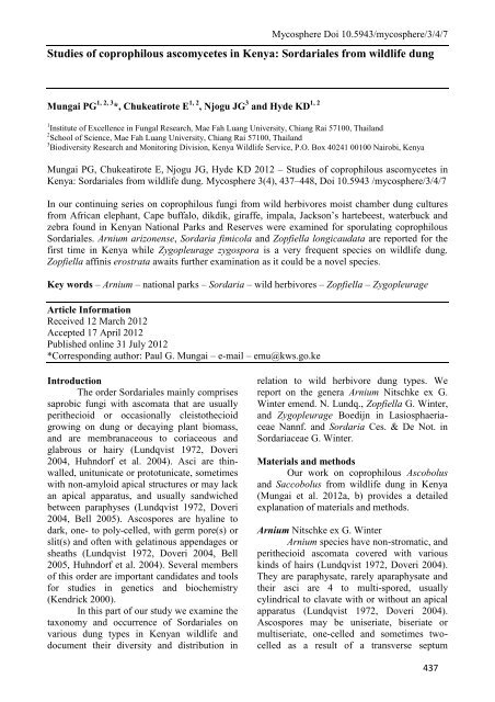
<strong>Mycosphere</strong> Doi 10.5943/mycosphere/3/4/7<br />
<strong>Studies</strong> <strong>of</strong> <strong>coprophilous</strong> <strong>ascomycetes</strong> <strong>in</strong> <strong>Kenya</strong>: Sordariales from wildlife dung<br />
Mungai PG 1, 2, 3 *, Chukeatirote E 1, 2 , Njogu JG 3 1, 2<br />
and Hyde KD<br />
1 Institute <strong>of</strong> Excellence <strong>in</strong> Fungal Research, Mae Fah Luang University, Chiang Rai 57100, Thailand<br />
2 School <strong>of</strong> Science, Mae Fah Luang University, Chiang Rai 57100, Thailand<br />
3 Biodiversity Research and Monitor<strong>in</strong>g Division, <strong>Kenya</strong> Wildlife Service, P.O. Box 40241 00100 Nairobi, <strong>Kenya</strong><br />
Mungai PG, Chukeatirote E, Njogu JG, Hyde KD 2012 – <strong>Studies</strong> <strong>of</strong> <strong>coprophilous</strong> <strong>ascomycetes</strong> <strong>in</strong><br />
<strong>Kenya</strong>: Sordariales from wildlife dung. <strong>Mycosphere</strong> 3(4), 437–448, Doi 10.5943 /mycosphere/3/4/7<br />
In our cont<strong>in</strong>u<strong>in</strong>g series on <strong>coprophilous</strong> fungi from wild herbivores moist chamber dung cultures<br />
from African elephant, Cape buffalo, dikdik, giraffe, impala, Jackson’s hartebeest, waterbuck and<br />
zebra found <strong>in</strong> <strong>Kenya</strong>n National Parks and Reserves were exam<strong>in</strong>ed for sporulat<strong>in</strong>g <strong>coprophilous</strong><br />
Sordariales. Arnium arizonense, Sordaria fimicola and Zopfiella longicaudata are reported for the<br />
first time <strong>in</strong> <strong>Kenya</strong> while Zygopleurage zygospora is a very frequent species on wildlife dung.<br />
Zopfiella aff<strong>in</strong>is erostrata awaits further exam<strong>in</strong>ation as it could be a novel species.<br />
Key words – Arnium – national parks – Sordaria – wild herbivores – Zopfiella – Zygopleurage<br />
Article Information<br />
Received 12 March 2012<br />
Accepted 17 April 2012<br />
Published onl<strong>in</strong>e 31 July 2012<br />
*Correspond<strong>in</strong>g author: Paul G. Mungai – e-mail – emu@kws.go.ke<br />
Introduction<br />
The order Sordariales ma<strong>in</strong>ly comprises<br />
saprobic fungi with ascomata that are usually<br />
perithecioid or occasionally cleistothecioid<br />
grow<strong>in</strong>g on dung or decay<strong>in</strong>g plant biomass,<br />
and are membranaceous to coriaceous and<br />
glabrous or hairy (Lundqvist 1972, Doveri<br />
2004, Huhndorf et al. 2004). Asci are th<strong>in</strong>walled,<br />
unitunicate or prototunicate, sometimes<br />
with non-amyloid apical structures or may lack<br />
an apical apparatus, and usually sandwiched<br />
between paraphyses (Lundqvist 1972, Doveri<br />
2004, Bell 2005). Ascospores are hyal<strong>in</strong>e to<br />
dark, one- to poly-celled, with germ pore(s) or<br />
slit(s) and <strong>of</strong>ten with gelat<strong>in</strong>ous appendages or<br />
sheaths (Lundqvist 1972, Doveri 2004, Bell<br />
2005, Huhndorf et al. 2004). Several members<br />
<strong>of</strong> this order are important candidates and tools<br />
for studies <strong>in</strong> genetics and biochemistry<br />
(Kendrick 2000).<br />
In this part <strong>of</strong> our study we exam<strong>in</strong>e the<br />
taxonomy and occurrence <strong>of</strong> Sordariales on<br />
various dung types <strong>in</strong> <strong>Kenya</strong>n wildlife and<br />
document their diversity and distribution <strong>in</strong><br />
relation to wild herbivore dung types. We<br />
report on the genera Arnium Nitschke ex G.<br />
W<strong>in</strong>ter emend. N. Lundq., Zopfiella G. W<strong>in</strong>ter,<br />
and Zygopleurage Boedijn <strong>in</strong> Lasiosphaeriaceae<br />
Nannf. and Sordaria Ces. & De Not. <strong>in</strong><br />
Sordariaceae G. W<strong>in</strong>ter.<br />
Materials and methods<br />
Our work on <strong>coprophilous</strong> Ascobolus<br />
and Saccobolus from wildlife dung <strong>in</strong> <strong>Kenya</strong><br />
(Mungai et al. 2012a, b) provides a detailed<br />
explanation <strong>of</strong> materials and methods.<br />
Arnium Nitschke ex G. W<strong>in</strong>ter<br />
Arnium species have non-stromatic, and<br />
perithecioid ascomata covered with various<br />
k<strong>in</strong>ds <strong>of</strong> hairs (Lundqvist 1972, Doveri 2004).<br />
They are paraphysate, rarely aparaphysate and<br />
their asci are 4 to multi-spored, usually<br />
cyl<strong>in</strong>drical to clavate with or without an apical<br />
apparatus (Lundqvist 1972, Doveri 2004).<br />
Ascospores may be uniseriate, biseriate or<br />
multiseriate, one-celled and sometimes twocelled<br />
as a result <strong>of</strong> a transverse septum<br />
437
438<br />
<strong>Mycosphere</strong> Doi 10.5943/mycosphere/3/4/7<br />
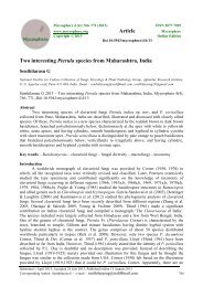
Fig. 1 – Arnium arizonense (KWSNNP017B-2010). A-B Asci and ascospores. C Immature<br />
<strong>in</strong>equilateral ascospores (arrows). D Apical chamber <strong>of</strong> asci with spores and free mature ascospores,<br />
note apex (green arrow). E Free mature ascospores show<strong>in</strong>g almost equal sized caudae (arrows). F-<br />
G Ascus show<strong>in</strong>g uniseriate spore arrangement (black arrows), paraphyses (red arrow) and stipe<br />
(green arrow). Scale bars: A, F = 200 µm B, C, D, G = 50 µm.<br />
develop<strong>in</strong>g after pigment formation, brownish<br />
black, smooth, ellipsoidal to broadly fusiform,<br />
with 1 or 2 germ pores and usually one<br />
gelat<strong>in</strong>ous cauda at each end (Lundqvist 1972,<br />
Doveri 2004). This genus is very close to<br />
Podospora Ces. but is easily dist<strong>in</strong>guished by<br />
hav<strong>in</strong>g ascospores lack<strong>in</strong>g a pedicel <strong>in</strong> addition<br />
to the characteristic ellipsoidal or fusiform<br />
immature ascospores. Arnium is usually<br />
cosmopolitan and <strong>coprophilous</strong> and grows on<br />
various herbivore dung types (Lundqvist 1972,<br />
Doveri 2004, Bell 2005).<br />
Arnium arizonense (Griffiths) N. Lundq. &<br />
J.C. Krug, Symb. bot. upsal. 20 (no.1): 232,<br />
1972. (Fig.1A–F)<br />
Pleurage arizonensis Griffiths, Mem. Torr.<br />
Bot. Cl. 11 (1): 57, 1901<br />
Sordaria arizonensis (Griffiths) Sacc.,<br />
Syll. Fung. 17: 601, 1905.<br />
Podospora arizonensis (Griffiths) Ca<strong>in</strong>,<br />
Can. J. Bot. 40: 549, 1962.<br />
Ascomata perithecioid, semi-immersed<br />
to superficial, 300–600 µm diam., scattered or<br />
gregarious, membranaceous to slightly<br />
coriaceous, black to translucent, pyriform.<br />
Neck, black, opaque sometimes curved,<br />
papilliform to cyl<strong>in</strong>drical, with long, one-sided<br />
tufts <strong>of</strong> rigid and septate hairs. Peridium<br />
pseudoparenchymatous, layered, olivaceous<br />
brown. Paraphyses numerous, simple, broadly<br />
filiform, septate, exceed<strong>in</strong>g the asci, variable<br />
width and taper<strong>in</strong>g. Asci 4-spored, 251–350 ×<br />
30–39 µm, unitunicate, clavate, with a<br />
thickened apical r<strong>in</strong>g, wall non-amyloid, with a<br />
slightly po<strong>in</strong>ted apex, apical membrane<br />
thickened, sub-apical chamber 5–7 µm broad<br />
long stipitate; stipe crooked, 100–150 × 25–35<br />
µm. Ascospores 43.5–48.5 × 22–29 µm,<br />
obliquely uniseriate, one-celled, ellipsoidal,
<strong>Mycosphere</strong> Doi 10.5943/mycosphere/3/4/7<br />
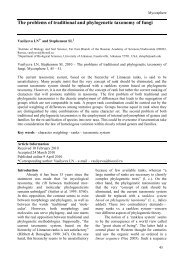
Fig. 2 – Zopfiella aff. erostrata (KWSTE005B-2009). A Cleistothecia on dung. B Squashed<br />
cleistothecium show<strong>in</strong>g long flexuous hairs. C Asci and ascospores at various stages <strong>of</strong> maturity D<br />
Ascus, note stipe (arrow). E Cleistothecial hairs, note septation (arrow). F Peridial wall. G Free<br />
ascospores amongst hyal<strong>in</strong>e swollen cells. H Free mature ascospores amongst immature asci. I Asci<br />
and ascospores show<strong>in</strong>g oil droplets (arrows). Scale bars: A = 500 µm, B = 200 µm, C-I = 20 µm.<br />
sometimes <strong>in</strong>equilateral, <strong>in</strong>itially hyal<strong>in</strong>e, later<br />
chang<strong>in</strong>g through yellowish, olivaceous to<br />
brown-black, smooth, thick walled, tipped at<br />
each end with a long gelat<strong>in</strong>ous cauda, almost<br />
equal <strong>in</strong> length and same morphology, lashlike,<br />
8–10 µm broad at base and over 100 µm<br />
long, persistent, not swell<strong>in</strong>g <strong>in</strong> water, solid,<br />
<strong>of</strong>ten densely transversely segmented,<br />
occasionally fa<strong>in</strong>tly longitud<strong>in</strong>ally and<br />
proximally furrowed; one cauda sub-apical, on<br />
the flattened side <strong>of</strong> the spore, not cover<strong>in</strong>g the<br />
germ pore; the other cauda somewhat eccentric<br />
on the same direction <strong>of</strong> opposite side <strong>of</strong> spore.<br />
Germ pore apical.<br />
Material exam<strong>in</strong>ed – (dung <strong>in</strong>cubated<br />
for 33 days) KENYA, Nairobi National Park,<br />
Nairobi Prov<strong>in</strong>ce, GPS 37M0257532 9848948,<br />
altitude 1647m, giraffe, 20 August 2010, Paul<br />
Mungai, KWSNNP017B-2010.<br />
Notes – Arnium arizonense is similar to<br />
Podospora australis (Speg.) Niessl, but P.<br />
australis has a prom<strong>in</strong>ent apiculum on each<br />
narrowly ovoid spore and does not have rigid,<br />
agglut<strong>in</strong>ated neck hairs (Lundqvist 1972,<br />
Doveri 2004). A.hirtum (E.C. Hansen) N.<br />
Lundq. & J.C. Krug is another similar taxon<br />
hav<strong>in</strong>g sometimes 4-spored asci, but it has nonfasciculate<br />
neck hairs (Doveri 2004) and<br />
differently placed and structured gelat<strong>in</strong>ous<br />
caudae. A. arizonense is a new record for<br />
<strong>Kenya</strong>.<br />
Zopfiella G. W<strong>in</strong>ter<br />
Zopfiella species usually have dark to<br />
olivaceous brown, non-stromatic, usually<br />
superficial cleistothecia with a peridium<br />
adorned with vary<strong>in</strong>g degrees <strong>of</strong> hair. They<br />
have highly evanescent asci that usually are 8spored,<br />
cyl<strong>in</strong>drical to clavate, lack<strong>in</strong>g an apical<br />
apparatus (Udagawa & Furuya 1974, Huhndorf<br />
et al. 2004, Bell 2005, Cai et al. 2006, Kirk et<br />
al. 2008). Ascospores lack a gelat<strong>in</strong>ous<br />
439
440<br />
<strong>Mycosphere</strong> Doi 10.5943/mycosphere/3/4/7<br />
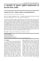
Fig. 3 – Zopfiella longicaudata (KWSTE005B-2009). A Ascomata on dung. B Squashed ascoma. C<br />
Immature ascus among free mature ascospores, note pedicels (arrow). D-E Details <strong>of</strong> peridial wall.<br />
F Immature and mature ascospores. G Ascomal wall section show<strong>in</strong>g hairs (arrow). H-I<br />
Ascospores, note germ pores (arrows). Scale bars: A = 500 µm, B = 200 µm, C-I = 20 µm.<br />
equipment, are 1-celled and hyal<strong>in</strong>e <strong>in</strong> the early<br />
stages, transversely septate and 2- sometimes<br />
3-celled at maturity, with an ellipsoidal,<br />
smooth, pigmented, <strong>of</strong>ten dark brown apical<br />
cell, and a hyal<strong>in</strong>e, basal pedicel. The apical<br />
cell (head) has an apical or subapical germ pore<br />
(Udagawa & Furuya 1974, Guarro et al. 1991,<br />
Huhndorf et al. 2004, Bell 2005, Kirk et al.<br />
2008).<br />
This genus is cosmopolitan (Guarro et<br />
al. 1991) and has been isolated from dung and<br />
soil. Zopfiella and Podospora are similar but<br />
the former can be dist<strong>in</strong>guished by its cleistothecial<br />
ascomata and ascospores without gelat<strong>in</strong>ous<br />
equipment and with short, easily<br />
collaps<strong>in</strong>g pedicels. The pedicel shape is a very<br />
important character <strong>in</strong> delimit<strong>in</strong>g species<br />
(Doveri 2004).<br />
Zopfiella aff. erostrata (Griffiths) Udagawa &<br />
Furuya, Trans. Mycol. Soc. Japan 15: 208,<br />
1974. (Fig. 2A–I)<br />
Ascomata cleistothecioid, superficial,<br />
280–300 µm diam., scattered or <strong>in</strong> small<br />
groups, black, globose to subglobose, with long<br />
flexuous, olivaceous brown to dark, robust and<br />
septate hairs evenly distributed, 2–5 µm broad,<br />
hair ends straight, smooth and po<strong>in</strong>ted; Peridial<br />
wall olivaceous brown, membranaceous,<br />
textura angularis <strong>of</strong> cells 5.5–8 × 4.5–6 µm.<br />
Paraphyses not observed. Asci 8-spored, 43–56<br />
× 10–14.5 µm, unitunicate, clavate, broadly<br />
rounded above and taper<strong>in</strong>g below <strong>in</strong>to 8–15<br />
µm long, very evanescent stipes, apical<br />
apparatus not dist<strong>in</strong>ct/observed, surrounded by<br />
hyal<strong>in</strong>e swollen cells, collaps<strong>in</strong>g <strong>in</strong> water<br />
mounts. Ascospores irregularly biseriate,<br />
hyal<strong>in</strong>e at first and one-celled, transversely<br />
septate later and two-celled. Upper cell 9.5–11<br />
× 6.5–8 µm, broadly limoniform, umbonate,<br />
with truncate base, greyish to black, guttulate;<br />
pedicel 4–6 × 1.5–4.5 µm, triangular, soon<br />
collaps<strong>in</strong>g. Germ pore apical.<br />
Material exam<strong>in</strong>ed – (dung <strong>in</strong>cubated<br />
for 14 days) KENYA, Tsavo East National<br />
Park, Coast Prov<strong>in</strong>ce, GPS S03 ° 02’29.7”<br />
E038 ° 41’35.8”, savannah, altitude 354 m,<br />
dikdik, 27 August 2009, Paul Mungai,
<strong>Mycosphere</strong> Doi 10.5943/mycosphere/3/4/7<br />
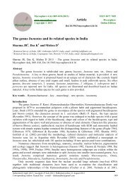
Fig. 4 – Zopfiella longicaudata (KWSTE005B-2009). A-C Details <strong>of</strong> asci and ascospores at various<br />
stages <strong>of</strong> development. Scale bars: A-B = 50 µm, C = 20 µm.<br />
KWSTE005B-2009.<br />
Notes – Apart from the triangular<br />
pedicel this collection has match<strong>in</strong>g features to<br />
descriptions <strong>of</strong> Zopfiella erostrata from<br />
Australia and Japan (Bell 2005, Udagawa &<br />
Furuya 1974). It is also close to Z.<br />
longicaudata but the latter has larger spore<br />
heads and pedicels and sporulates late <strong>in</strong><br />
<strong>in</strong>cubation. This specimen does fully fit the<br />
exist<strong>in</strong>g keys on account <strong>of</strong> the triangular and<br />
small ascospores, therefore it is identified as<br />
Zopfiella "aff<strong>in</strong>is" erostrata. Further<br />
collections will be made to determ<strong>in</strong>e whether<br />
this is a new species.<br />
Zopfiella longicaudata (Ca<strong>in</strong>) Arx, Proc.<br />
Konik. Nederl. Akad. van Wetensch. 76(3):<br />
291, 1973. (Figs. 3A–I, 4A–C, 5A–H)<br />
Tripterospora longicaudata Ca<strong>in</strong>, Can. J.<br />
Bot. 34: 703, 1956.<br />
Ascomata cleistothecioid, superficial,<br />
170–220 µm diam., scattered or <strong>in</strong> small<br />
groups, brown, globose to subglobose, with<br />
olivaceous brown to light grey, septate,<br />
unbranched hairs evenly distributed; hairs<br />
10.5–18 × 2–3 µm, hair ends almost straight,<br />
smooth and blunt. Peridial wall semitransparent,<br />
olivaceous brown, textura<br />
angularis <strong>of</strong> polygonal cells 7–11.5 × 4.5–10<br />
µm. Paraphyses not observed. Asci 8-spored,<br />
61–82 × 13.5–16 µm, unitunicate, clavate to<br />
cyl<strong>in</strong>drical, broadly rounded above and<br />
taper<strong>in</strong>g below <strong>in</strong>to evanescent long stipes<br />
measur<strong>in</strong>g 10–17 × 3–4 µm, lack<strong>in</strong>g apical<br />
apparatus, surrounded by hyal<strong>in</strong>e swollen cells,<br />
collaps<strong>in</strong>g <strong>in</strong> water mounts. Ascospores<br />
irregularly biseriate, hyal<strong>in</strong>e at first and one-<br />
celled, transversely septate later and twocelled.<br />
Upper cell 13–17.5 × 9.5–11 µm,<br />
broadly limoniform, slightly <strong>in</strong>equilateral,<br />
<strong>in</strong>itially light greyish to black, with a truncate<br />
base; pedicel 8–11 × 3–4 µm, hyal<strong>in</strong>e, slightly<br />
curved, cyl<strong>in</strong>drical with rounded ends,<br />
collaps<strong>in</strong>g with maturity, umbonate at the apex,<br />
441
442<br />
<strong>Mycosphere</strong> Doi 10.5943/mycosphere/3/4/7<br />
Fig. 5 – Zopfiella longicaudata (KWSTE005B-2009). A Immature asci and free ascospores<br />
show<strong>in</strong>g oil droplets (green arrows). B Details <strong>of</strong> peridial <strong>in</strong> section, note double layered polygonal<br />
cells (arrow). C-D Ascoma squash show<strong>in</strong>g vestiture (arrow). E-H Mature and immature asci and<br />
ascospores show<strong>in</strong>g stipe, pedicel and germ pore (arrows). Scale bars: A-C = 20 µm, D-H = 50<br />
µm.<br />
immature ascospores guttulate. Germ pore<br />
conspicuous, sub-apical.<br />
Material exam<strong>in</strong>ed – (2 ascomata on<br />
dung <strong>in</strong>cubated for 80 days) KENYA Tsavo<br />
East National Park, Coast Prov<strong>in</strong>ce, GPS<br />
S03 ° 02’29.7” E038 ° 41’35.8”, savannah,<br />
altitude 354 m, dikdik, 27 August 2009, Paul<br />
Mungai, KWSTE005B-2009; GPS<br />
S03 ° 21’666”, E038 ° 38’772”, altitude 514 m,<br />
river<strong>in</strong>e bushed-grassland, African elephant, 23<br />
September 2008, Paul Mungai, KWSTE005A-<br />
2009.<br />
Notes – The ascospores <strong>of</strong> Z.<br />
longicaudata measur<strong>in</strong>g 9.5–11.5 × 6.5–8 µm<br />
(<strong>in</strong> this study) are larger than those <strong>of</strong> Z.<br />
erostrata but smaller than Z. flammifera L.H.<br />
Huang, which measure 16–21.5 × 9.5–13 µm<br />
(Doveri 2004). Zopfiella longicaudata is<br />
apparently more frequent than Zopfiella aff<strong>in</strong>is<br />
erostrata and sporulates very late on <strong>in</strong>cubated<br />
wildlife dung. Z. longicaudata is a new record<br />
for <strong>Kenya</strong>.<br />
Zygopleurage Boedijn<br />
The ma<strong>in</strong> characteristics dist<strong>in</strong>guish<strong>in</strong>g<br />
Zygopleurage from other Lasiosphaeraceae is<br />
the unique ascospore morphology which<br />
consists <strong>of</strong> two dark ellipsoidal cells connected<br />
by an elongated cyl<strong>in</strong>drical hyal<strong>in</strong>e cell. The<br />
hyal<strong>in</strong>e, <strong>in</strong>tercalary cells are <strong>of</strong>ten coiled <strong>in</strong> the<br />
central part <strong>of</strong> ascus before spore discharge and<br />
separate two sets <strong>of</strong> polar pigmented cells. The<br />
size and shape <strong>of</strong> the <strong>in</strong>tercalary cells and<br />
pigmented cells (spore heads), with their<br />
gelat<strong>in</strong>ous sheath, and claw-shaped appendages<br />
vary <strong>in</strong> Zygopleurage and are very useful <strong>in</strong><br />
species delimitation.<br />
Currently there are only three described<br />
species <strong>of</strong> Zygopleurage: Z. faiyumensis N.<br />
Lundq., Z. multicaudata Mirza and Z.<br />
zygospora (Speg.) Boedijn.<br />
This unique <strong>coprophilous</strong> and<br />
cosmopolitan genus was reported by Lundqvist<br />
(1969) from Europe, North America, South<br />
America and Africa. Records from other parts
<strong>Mycosphere</strong> Doi 10.5943/mycosphere/3/4/7<br />
Fig. 6 – Zygopleurage zygospora (KWSNNP002-2009). A-B Squashed ascomata. C Asci with<br />
ascospores. D Free mature ascospores, note <strong>in</strong>tercalary cell and short caudae (arrows). E Ascus<br />
stipe (arrowed) and free ascospores. F Immature asci with young filamentous ascospores amongst<br />
paraphyses. Scale bars: A-B = 500 µm, C-F = 50 µm.<br />
<strong>of</strong> the world <strong>in</strong>clude Thailand <strong>in</strong> South East<br />
Asia (Mungai et al. 2011), South America<br />
(Richardson 2001), the Middle East (Abdullah<br />
& Rattan 1978) and Australia (Bell 2005).<br />
Zygopleurage zygospora (Speg.) Boedijn,<br />
Persoonia 2: 316, 1962. (Figs. 6A–F, 7A–F)<br />
Sordaria zygospora Speg., Michelia 1: 227,<br />
1878.<br />
= Philocopra zygospora (Speg.) Sacc., Syll.<br />
fung. (Abell<strong>in</strong>i) 1: 251, 1882.<br />
= Pleurage zygospora (Speg.) Kuntze 3: 1–<br />
576, 1898.<br />
= Podospora zygospora (Speg.) Niessl,<br />
Hedwigia 22: 156, 1883.<br />
Ascomata perithecioid, immersed to<br />
semi-immersed, 600–1340 µm high, 400–760<br />
µm diam., scattered or <strong>in</strong> small groups,<br />
olivaceous brown, pyriform, with a venter<br />
usually covered with long, brown, septate,<br />
flexuous hairs. Neck 200–530 × 120–370 µm,<br />
cyl<strong>in</strong>drical, covered with short hair-like cells,<br />
darker, ostiole 105–115 µm diam. Peridium 3-<br />
layered; exoperidium th<strong>in</strong>, semi-translucent <strong>of</strong><br />
textura angularis cells, 65 µm thick,<br />
mesoperidium <strong>of</strong> smaller vertically elongated<br />
cells, endoperidium consist<strong>in</strong>g <strong>of</strong> subhyal<strong>in</strong>e to<br />
light brown textura angularis cells. Paraphyses<br />
simple, hyal<strong>in</strong>e, septate, evanescent. Asci 8spored,<br />
250–322 × 40–49.5 µm, clavate,<br />
unitunicate, long-stipitate, rounded apex.<br />
Ascospores filamentous, one-celled and hyal<strong>in</strong>e<br />
when young, loosely coiled <strong>in</strong> the ascus, 3celled<br />
at maturity, composed <strong>of</strong> two dark<br />
brown end cells, 29–37 × 17.5–23 µm, usually<br />
smooth, ellipsoidal, each with an apical germ<br />
pore, jo<strong>in</strong>ed by a long subhyal<strong>in</strong>e <strong>in</strong>tercalary<br />
cell, cyl<strong>in</strong>drical, 211– 228 × 5.5–7.5 µm; 7–9<br />
µm broad at the po<strong>in</strong>t <strong>of</strong> <strong>in</strong>sertion to dark cell,<br />
sta<strong>in</strong><strong>in</strong>g blue <strong>in</strong> lactophenol cotton blue,<br />
usually parallel or coiled, each dark end cell<br />
with 4 dist<strong>in</strong>ct, short, claw-shaped, hyal<strong>in</strong>e,<br />
apical, gelat<strong>in</strong>ous appendages, 11–15 × 3–4 µm<br />
long and 4 short gelat<strong>in</strong>ous caudae aris<strong>in</strong>g at<br />
the septa <strong>of</strong> the <strong>in</strong>tercalary cell, 10–13 × 3–4<br />
µm.<br />
443
444<br />
<strong>Mycosphere</strong> Doi 10.5943/mycosphere/3/4/7<br />
Fig. 7 – Zygopleurage zygospora (KWSNNP002-2009). A Squashed ascoma. B Details <strong>of</strong> peridial<br />
wall. C Free mature ascospores. D Mature asci with ascospores show<strong>in</strong>g spore arrangement,<br />
<strong>in</strong>tercalary cell and short caudae. E Mature ascus with ascospores. F Free mature ascospores. Scale<br />
bars: A = 500 µm, B = 20 µm, C-F = 50 µm.<br />
Material exam<strong>in</strong>ed – (10 ascomata from<br />
dung <strong>in</strong>cubated for between 40 and 79 days)<br />
KENYA, Tsavo East National Park, Coast<br />
Prov<strong>in</strong>ce, GPS S03 ° 02’29.7” E038 ° 41’35.8”,<br />
altitude 354 m, African elephant, 27 August<br />
2009, Paul Mungai, KWSTE003A-2009; GPS<br />
S03 ° 21’064” E038 ° 37’501”, altitude 514 m,<br />
Cape buffalo, 27 August 2009 Paul Mungai,<br />
KWSTE008B-2009; GPS S03 ° 02’52.3”,<br />
E038 ° 54’37.0”, altitude 354 m, African<br />
elephant, 27 August 2009, Paul Mungai,<br />
KWSTE003B-2009; Aberdare Country Club<br />
Game Sanctuary, Central Prov<strong>in</strong>ce, GPS<br />
S00 ° 19’28.1”, E036 ° 55’54.3”, altitude 2161 m,<br />
zebra, 30 August 2009, Paul Mungai<br />
KWSACC002-2009; Aberdare National Park,<br />
Central Prov<strong>in</strong>ce, GPS S00 ° 20’23.2”,<br />
E036 ° 47’11.1”, altitude 2075 m, waterbuck, 29<br />
August 2009, Paul Mungai, KWSANP005-<br />
2009; Shimba Hills National Reserve, Coast<br />
Prov<strong>in</strong>ce, GPS S04 ° 14’22.4”, E039 ° 26’07.2”,<br />
altitude 374 m, impala, 24 September 2008,<br />
Paul Mungai, KWSSH005B-2008; GPS<br />
S04 ° 14’35.6”, E039 ° 26’07.1”, altitude 360 m,<br />
Cape buffalo, 26 August 2009, Paul Mungai,<br />
KWSSH004-2009; GPS S04 ° 14’14 4”,<br />
E039 ° 26’01.0”, altitude 361 m, Jackson’s<br />
hartebeest, 26 August 2009, Paul Mungai,<br />
KWSSH003-2009; Nairobi National Park,<br />
Nairobi Prov<strong>in</strong>ce, GPS 37M0255191, 9849808,<br />
altitude 1693 m, Cape buffalo, 20 August 2010,<br />
Paul Mungai, KWSNNP015-2010; GPS<br />
UTM370253715, M9849130, altitude 1876 m,<br />
zebra, 18 August 2009, Paul Mungai,<br />
KWSNNP002-2010.<br />
Notes – Zygopleurage zygospora is<br />
characterized by ascospores with longer<br />
<strong>in</strong>tercalary cells always coiled <strong>in</strong>side the asci<br />
and four dist<strong>in</strong>ct, short, claw-shaped, hyal<strong>in</strong>e<br />
gelat<strong>in</strong>ous processes on the polar cells<br />
(Abdullah & Rattan 1978). These characters<br />
vary with<strong>in</strong> the taxa <strong>of</strong> Zygopleurage and are<br />
very useful <strong>in</strong> delimitation (Lundqvist 1969,<br />
1972, Abdullah & Rattan 1978). In addition, Z.<br />
zygospora has an olivaceous brown<br />
perithecium. The ascospores <strong>of</strong> Z. zygospora<br />
are <strong>in</strong>termediate <strong>in</strong> size between those <strong>of</strong> Z.<br />
multicaudata, which are smaller and Z.
<strong>Mycosphere</strong> Doi 10.5943/mycosphere/3/4/7<br />
Fig. 8 – Sordaria fimicola (KWSKIN004-2009). A Ascoma on dung. B squashed ascoma. C Free<br />
mature ascospores show<strong>in</strong>g gelat<strong>in</strong>ous sheath and germ pore (arrows). D Asci apex and paraphyses.<br />
E Mature asci with ascospores. Scale bars: A = 500 µm, B = 200 µm, C = 20 µm, D = 20 µm, E =<br />
50 µm.<br />
faiyumensis, which are larger (Abdullah &<br />
Rattan 1978). Z. zygospora is a very unique<br />
species and therefore not easy to confuse with<br />
any other known species (Lundqvist 1969,<br />
1972, Abdullah & Rattan 1978).<br />
Sordaria Ces. & De Not.<br />
Sordaria is characterized by dark,<br />
superficial or semi-immersed, non-stromatic<br />
perithecia and a layered, pseudoparenchymatous<br />
peridium (Ca<strong>in</strong> 1934, Lundqvist 1972,<br />
Doveri 2004, Bell 2005). Asci are unitunicate,<br />
non-amyloid, cyl<strong>in</strong>drical, usually 8-spored,<br />
each with a well developed apical apparatus<br />
(Lundqvist 1972, Doveri 2004, Bell 2005).<br />
Ascospores are one-celled, broadly ovoid to<br />
ellipsoidal, sometime subglobose or subfusiform,<br />
dark pigmented at maturity, with a basal<br />
germ pore and usually surrounded by a hyal<strong>in</strong>e<br />
mucilag<strong>in</strong>ous sheath (Lundqvist 1972, Doveri<br />
2004, Bell 2005). Sordaria species have very<br />
similar morphological features thus creat<strong>in</strong>g a<br />
challenge <strong>in</strong> species delimitation. Accord<strong>in</strong>g to<br />
Lundqvist (1972) and Guarro & von Arx<br />
(1987) analysis <strong>of</strong> the perithecial structure,<br />
ascus and spore size is a very reliable way <strong>of</strong><br />
delimit<strong>in</strong>g Sordaria species.<br />
This genus is composed <strong>of</strong> ma<strong>in</strong>ly<br />
fimicolous species. However, several Sordaria<br />
species have been isolated from rema<strong>in</strong>s <strong>of</strong><br />
plant biomass, live plants, seeds and from soil<br />
(Doveri 2004). Sordaria has been recorded<br />
worldwide (Lundqvist 1972, Khan & Krug<br />
1989, Doveri 2004, Bell 2005, Jeamjitt et al.<br />
2007, Richardson 2008).<br />
Sordaria fimicola (Roberge) Ces. & De Not.,<br />
Comm. Soc. Critt. Ital. 1: 226, 1863. (Figs.<br />
8A–E, 9A–G)<br />
Sphaeria fimicola Roberge <strong>in</strong> Desm., Ann.<br />
445
446<br />
<strong>Mycosphere</strong> Doi 10.5943/mycosphere/3/4/7<br />
Fig. 9 – Sordaria fimicola (KWSKIN004-2009). A Squashed ascoma. B Mature ascus with spores,<br />
note uniseriate arrangement (arrow). C Free mature spores. D Asci stipes (arrows). E Asci apex<br />
show<strong>in</strong>g apical r<strong>in</strong>g (arrow). F Paraphyses. G Details <strong>of</strong> peridial wall. H Mature asci with spores.<br />
Scale bars: A = 200 µm, B = 50 µm, C = 20 µm, D = 20 µm, E = 20 µm, F = 20 µm, G = 20 µm,<br />
H = 50 µm.<br />
Sci. Nat. 3 sér. Bot. 11: 353, 1849.<br />
An extensive list <strong>of</strong> synonyms is<br />
provided by Doveri (2004). Ascomata<br />
perithecioid, semi-immersed to superficial,<br />
550–620 µm, high, 450–500 µm, diam.,<br />
scattered or more <strong>of</strong>ten gregarious or even<br />
crowded, membranaceous, dark brown,<br />
sparsely covered with hyphoid hairs, ovoid to<br />
pyriform. Neck conical or subcyl<strong>in</strong>drical, 100–<br />
120 × 120–150 µm. Peridial wall layered,<br />
pseudoparenchymatous; exostratum a textura<br />
angularis <strong>of</strong> polygonal cells <strong>in</strong> the venter, a<br />
textura globulosa <strong>in</strong> the neck, 10.5–17 × 8.5–<br />
13 µm. Paraphyses moniliform, septate, with<br />
segments 4.5–12.5 µm broad, abundant,<br />
conta<strong>in</strong><strong>in</strong>g hyal<strong>in</strong>e vacuoles. Asci 8-spored,<br />
111–163 × 10.5–14 µm, cyl<strong>in</strong>drical, flattened<br />
at apex, short stipitate, with a lobate stipe, and<br />
prom<strong>in</strong>ent apical apparatus. Ascospores 15.5–<br />
18.5 × 9.5–11.5 µm, obliquely to vertically<br />
uniseriate, dark brown, ellipsoidal,<br />
occasionally ovoid, smooth, slightly po<strong>in</strong>ted<br />
and apiculate at the base, surrounded by a<br />
gelat<strong>in</strong>ous sheath usually <strong>in</strong>vag<strong>in</strong>ated at the<br />
apiculum. Germ pore s<strong>in</strong>gle and basal.<br />
Material exam<strong>in</strong>ed – (dung <strong>in</strong>cubated<br />
for 14 days) KENYA, K<strong>in</strong>ondo Forest Reserve,<br />
Coast Prov<strong>in</strong>ce, GPS S04 ° 25’197”<br />
E039 ° 32’602”, coastal forest, altitude 18m,<br />
dikdik, 19 April 2009, Paul Mungai,<br />
KWSKIN004-2009.<br />
Notes – Sordaria species are very<br />
homogenous and therefore are very difficult to<br />
delimit. Sordaria fimicola differs from S.<br />
superba De Not. and S. macrospora Auersw. <strong>in</strong><br />
hav<strong>in</strong>g smaller spores, ellipsoidal rather than<br />
broadly ellipsoidal and smaller perithecia and<br />
asci (Doveri 2004). Other similar taxa namely<br />
S. sibutii Cailleux and S. conoidea Cailleux<br />
lack a gelat<strong>in</strong>ous perisporium on their spores.<br />
S. fimicola is homothallic with four hardly<br />
differentiated heterothallic relatives, namely, S.<br />
thermophila Fields, S. sclerogenia Fields &<br />
Grear, S. tomentoalba Cailleux and S.
evicollis L.S. Olive & Fant<strong>in</strong>i (Doveri 2004).<br />
Although reported as a very common<br />
cosmopolitan pyrenomycete by other<br />
<strong>in</strong>vestigators (Lundqvist 1972, Doveri 2004), S.<br />
fimicola was isolated only once <strong>in</strong> this study.<br />
This is a new record for <strong>Kenya</strong>.<br />
Discussion<br />
Ecology<br />
Zygopleurage zygospora on 72% <strong>of</strong> dung<br />
samples was the most common species and<br />
occurred on the widest range <strong>of</strong> dung types.<br />
Ten isolates <strong>of</strong> Z. zygospora sporulated on six<br />
dung types. Arnium arizonense, Sordaria<br />
fimicola and Zopfiella aff. erostrata (7% each)<br />
were the least common.<br />
Sordaria fimicola and Zopfiella<br />
‟aff<strong>in</strong>is” erostrata sporulated quite early on<br />
<strong>in</strong>cubation. Arnium arizonense sporulated <strong>in</strong><br />
the mid-<strong>in</strong>cubation period, while Zopfiella<br />
longicaudata and Zygopleurage zygospora<br />
sporulated <strong>in</strong> the last period <strong>of</strong> <strong>in</strong>cubation.<br />
The age <strong>of</strong> dung at sampl<strong>in</strong>g and the<br />
time taken from sampl<strong>in</strong>g to <strong>in</strong>cubation had a<br />
notable <strong>in</strong>fluence on the composition <strong>of</strong><br />
Sordariales sporulat<strong>in</strong>g with most <strong>of</strong> the early<br />
sporulat<strong>in</strong>g species be<strong>in</strong>g less common on old<br />
or preserved dung.<br />
Accord<strong>in</strong>g to dung types, zebra, Cape<br />
buffalo, giraffe and impala, all exhibit<strong>in</strong>g<br />
different feed<strong>in</strong>g habits, had the highest<br />
number <strong>of</strong> specimens and species.<br />
A s<strong>in</strong>gle isolate <strong>of</strong> Arnium arizonense<br />
and Sordaria fimicola sporulated on just one<br />
dung type each. This may be due to a taxon<br />
substrate preference or rarity.<br />
Acknowledgements<br />
This study was undertaken with funds<br />
provided by Novozymes A/S <strong>of</strong> Denmark,<br />
<strong>Kenya</strong> Wildlife Service (KWS) and the<br />
Mushroom Research Foundation. We are<br />
grateful to the Director <strong>Kenya</strong> Wildlife Service<br />
and Deputy Director, Biodiversity Research<br />
and Monitor<strong>in</strong>g Division, <strong>Kenya</strong> Wildlife<br />
Service for allow<strong>in</strong>g the study with<strong>in</strong> <strong>Kenya</strong>n<br />
National Parks and Reserves. We wish to<br />
appreciate and thank Dr Francesco Doveri for<br />
his never falter<strong>in</strong>g help <strong>in</strong> taxonomic<br />
corrections to this paper and advice on fungi<br />
taxonomy <strong>in</strong> general through numerous email<br />
<strong>Mycosphere</strong> Doi 10.5943/mycosphere/3/4/7<br />
exchanges. It is noted that without his advice<br />
this work would not have been possible. We<br />
also feel highly <strong>in</strong>debted to staff and students<br />
<strong>of</strong> the Institute <strong>of</strong> Excellence <strong>in</strong> Fungal<br />
Research, Mae Fah Luang University, Thailand<br />
and <strong>Kenya</strong> Wildlife Service colleagues<br />
especially Mr. Patrick Omondi, Dr Samuel<br />
Andanje and Mr. Moses Otiende for giv<strong>in</strong>g<br />
very useful suggestions dur<strong>in</strong>g this study. Ms<br />
Asenath Ak<strong>in</strong>yi is thanked for grammar and<br />
spell-check<strong>in</strong>g the text and her assistance <strong>in</strong> the<br />
laboratory.<br />
References<br />
Abdullah SK and Rattan SS. 1978 –<br />
Zygopleurage, Tripterosporella and<br />
Podospora (Sordariaceae: Pyrenomycetes)<br />
<strong>in</strong> Iraq. Mycotaxon 7, 102–116.<br />
Bell A. 2005 – An Illustrated Guide to the<br />
Coprophilous Ascomycetes <strong>of</strong><br />
Australia. CBS Biodiversity Series 3.<br />
Centraalbureau voor Schimmelcultures,<br />
Utrecht, The Netherlands.<br />
Cai L, Jeewon R, Hyde KD. 2006 – Molecular<br />
Systematics <strong>of</strong> Zopfiella and allied<br />
Genera: Evidence from multi-gene<br />
sequence and analyses. Mycological<br />
Research 110, 359–368.<br />
Ca<strong>in</strong> RF. 1934 – <strong>Studies</strong> <strong>of</strong> <strong>coprophilous</strong><br />
sphaeriales <strong>in</strong> Ontario. University <strong>of</strong><br />
Toronto <strong>Studies</strong> <strong>in</strong> Mycology Series 38,<br />
1–126.<br />
Doveri F. 2004 – Fungi Fimicoli Italici.<br />
Associazione Micologica Bresadola,<br />
Trento, Italy.<br />
Guarro J and Arx JA, von. 1987 – The<br />
Ascomycetes genus Sordaria.<br />
Persoonia 13, 301–313.<br />
Guarro J, Cannon PF, van der Aaa. 1991 – A<br />
synopsis <strong>of</strong> the genus Zopfiella<br />
(<strong>ascomycetes</strong>, Lasiosphaeraceae). Systema<br />
Ascomycetum 10, 79–112.<br />
Huhndorf SM, Miller AN, Fernández FA. 2004<br />
– Molecular systematics <strong>of</strong> the<br />
Sordariales: the order and the family<br />
Lasiosphaeriaceae redef<strong>in</strong>ed.<br />
Mycologia 96 2, 368–387.<br />
Jeamjitt O, Manoch L, Visarathanonth N,<br />
Chamswarng C, Watl<strong>in</strong>g R, Sharples<br />
GP, Kijjoa A. 2007 – Coprophilous<br />
<strong>ascomycetes</strong> <strong>in</strong> Thailand. Mycotaxon<br />
447
100, 115–136.<br />
Kendrick B. 2000 – The Fifth K<strong>in</strong>gdom.<br />
Mycologue Publications. pp 373.<br />
Khan RS and Krug JC. 1989 – New records <strong>of</strong><br />
the Sordariaceae from East Africa.<br />
Mycologia 81, 862–869.<br />
Kirk PM, Cannon PF, David JC and Stalpers<br />
JA. (eds) 2008 – A<strong>in</strong>sworth & Bisby’s<br />
Dictionary <strong>of</strong> the Fungi. CABI,<br />
Wall<strong>in</strong>gford.<br />
Lundqvist N. 1969 – Zygopleurage and<br />
Zygospermella (Sordariaceae s.lat.<br />
Pyrenomycetes). Botaniska Notiser<br />
122, 353–374.<br />
Lundqvist N. 1972 – Nordic Sordariaceae s.<br />
lat. Symbolae Botanicae Upsalliensis<br />
20, 1–374.<br />
Mungai P, Hyde KD, Cai L, Njogu J,<br />
Chukeatirote K. 2011 – Coprophilous<br />
<strong>ascomycetes</strong> <strong>of</strong> northern Thailand.<br />
Current Research <strong>in</strong> Environmental and<br />
Applied Mycology 1, 135–159.<br />
448<br />
<strong>Mycosphere</strong> Doi 10.5943/mycosphere/3/4/7<br />
Mungai PG, Njogu JG, Chukeatirote E, Hyde<br />
KD. 2012a – <strong>Studies</strong> <strong>of</strong> <strong>coprophilous</strong><br />
<strong>ascomycetes</strong> <strong>in</strong> <strong>Kenya</strong>. Ascobolus<br />
species from wildlife dung. Current<br />
Research <strong>in</strong> Environmental and<br />
Applied Mycology 2, 1–16.<br />
Mungai PG, Njogu JG, Chukeatirote E, Hyde<br />
KD. 2012b – <strong>Studies</strong> <strong>of</strong> <strong>coprophilous</strong><br />
<strong>ascomycetes</strong> <strong>in</strong> <strong>Kenya</strong>. Saccobolus<br />
species from wildlife dung.<br />
<strong>Mycosphere</strong> 3(2), 111–129.<br />
Richardson MJ. 2001 – Coprophilous fungi<br />
from Brazil. Brazilian Archives <strong>of</strong> Biology<br />
and Technology (44) 3, 283–289.<br />
Richardson MJ. 2008 – Records <strong>of</strong><br />
<strong>coprophilous</strong> fungi from the Lesser<br />
Antilles and Puerto Rico. Caribean<br />
Journal <strong>of</strong> Science 44, 206–214.<br />
Udagawa S and Furuya K. 1974 – Notes on<br />
some Japanese <strong>ascomycetes</strong> XIII.<br />
Transactions <strong>of</strong> the Mycological<br />
Society <strong>of</strong> Japan 15, 206–214.