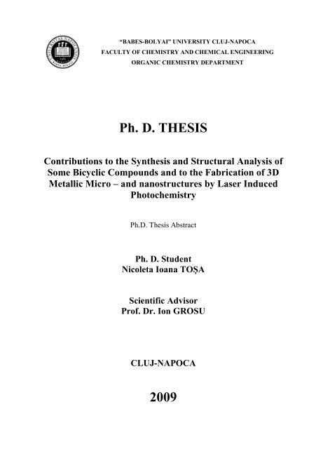
Ph. D. THESIS 2009
Ph. D. THESIS 2009
Ph. D. THESIS 2009

You also want an ePaper? Increase the reach of your titles
YUMPU automatically turns print PDFs into web optimized ePapers that Google loves.
“BABES-BOLYAI” UNIVERSITY CLUJ-NAPOCA<br />
FACULTY OF CHEMISTRY AND CHEMICAL ENGINEERING<br />
ORGANIC CHEMISTRY DEPARTMENT<br />
<strong>Ph</strong>. D. <strong>THESIS</strong><br />
Contributions to the Synthesis and Structural Analysis of<br />
Some Bicyclic Compounds and to the Fabrication of 3D<br />
Metallic Micro – and nanostructures by Laser Induced<br />
<strong>Ph</strong>otochemistry<br />
<strong>Ph</strong>.D. Thesis Abstract<br />
<strong>Ph</strong>. D. Student<br />
Nicoleta Ioana TOŞA<br />
Scientific Advisor<br />
Prof. Dr. Ion GROSU<br />
CLUJ-NAPOCA<br />
<strong>2009</strong>
JURY<br />
“BABES-BOLYAI” UNIVERSITY CLUJ-NAPOCA<br />
FACULTY OF CHEMISTRY AND CHEMICAL ENGINEERING<br />
ORGANIC CHEMISTRY DEPARTMENT<br />
<strong>Ph</strong>. D. <strong>THESIS</strong><br />
Contributions to the Synthesis and Structural Analysis of<br />
Some Bicyclic Compounds and to the Fabrication of 3D<br />
Metallic Micro – and nanostructures by Laser Induced<br />
<strong>Ph</strong>otochemistry<br />
<strong>Ph</strong>.D. Thesis Abstract<br />
<strong>Ph</strong>. D. Student<br />
Nicoleta Ioana TOŞA<br />
President Prof. Dr. Ioan Silaghi-Dumitrescu, Dean of the<br />
Faculty of Chemistry and Chemical Engineering,<br />
Cluj-Napoca<br />
Scientific advisor Prof. Dr. Ion Grosu, Faculty of Chemistry and<br />
Chemical Engineering, Cluj-Napoca<br />
Reviewers Prof. Dr. Ionel Mangalagiu, University “Al.I. Cuza”,<br />
Iasi<br />
D. R. Dr. Patrice Baldeck, University “Joseph<br />
Fourier”, Grenoble, France<br />
Prof. Dr. Simion Astilean, University “Babes-<br />
Bolyai”, Cluj-Napoca<br />
Defense on 7 th October <strong>2009</strong><br />
1
General Introduction<br />
OUTLINE<br />
Part A<br />
Synthesis and Structural Analysis of Some<br />
Bicyclo[3.3.1]nonane Derivatives<br />
1. Bicyclo[3.3.1]nonane-3,7-dione 11<br />
1.1. Introduction 11<br />
1.1.2. Synthesis of bicyclo[3.3.1]nonane-3,7-dione 11<br />
1.3. Structural aspects in solution 13<br />
1.3.1. NMR investigations 13<br />
1.3.1.1. Tetramethyl 3, 7-dihydroxybicyclo[3.3.1] nona-2, 6-diene<br />
-2, 4, 6, 8-tetracarboxylate 13<br />
1.3.1.1. Bicyclo[3.3.1]nonane-3,7-dione 17<br />
1.3.2. FT-IR investigation of tetramethyl 3, 7-dihydroxybicyclo[3.3.1] nona-<br />
2, 6-diene-2, 4, 6, 8-tetracarboxylate 20<br />
1.4. Solid state investigations 23<br />
1.4.1. FT-IR and Raman investigations 23<br />
1.4.1.1. Tetramethyl 3, 7-dihydroxybicyclo[3.3.1] nona-2, 6-diene-<br />
2,4,6,8-tetracarboxylate 24<br />
1.4.1.2. Bicyclo[3.3.1]nonane-3,7-dione 30<br />
1.4.2. Mass spectrometry study of bicyclo[3.3.1]nonane-3,7-dione 35<br />
1.4.3. Single crystal X-ray diffraction investigations 38<br />
1.4.3.1. Tetramethyl 3,7-dihydroxybicyclo [3.3.1] nona-2,6-<br />
diene-2,4,6,8-tetracarboxylate 38<br />
1.4.3.2. Bicyclo[3.3.1]nonane-3,7-dione 40<br />
1.5. Molecular modeling: structure and IR spectra 42<br />
1.5.1. Tetramethyl 3,7-dihydroxybicyclo[3.3.1]nona-2,6-diene-<br />
2,4,6,8-tetracarboxylate structure 42<br />
1.5.2. Bicyclo[3.3.1]nonane-3,7-dione 48<br />
1.6. Attempts of synthesis of spiro-bis(dioxane) bicyclic compounds type 50<br />
1.6.1. Synthesis of spiro-monodioxane compounds 50<br />
1.6.2. NMR spectra in solution 51<br />
1.6.3. Mass spectra of monospirane 56<br />
1.6.4. FT-IR measurements 60<br />
2. Bicyclo[3.3.1]nonane-3,7-dione dioxime<br />
62<br />
2.1. Introduction 62<br />
2.2. Synthesis of syn and anti isomers 63<br />
2.3. Investigations in solution 66<br />
2.3.1. NMR spectra of syn and anti isomers 66<br />
2.3.2. NMR study of kinetics of the synanti isomerisation 70<br />
2.3.3. Chiral HPLC studies 74<br />
2.3.3.1. Investigation of a mixture of syn and anti isomers 74<br />
2.3.3.2. Investigation of the isolated anti isomer 76<br />
2.3.4. FT-IR study 77<br />
2.4. Investigation of bicyclo[3.3.1]nonane-3,7-dione dioxime in solid state 80<br />
2.4.1. FT-IR 80<br />
2.4.2. Single crystal X-ray diffraction 83<br />
2
2.5. Mass spectrometry of bicyclo[3.3.1]nonane-3,7-dione dioxime 87<br />
2.6. Molecular modeling for the supramolecular architectures of syn and anti isomer 90<br />
2. Conclusions of the part A<br />
96<br />
3. Experimental part<br />
97<br />
4.1. General experimental 97<br />
4.2. Synthesis and characterization of compounds 100<br />
References 101<br />
Part B<br />
Fabricatio n o f 3 D Metallic Micro - an d n an o stru ctu res by<br />
Laser In d u ced <strong>Ph</strong> o to ch em istry<br />
1. Principle of the micro/nanostructuration by two-photon induced<br />
photochemistry. State of the art 1 4 6<br />
2. Experimental part: fabrication and characterization 159<br />
2.1. Sample conditioning 159<br />
2 1.1.Composition 159<br />
2.1.2. Preparation 161<br />
2.1.3. Thin film deposition 163<br />
2.1.3.1 The key stages in spin coating 163<br />
2.1.3.2.Polyimide underlayer spin-coating 163<br />
2.1.4. Washing process 166<br />
2.2. The metallic fabrication process 168<br />
2.2.1. Experimental set-up 168<br />
2.2.2. Laser source 168<br />
2.2.3. The control of the microscope motorized stage 169<br />
2.3. Characterization of the metallic structures 170<br />
2.3.1. Optical and spectroscopical microscopy 171<br />
2.3.2. Scanning electronic microscopy 171<br />
2.3.3. Atomic force microscopy 172<br />
3. Optimization of silver micro/nanostructures fabrication<br />
175<br />
3.1.Introduction 175<br />
3.2. The approach of silver photographic process 177<br />
3.2.1. The principle of the silver photographic process 177<br />
3.2.2. Our chemical system 179<br />
3.3. TPA fabrication of silver wires 180<br />
3.3.1. Processing parameters 180<br />
3.3.2. Thermal effect limitation 181<br />
3.4. Chemical nature of the silver deposition 183<br />
3.5. TPA approach induced by silver nanoparticles 185<br />
3.5.1. General mechanism 185<br />
3.5.2. The reduction mechanism by thermal effect 185<br />
3.5.3. Our chemical system 187<br />
3.6. TPA fabrication of 2D and 3D silvermicro/nanostructures 188<br />
3
3.6.1. Dots 188<br />
3.6.2. 3D structures 192<br />
3.6.2.1. Fabrication 192<br />
3.6.2.2. Silver porosity due to the corosion 196<br />
3.6.2.3. Electroless plating process 199<br />
4. Optimization of gold micro/nanostructures fabrication<br />
207<br />
4.1. Introduction 207<br />
4.2. UV-Vis investigation 208<br />
4.3. The gold structures fabrication process 211<br />
4.3.1. PSS- matrix for gold microfabrication 211<br />
4.3.2. Double-line effect in thin PSS matrix 214<br />
4.3.3. The influence of the operating parameters 219<br />
4.3.3.1. The laser power 219<br />
4.3.3.2. The substrate 222<br />
4.3.3.2.1. Glass 222<br />
4.3.3.2.2. Polyimide underlayered glass 223<br />
4.3.3.3. Gold salt concentration 224<br />
4.4. The thermal effect 227<br />
4.4.1. Theoretical model 230<br />
4.5 The overcoming of the thermal effect 232<br />
4.5.1 Nanodots 230<br />
4.5.2. Single wires 234<br />
4.5.3. 3D structures 237<br />
5. Optical properties of the metallic structures<br />
239<br />
5.1. Introduction 239<br />
5.2. Diffraction properties 239<br />
5.3. 3D photonic crystals 243<br />
6. Conclusions of the part B<br />
247<br />
References 248<br />
Keywords: intra- and intermolecular H-bonds, keto-enol tautomerism, syn and anti<br />
stereoisomers, self-assembly, supramolecular chemistry, laser induced photochemistry,<br />
two-photon absorption, thermal effect, 3D structures.<br />
4
General Introduction<br />
The chemistry of the bicyclic compounds and their derivatives is a<br />
challenging domain. Particularly, bicyclo[3.3.1]nonane derivatives represent<br />
important intermediates in the synthetic routes to alkenes containing<br />
piramidalized carbon atoms, to derivatives of potential interest for the treatment<br />
of Alzheimer’s disease as well as to derivatives of dioximes type which can<br />
form self-assembled supramolecular structures through intermolecular<br />
hydrogen bonds. The fabrication of metallic structures based on two-photon<br />
absorption represents a new and promising domain due to the innovative<br />
technique and to the multiple applications in microelectronics, biosensing and<br />
spectral filtering devices.<br />
This work reflects my activity accomplished within the framework of<br />
two powerful research groups. Even if the subjects developed seem to be<br />
disconnected, at the first sight, they belong to the field of the chemical reactions<br />
assisted by the electromagnetic radiation. The aim of this thesis is to bring a<br />
contribution within these fields.<br />
The thesis is structured in two parts. The first part – Part A – refers to<br />
the contributions to the synthesis of some bicyclo[3.3.1]nonane derivatives and<br />
their structural analysis. The second part –Part B – presents innovative issues<br />
for the engineering of 3D metallic micro- and nanostructures by laser induced<br />
photochemistry. The second part presents also some aspects concerning optical<br />
and spectroscopic characteristics of such metallic structures.<br />
5
Part A: Synthesis and Structural Analysis of Some<br />
Bicyclo[3.3.1]nonane Derivatives<br />
1. Bicyclo[3.3.1]nonane-3,7-dione<br />
Due to its special characteristics, which involve a preorganisation of the<br />
molecular structure, bicyclo[3.3.1]nonane-3,7-dione 2 is of interest for the<br />
synthesis of some bicyclic derivatives and thus, for macrocycles as a<br />
supramolecular connector. Thus, a series of macrocyclic compounds containing<br />
bicyclic units have been already obtained [1]. The properties of synthesized<br />
macrocyclic compounds are significantly dependent on the spatial arrangement<br />
of the synthon at molecular level.<br />
In this respect we focused on the synthesis and structural aspects of compound<br />
2, as well as on the structural analysis of its precursor, namely tetramethyl 3,7dihydroxybicyclo[3.3.1]<br />
nona-2,6-diene-2,4,6,8-tetracarboxylate 1.<br />
1 .2 . Sy n th esis o f bicy clo [3 .3 .1 ]n o n an e -3 ,7 -d io n e<br />
Sy n t h esis o f b icy clo [3 .3 .1 ]n o n a n e -3 ,7 -d io n e st a r in g fr o m<br />
t etr a m eth y l 3 ,7 -d ih y d r o xy b icy clo [3 .3 .1 ] n o n a -2 ,6 -d ien e -<br />
2 ,4 ,6 ,8 -t e t r a ca r b o xy la t e 1 r ep r esen t ed a ch a llen ge d u e t o t h e<br />
h a r d co n d it io n s o f r e a ct io n . Th e lit er a t u r e d a t a in d ica t e a<br />
t wo -st ep s h y d r o ly sis-d eca r b o xy la t io n r o u t e, in vo lv in g<br />
gen er a t io n o f b icy clic β -d ik etotetr a est er s a s in t er m ed ia r , a n d<br />
t h en it s co n v er sio n in t o b icy clic d ik eton e m a in ly u sin g<br />
a cid ic ca t a ly st s (e.g. h y d r o ch lo r ic a cid [2 ] o r a m ixt u r e o f<br />
h y d r o ch lo r ic a cid a n d a cet ic a cid [3 ]).<br />
In t h e ligh t o f t h e se a sp ect s we p r o p o se a st r a igh t fo r wa r d<br />
co n ver sio n o f co m p o u n d 1 in t o co m p o u n d 2 sim p ly h ea t in g<br />
a t h igh t em p er a t u r e a m ixt u r e o f t etr a est er 1 a n d a sm a ll<br />
a m o u n t o f wa t er , p la ce d in t o in a sea led b o r o silica t e t u b e.<br />
H 3COOC<br />
HO<br />
H 3COOC<br />
COOCH 3<br />
(a)<br />
OH<br />
O<br />
(b) H2O, K-10<br />
MW irrad.<br />
1<br />
COOCH3 2<br />
6<br />
H 2O<br />
O
Sch e m e 1 . Th e co n ve r sio n o f 1 in t o 2 via h y d r o ly sisd<br />
e ca r b o xy la tio n r o u t e .<br />
The reaction underwent with the complete hydrolysis and decarboxylation<br />
of all four ester groups. The total transformation is supported by TLC<br />
chromatography as well as by the melting point of the crude product, which is<br />
0.5°C smaller than that of the pure compound 2.We have to highlight the<br />
novelty of this method through its simplicity (one-step), high efficiency (yields<br />
over 96%) and because it requires no catalyst.<br />
We also developed an alternative way to the above proposed method. Thus,<br />
compound 1 was deposited on a solid support, placed into a teflon sealed tube<br />
and irradiated in microwave field.<br />
The results of these two methods can be summarized in Table 1:<br />
Table 1 Comparisson between classic and microwave irradiation methods<br />
Method Yield (%) M.p.<br />
7<br />
(ºC)<br />
classic Method (a) 98 250<br />
microwave<br />
irrad.<br />
Method (b) 90 249-250<br />
The advantages of these syntheses over the previous ones lie in the<br />
direct transformation of 1 into 2, as well as in the significantly increased yields,<br />
due to the absence of the acidic conditions and to the facile work-up procedure.<br />
1.3. Structural aspects in solution<br />
The bicyclic tetraester 1 and the bicyclic diketone 2 were both investigated<br />
by NMR spectroscopy in solution at room temperature in order to establish<br />
their structure and thereby to evidence the complete hydrolysis-decarboxylation<br />
of the precursor 1. This may present as most of β-ketoesters two tautomeric<br />
forms, enol and ketone, in equilibrium.<br />
Keto-enol tautomerization of compound 1 (Scheme 2) in CDCl3 solution<br />
has been investigated by NMR spectroscopy.
H 3C<br />
H 3C<br />
O<br />
O<br />
O<br />
C<br />
C<br />
O<br />
O<br />
1a<br />
O<br />
O<br />
C<br />
C<br />
O<br />
O<br />
O<br />
CH 3<br />
CH 3<br />
8<br />
H 3C<br />
H<br />
O<br />
O<br />
O<br />
C<br />
C<br />
H 3C<br />
O<br />
O<br />
O<br />
O<br />
CH 3<br />
Scheme 2. The keto-enol tautomeric equilibrium<br />
In Figure 1 is presented a fragment of the 1 H-NMR spectrum (CDCl3, 500<br />
MHz) of compound 1. The 1 H NMR spectrum of 1 consists of 2H triplet at δ=<br />
1.93(J=3.0 Hz), another 2H triplet at δ= 3.22(J=3.0 Hz), singlets at δ= 3.29<br />
(2H), δ= 3.76 (6H), δ= 3.83 (6H), and δ= 12.33 (2H) ppm.<br />
Figu re 1 . Th e 1<br />
H-NMR sp e ct r u m (CDCl , 5 0 0 MHz, fr a gm e n t )<br />
3<br />
o f co m p o u n d 1<br />
The fact that the hydrogen atoms at 1-C/5-C and those at 9-C give rise to<br />
triplets indicate that the bridgehead hydrogen atoms are equivalent and<br />
establishes the C2 symmetry of the molecule. Also, the broadening of both<br />
peaks suggests a weak coupling between the protons at 2-C /6-C and those at 9-<br />
C, which are connected in W path [7].<br />
1b<br />
C<br />
C<br />
O<br />
O<br />
O<br />
H<br />
CH 3
Figu re 1 . Th e<br />
co m p o u n d 1<br />
1<br />
H-NMR sp ectr u m (CDCl , 5 0 0 MHz, fr a gm en t) o f<br />
3<br />
The signal at δ= 3.29 from hydrogen atoms at 2-C/6-C is a singlet. As time<br />
as the dihedral angle between these protons and those at 1-C/5-C is close to 90 0<br />
[8], the vicinal coupling between them is not possible. Also, this is possible to<br />
have a singlet if the ester groups located in position 2-C/6-C have the exo<br />
configuration, and the conformations of the two six-membered rings are closed<br />
to the half-chair [9], expected for a cyclohexene derivative.<br />
The two singlets at δ= 3.76 and δ= 3.83 correspond to the protons of ester<br />
methyl groups at 15-C/17-C and 14-C/16-C, respectively. The small shift<br />
appears because the ester groups are not equivalent, those located at position 4-<br />
C/8-C being involved in intramolecular hydrogen bonding.<br />
Compound 1 exhibits two possible enol forms, both of them involving the<br />
formation of hydrogen bonds (Scheme 3).<br />
CH3<br />
H<br />
O<br />
O<br />
O<br />
C<br />
C<br />
O<br />
O<br />
CH 3<br />
1a<br />
O<br />
O<br />
CH 3<br />
C<br />
C<br />
O<br />
O<br />
O<br />
H<br />
CH 3<br />
9<br />
CH 3<br />
H<br />
O<br />
O<br />
O<br />
CH 3<br />
Scheme 3. Theoretical enol forms for compound 1<br />
C<br />
C<br />
O<br />
O<br />
1b<br />
O<br />
O<br />
C<br />
C<br />
O<br />
O<br />
O<br />
CH 3<br />
H<br />
CH 3
The two forms are in equilibrium and because the equilibration is fast, the<br />
NMR spectrum does not show different signals for the two isomers. The spectra<br />
exhibit unique signals at mean values of chemical shifts.<br />
The strong deshielded signal at δ= 12.32 ppm indicate the presence of the enol<br />
form and can be assigned to the hydrogen atom involved in the formation of<br />
intramolecular hydrogen bonds. The enol structure is supported by the<br />
interactions (observed in HMQC spectrum) of the signal belonging to the<br />
carbon atoms at positions 3 and 7 and the signal pertaining to the enol hydrogen<br />
atom.<br />
The 13 C NMR spectrum is presented in Figure 2. The presence of two very<br />
deshielded signals (Figure 2) located at δ= 171.77ppm and δ= 170.76 ppm,<br />
characteristic for the C=O of an ester group, as well as the absence of a C=O,<br />
characteristic for ketone (close to 208 ppm) come to support the bis(enol)<br />
structure, predicted by the 1 H NMR data.<br />
The spectrum also shows two peaks at δ= 168.03 ppm and δ= 101.99 ppm,<br />
characteristic for quaternary atoms involved in carbon-carbon double bonds,<br />
ascribed to the 3-C/7-C and 4-C/8-C, respectively.<br />
Figure 2. The 13 C-NMR (spectrum CDCl 3, 125 MHz) of compound 1<br />
The 13 C NMR data combined with the evidences of the hydrogen bonds from<br />
1<br />
H-NMR spectrum reveal no keto form but exclusively a bis(enol) form,<br />
stabilized by intermolecular hydrogen bonds, for compound 1.<br />
The 1<br />
H-NMR and 13<br />
C-NMR spectra of compound 2 are presented in Figure 3<br />
and Figure 4 and present some characteristic patterns observed at compound 1.<br />
10
Figure 3. The 1 H-NMR spectrum (CDCl 3, 500 MHz, fragment) of compound 2<br />
Thus, the 2H multiplet at resonance δ= 2.20 ppm(J=3.0 Hz) is ascribed to the<br />
protons located at 9-C, whereas the most deshielded 2H multiplet at δ= 2.86<br />
ppm (J=3.0 Hz) corresponds to the bridgehead protons belonging to 1-C/5-C.<br />
The bridge protons 9-H2 are splitted by the 1-H and 5-H protons due to a vicinal<br />
coupling. The broadening of these peaks suggests an additional weak coupling<br />
in W path between the equatorial protons at 2,4,6,8-C and those at 9-C, whereas<br />
the protons 1-H and 5-H are coupled with the axial protons at 2,4,6,8-C. These<br />
two triplets suggest also the equivalence of the bridgehead protons and the C2<br />
symmetry of the molecule.<br />
The 4H doublet at δ= 2.41 ppm is attributed to the equatorial protons at<br />
2,4,6,8-C , which are connected with the corresponding axial protons through<br />
an AB geminal coupling (J=15.5 Hz). The lack of supplementary splitting of<br />
the doublet indicates that an equatorial-equatorial vicinal coupling with the<br />
bridgehead protons at 1-C/5-C is not possible because a dihedral angle close to<br />
90. The axial protons at 2,4,6,8-C are more deshielded and appear as a<br />
doublet of doublets at δ= 2.58 ppm due to the both splitting originating from<br />
the AB geminal coupling with the equatorial protons and the vicinal coupling<br />
with the protons 1-H/5-H.<br />
The most deshielded signal in the spectrum (Figure 4) has low intensity and is<br />
situated at δ= 208.59 ppm, very close to the chemical shift of the carbonyl in<br />
the cyclohexanone.<br />
11
Figu re 4 . Th e 13<br />
C-NMR APT (CDCl , 1 2 5 MHz) sp e ct r u m o f<br />
3<br />
co m p o u n d 2<br />
The bridgehead tertiary carbon atoms at 1-C/5-C are located in the 13 C-<br />
NMR APT spectrum at resonance δ= 32.64 ppm.<br />
In conclusion, the missing of a deshielded signal pertaining to the enol<br />
protons in 1 H NMR spectrum, as well as the presence of a highly deshielded<br />
signal at δ= 208.59 ppm in the 13 C NMR spectrum, characteristic to the<br />
quaternary carbonyl atom, suggests the preference for the ketone form in the<br />
case of compound 2. Also, the conversion of 1 to 2 and the common elements<br />
of the NMR spectra of these compound are stated to confirm the presence of<br />
the bicyclo[3.3.1]nonane skeleton into the structure of compound 2.<br />
1 .3 .2 . FT-IR in ve stigatio n o f tetram eth y l 3 , 7 -<br />
d ih y d ro xy bicy clo [3 .3 .1 ] n o n a -2 , 6 -d ien e -2 , 4 , 6 , 8 -<br />
tetracarbo xy late<br />
One of the most suitable tools to investigate the nature of the hydrogen bonds<br />
in the bicyclic tetraester 1 is the FT-IR spectroscopy in solution. The<br />
experimental study consisted of the IR spectra registration in solution for<br />
compound 1 [10] in order to investigate the behavior of substance in terms of<br />
maintaining the hydrogen bonds and the way in which specific peaks of the<br />
form enol changes with solvent polarity.<br />
Theoretical spectra of conf_A and conf_B have been modeled to be compared<br />
with experimental spectrum in order to identify which is the preferred<br />
12
conformation of the enol form, stabilized by hydrogen bonds (the best<br />
agreement between spectra). For theoretical modeling of IR spectra of the enol<br />
form were considered two tautomeric structures, namely conf_A, where the Hbonds<br />
are established between the enol OH group and the oxygen belonging to<br />
the methoxy fragment, and conf-B, where the H-bonds are established between<br />
the enol OH group and the carbonyl oxygen of the ester group. Both structures<br />
are presented in Figure 5. Therefore, for the sake of clarity we will still use<br />
these names in order to denote the corresponding structures during this study.<br />
a b<br />
Figure 5. Two possible conformations of the enol form: a) conf_A; b) conf_B.<br />
In Figure 6 are presented comparatively the experimental spectrum in solution<br />
and the theoretical spectra for both conformations modeled for compound 1.<br />
Absorbance [a.u.]<br />
100<br />
80<br />
60<br />
40<br />
20<br />
0<br />
1628<br />
1663<br />
1600 1650 1700 1750 1800 1850<br />
Wavenumber [cm -1 ]<br />
1739<br />
13<br />
Conf_A<br />
Conf_B<br />
Exp<br />
Figure 6. Theoretical and experimental IR spectra (1600 – 1800 cm-1)<br />
in the C=O and C=C stretching region, using dichloromethane as solvent.<br />
The presence of carbon-carbon double bond peak around 1630 cm -1<br />
associated with the carbon-oxygen double bond peak around 1660 cm -1 ,<br />
characteristic to the carbonyl stretching vibration in enols, comes to support the<br />
preference of compound 1 for enol form. The partial unagreement between the<br />
theoretical and experimental spectra originates from the dimension of the basis
set used , the anarmonicities that occurs and the fact that the molecule is<br />
considered as being isolated.<br />
1.4. Solid state investigations<br />
The experimental FT-IR and FT-Raman spectra of the 1 are presented<br />
in Figure 7, in order to draw conclusions about the vibrational behaviour of<br />
compound 1.<br />
Absorbance (a.u.)<br />
0.7<br />
0.6<br />
0.5<br />
0.4<br />
0.3<br />
0.2<br />
0.1<br />
(2)<br />
(1)<br />
706<br />
839<br />
757<br />
809<br />
840<br />
923<br />
947<br />
992<br />
1031<br />
FT-IR (1)<br />
FT-Raman (2)<br />
1171<br />
1101<br />
1168<br />
1195<br />
1278<br />
1311<br />
1353<br />
1438<br />
1447<br />
0.0<br />
600 800 1000 1200 1400 1600 1800<br />
Wavenumber (cm-1 )<br />
1241<br />
1256<br />
1357<br />
1446<br />
1630<br />
1627<br />
1660<br />
1729<br />
1740<br />
1661<br />
1734<br />
1743<br />
14<br />
Absorbance (a.u.)<br />
0.20<br />
0.15<br />
0.10<br />
0.05<br />
0.00<br />
839<br />
2844<br />
757<br />
2844<br />
2871<br />
2870<br />
2947<br />
2959<br />
2938<br />
2956<br />
454<br />
368<br />
233<br />
3003<br />
3029<br />
3005<br />
3025<br />
3050<br />
(2)<br />
2900 3000 3100<br />
Wavenumber (cm -1 )<br />
a b<br />
Figure 7. a) FT-IR (ATR) and FT-Raman spectra of the compound 1; b) The<br />
corresponding high wavenumber spectral regions. Excitation: 1064 nm, 380 mW<br />
Th e e xp er im en t a l FT-IR a n d FT-Ra m a n sp ect r a o f t h e<br />
co m p o u n d 2 a r e p r e se n t ed in Figu r e 8 , in o r d er t o est a b lish<br />
co m m o n a s well a s d iffer en t elem en t s a b o u t t h e v ib r a t io n a l<br />
b eh a vio u r o f 2 r e la t iv ely t o co m p o u n d 1 . Due to the molecular<br />
symmetry, a considerable number of frequency lines are missing both from the<br />
IR and Raman spectra (Figure 8), while the other lines have very small<br />
intensity. Both absorption spectra are simpler than in the case of structure 1.<br />
(1)
Absorbance (a.u.)<br />
1.0<br />
0.8<br />
0.6<br />
0.4<br />
0.2<br />
0.0<br />
706<br />
708<br />
792<br />
816<br />
938<br />
1075<br />
1092<br />
872<br />
979<br />
1017<br />
1093<br />
1101<br />
1231<br />
1239<br />
1251<br />
1301<br />
1337<br />
1423<br />
FT-IR (1)<br />
FT-Raman (2)<br />
800 1000 1200 1400 1600 1800<br />
Wavenumber (cm -1 )<br />
1294<br />
1358<br />
1456<br />
1700<br />
1698<br />
1705<br />
(2)<br />
(1)<br />
15<br />
Absorbance (a.u.)<br />
0.30<br />
0.25<br />
0.20<br />
0.15<br />
0.10<br />
0.05<br />
1093<br />
2807<br />
979 1017<br />
2856<br />
2882<br />
2856<br />
0.00<br />
2800 2900 3000<br />
872<br />
2902<br />
2938<br />
2949<br />
792<br />
2931<br />
2948<br />
706<br />
(1)<br />
Wavenumber (cm -1 )<br />
a b<br />
Figure 8. FT-IR (ATR) and FT-Raman spectra of compound 2; b) The high<br />
wavenumber spectral region. Excitation: 1064 nm, 380 mW<br />
Th e sp ect r a h igh ligh t t h e C 2 sy m m etr y o f t h e<br />
co m p o u n d s a s well a s t h e p r e sen ce o f t h e co m m o n p ea k s<br />
co r r esp o n d in g t o t h e r igid b icy clic sk e le t o n in t h e r a n ge<br />
7 0 0 -1 8 0 0 cm -1 a n d 2 7 5 0 -3 1 0 0 . Th e Ra m a n sp ect r a su p p o r t<br />
evid en t ly t h e en o l fo r m fo r co m p o u n d 1 (sy m m etr ic<br />
vib r a t io n ν C=C a t 1 6 2 7 cm -1 ) a n d k e t o n e fo r m (sy m m etr ic<br />
vib r a t io n ν C=O a t 1 7 0 0 cm -1)<br />
1.4.3. Single crystal X-ray diffraction investigations<br />
The molecular structure in solid state for compound 1 was obtained by<br />
single crystal X ray diffractometry. These investigations revealed exclusively<br />
the presence of enol tautomer which displays intramolecular H-bonds forming<br />
between OH and carbonyl belonging to the ester group.<br />
The internal torsion angles of the carbobcyclic rings are listed in Table 3.<br />
Both six-membered rings (cyclohexene) adopt a flattened half-chair (C2)<br />
conformation as indicated by the torsion angle C1-C2-C3-C4. The length<br />
values of C3-C4 and C7-C8 bonds are situated around 1.33 Å indicating two<br />
carbon-carbon double bonds. These presences reinforce the bicycle and induce<br />
locally a planarity which brings forward the intramolecular H-bond O2-H O9<br />
and O1-H O5.<br />
The molecule adopts a chair-chair conformation with the ester groups<br />
substituents situated symmetrically relative to the bridge.<br />
2806<br />
2902<br />
2962<br />
(2)
Figure 9. ORTEP diagram for the bicyclic tetraester 1<br />
In fact, the six-membered rings of the bicyclic skeleton are cyclohexene rings,<br />
which adopt a flattened half-chair conformation.<br />
Figure 10. X-ray structure for the bicyclic tetraester 1 displayed by Mercury program<br />
The distances from the hydrogen atoms of OH groups to the carbonyl<br />
O atoms are shown as a dashed line and are d = 1.851-1.860 Å. These low<br />
values suggest strong interactions by hydrogen bonds and a high stability of the<br />
cyclic structures.<br />
The investigations of the molecular structure of compound 2 by single<br />
crystal X-ray diffractometry indicated exclusively the presence of ketone form,<br />
which contains two carbonyl groups located at the positions 3 and 3a of the<br />
molecule (Figure 11).<br />
16
The molecule adopts a chair-chair conformation with the carbonyl groups<br />
arranged symmetrically in endo relative to the bridge.<br />
Figure 11. ORTEP diagram for the bicyclic dione 2<br />
The six-membered rings of the bicyclic skeleton adopt a slightly flattened<br />
chair-chair conformation due to the sp 2 hybridization of the carbon atoms C3<br />
and C3a, and the molecule displays a C2 symmetry. This fact is evidenced by<br />
the torsion angle C5-C1-C2-C3 as well as the bond angles situated around the<br />
C3: O1-C3-C2 and O1-C3-C4a, specific for a carbonyl group atom (around<br />
120º). The C3-C2-C4a bond angle is diminished due to the imposed constrains<br />
by the geometry of the six-membered ring containing a sp 2 carbon atom.<br />
Th e len gt h va lu es o f C3 -O1 a n d C3 a -O1 a b o n d s a r e sit u a t ed<br />
a r o u n d 1 .2 2 Å in d ica t in g t wo ca r b o n -o xy gen d o u b le b o n d s,<br />
wh ich su p p o r t t h e e xclu siv e p r esen ce o f k eto n e fo r m .<br />
1.5. Molecular modeling<br />
The goal of the theoretical study was the elucidation of the structure that<br />
reproduces in the best manner the experimental IR and Raman spectra of the<br />
compounds 1 and 2. The enolisable β-keto esters exhibit the phenomenon of<br />
chelate ring formation through the intramolecular hydrogen bonds, similar<br />
effects being observed in the case of the corresponding diketones [22].<br />
17
1.5.1. Tetramethyl 3,7-dihydroxybicyclo[3.3.1]nona-2,6-diene-<br />
2,4,6,8-tetracarboxylate structure<br />
Two hypothetical structures for the investigated β-ketoester [23] were<br />
theoretically calculated by DFT. One of them has the intramolecular hydrogen<br />
bonds established between the enol hydrogen atom and the carbonyl oxygen<br />
atom of the methoxycarbonyl group (Figure 5b). The second structure has the<br />
intramolecular hydrogen bonds established between the enol hydrogen atom<br />
and the methoxy oxygen atom of the methoxycarbonyl group (Figure 5a).<br />
a b<br />
Figure 5. Two possible conformations of the enol form: a) conf_A; b) conf_B.<br />
The strength of the hydrogen bond shows a steady increase of the stability of βketoester<br />
chelate, which closes a stable six-membered ring through such of<br />
intramolecular weak interactions.<br />
Figure 12. The optimized energetic state transition diagram for both conformations of 1<br />
These two conformations were found to have an energetic barrier of only<br />
0.2204 eV, which suggests a very fast interconversion between each other.<br />
18
It can be concluded that the geometry of the conf_B structure is in better<br />
agreement with the experimental data than that of the conf_A structure, on the<br />
contrary with the single crystal X-ray diffraction.<br />
1.5.2. Bicyclo[3.3.1]nonane-3,7-dione<br />
The geometry structure of compound bicyclo[3.3.1]nonane-3,7-dione<br />
was optimized using 6-31G Pople’s basis set by B3LYP with analytical<br />
gradient method. The molecular structure of compound 2 shows two typical<br />
C=O bonds and a bicyclic structure which exhibits a chair-chair conformation.<br />
The molecular backbone conformation is identical with that of the compound 1.<br />
The selected geometrical parameters (bond lengths and bond angles)<br />
obtained at 6-31G** basis set are presented in the Table 7 (the index of atoms<br />
is shown in Figure 21).<br />
Figure 13. The optimized geometry of the diketone compound 2 obtained by<br />
DFT method using B3LYP exchange-correlation functional<br />
and 6-311++G** basis set.<br />
19
Table 7. The selected molecular parameters of bicyclo[3.3.1]nonane-3,7-dione<br />
calculated by DFT method using B3LYP exchange-correlation function with 6-<br />
31G** basis set.<br />
O1-C3<br />
C3-C4<br />
C4-H4<br />
C4-H5<br />
C4-C5<br />
C5-C9<br />
C5-C6<br />
C3-C2<br />
C9-C1<br />
C9-H12<br />
C4-C3-C2<br />
Coord. B3LYP/6-31G**<br />
Bond lengths (Å)<br />
Angle (Degree)<br />
20<br />
1.2182<br />
1.5252<br />
1.1007<br />
1.0941<br />
1.5467<br />
1.5396<br />
1.5467<br />
1.5252<br />
1.5396<br />
1.0977<br />
116.466<br />
1.6. Attempts of synthesis of bicyclic compounds of spiro-bis(1',5'-dioxane)<br />
type<br />
The synthesis of spiro-1,3-dioxane bicyclic compounds through acetalization<br />
process starting from bicyclo[3.3.1]nonane-3,7-dione dione 2 and different<br />
symmetrically 2,2-substituted 1,3-propanediols was an important step to create<br />
a synthon bearing two functionalized sites, critically required in<br />
macrocyclization reactions.<br />
O O + 2<br />
HO<br />
HO<br />
R 1<br />
R 2<br />
7 R 1, R 2: CH 3<br />
8 R 1, R 2: CH 2Br<br />
1. PTSA, C 6H 6, reflux<br />
2. K10, MW<br />
R 1<br />
R 2<br />
R 1, R 2: CH 3<br />
R 1, R 2: CH 2Br<br />
R 1<br />
R 2
All the attempts to obtaine the desired bicyclo spiro- bis(1',5'-dioxane) failed<br />
due to the strained geometry of the starting diketone 2, the mass spectrometry<br />
analysis indicating only the presence of spiro-mono(1',5'-dioxane) compounds.<br />
As depicted in Scheme 5, the acetalization reaction was carried out using two<br />
different routes: the classic one (method a), consisting of the azeotropic<br />
distillation, at reflux in benzene, catalyzed by PTSA [24] and under microwave<br />
irradiation (method b), deposited on a solid support of Montmorillonite clay<br />
K10 [25], followed by extraction with solvent in microwave oven.<br />
O O +<br />
OH<br />
OH<br />
R1 a. pTSA, C6H6, reflux<br />
O<br />
b. K10, microwave irrad.<br />
R2 O<br />
O<br />
R1 R2 2 7 R1, R2: CH3 8 R1, R2: CH2Br 3 R1, R2: CH3 4 R1, R2: CH2Br Scheme 5. Acetalization reaction of compound 2 with 2.2-disubstituted-propane-1,3-diols<br />
Table 8. Yields and reaction time for compounds 3 and 4 using method (a) and method (b)<br />
Compound Yield (%) Reaction time Yield (%)<br />
Reaction<br />
Meth. (a)<br />
(day) Meth. (b) time (min)<br />
3 25 8 60 3<br />
4 30 6 65 3<br />
The advantages of the synthesis using method (b) over the method (a) lie in a<br />
remarkably reduced reaction time, less toxic and inexpensive conditions of<br />
reaction, as well as in the significantly increased yields. Also, it is worth<br />
mentioning the absence of harmful solvents such as benzene or toluene, the<br />
PTSA catalyst, and the facile work-up procedure.<br />
1.6.2. NMR spectra in solution<br />
The 1 H NMR spectra (Figure 14 a, b) of spiro compounds 3 and 4 revealed a<br />
semi-flexible structure consisting of an anancomeric carbobicycle and a flexible<br />
1',5'-dioxanic ring.<br />
They display unique signals, with mean values of the chemical shifts, for the<br />
axial and equatorial protons of the heterocycle (positions 2' and 4').<br />
Thus, the spectrum of compound 3 (Figure 14a) exhibits two singlets<br />
(δ=3.31 and δ=3.46 ppm) for axial and equatorial protons at 2'-C and 4'-C as a<br />
consequence of the identical magnetic environment of the two protons and of<br />
the heterocycle flipping. The 6 H singlet at δ=0.91 ppm is ascribed to the<br />
protons of the equivalent methyl groups located at the position 3' of the<br />
dioxanic ring, which exhibit a unique signal due to the flexibility of the ring.<br />
21
a b<br />
Figure 14. 1 H NMR spectrum (CDCl3, fragment) of: a)compound 3 (300<br />
MHz); b)compound 4 (500 MHz).<br />
The overlapped signals corresponding to the protons of the carbobicycle are<br />
located in the range 1.5-2.5 ppm of the spectrum, their assignment being<br />
partially elucidated yet.<br />
The 1 H NMR spectrum of compound 4 (Figure 14b) displays unique signals<br />
(two singlets) for the axial and equatorial protons of the flexible dioxanic ring<br />
(δ==3.66 ppm at position 2' and δ==3.78 ppm at position 4').<br />
Despite of the flexibility of the heterocycle, the bromomethyl substituents<br />
located at position 3' exhibits two doublets characteristic for an AB system<br />
instead of a singlet as in the case of the methyl substituents in compound 3.<br />
This fact is due to the diastereotopicity of the Ha and Hb protons belonging to<br />
the CHaHbBr groups, which give different signals, so they are not rendered<br />
equivalent by conformational equilibrium. The correct assignment was possible<br />
only in the 2D Heteronuclear spectrum, where the 13 C NMR spectrum signal at<br />
δ=35.66ppm (3'-CH2Br secondary prochiral carbon atom) is correlated with the<br />
both doublets ascribed to the methylenic diastereotopic protons (δa=3.56 ppm<br />
and δb=3.47 ppm) of the bromomethyl groups.<br />
The 13 C NMR spectrum of compound 4 (Figure 15) shows ten well<br />
separated signals, attributed by the means of the 13 C NMR ATP and 2D<br />
heteronuclear NMR (HMBC and HSQC) spectra.<br />
22
At down-field appear two quaternary carbon atoms: the acetalic one (δ==98.05<br />
ppm, 7-C) and that of the carbonyl group (δ==207.10 ppm, 3-C).<br />
Figure 15. 13 C NMR spectrum APT (CDCl 3, 500 MHz, fragment) of compound 4<br />
The secondary dioxanic carbon atoms at the position 2' and 4' are diastereotopic<br />
and display different but very close signals (δ==63.42 ppm and δ==63.85 ppm).<br />
The signal at δ=37.86 ppm is ascribed to the quaternary carbon situated in<br />
position 3', whereas the bromomethyl substituents located at the 3'-C display a<br />
unique signal at δ==35.66 ppm.<br />
The spectrum also revealed the diastereotopicity of the secondary carbon atoms<br />
2-C/4-C and 6-C/8-C, which exhibit two different signals (δ==37.11 ppm and<br />
δ==45.54ppm). The bridgehead carbon atoms (1-C/5-C, tertiary) are located at<br />
δ=28.59 ppm and the bridge carbon (9-C, secondary) at δ==63.85 ppm.<br />
23
Figure 16. 13 C NMR spectrum APT (CDCl3, 75 MHz, fragment) of 3<br />
The 13 C NMR spectrum of compound 3 (Figure23) displays different signals<br />
for the secondary carbon atoms of the positions 2' and 4' (δ=68.22 ppm and<br />
δ=70.09 ppm) of the dioxane ring, their diastereotopicity being correlated with<br />
the presence of the rigid bicyclic fragment in the ketalic part of the heterocycle.<br />
Also the primary carbon atoms belonging to the methyl groups at 3'-C exhibits<br />
a unique signal very shielded at δ=22.64 ppm due to the flipping of the<br />
heterocycle.<br />
The peaks located at δ=32.59 and 96.84 ppm are assigned to the quaternary<br />
carbon atoms at the position 3' and 7 of the flexible ring, the last one being<br />
more deshielded due to the vicinity of the oxygen atoms from position 1' and 5'.<br />
The signals corresponding to the carbon atoms of the carbocycle are situated in<br />
the range 25-50 ppm of the spectrum as follows: δ==28.69 for 1-C/5-C,<br />
δ==30.20 for 9-C, δ==37.27 for 6-C/8-C and δ==45.53 for 2-C/4-C. The small<br />
signal at δ==207.11 ppm corresponds to the ketone carbonyl carbon at position<br />
3 of the carboxycle and prove that the acetalization of diketone 2 underwent<br />
exclusively with obtaining of the monospirane instead dispirane compound.<br />
1.6.3. Mass spectra of monospirane<br />
The 70 eV mass spectra of 5',5'-dimethyl-spiro{bicyclo[3.3.1]nonane-3,2'-<br />
[1',3'-dioxane]}-7-one 3 and 5',5'-bis(bromomethyl)spiro{bicyclo[3.3.1]nonane-<br />
3,2'-[1',3'-dioxane]}-7-one 4 (Figure 17) reveal high abundance peaks for the<br />
molecular ions [M] +· , 238 and 394, respectively. This fact indicate indicating<br />
very stable molecules, unlike the data found for other highly ramified<br />
molecules with dioxane sequence [16-18]. Also, there are proofs that support<br />
the fact that only spiro-monodioxane compounds have been obtained through<br />
acetalization reaction of compound 2.<br />
24
a<br />
b<br />
Figure 17. 70 eV EI mass spectra of: a) compound 3, b) compound 4<br />
1.6.4. FT-IR measurements<br />
Beside the NMR spectroscopy, especially the 13 C NMR one, FT-IR<br />
spectroscopy is one of the most powerful tool that can establish the nature of<br />
the spiro-compounds obtained in the acetalization reactions of compound 2:<br />
mono- or dispiro 1,3-dioxane derivatives.<br />
FT-IR spectra in solid state, recorded in KBr matrix for both compounds 3 and<br />
4 (Figure 18), revealed the presence of carbonyl stretching frequency at about<br />
1724 and 1684, respectively [27].<br />
25
Absorbance (a. u.)<br />
Absorbance (a.u.)<br />
0.12<br />
0.09<br />
0.06<br />
0.03<br />
0.00<br />
0.20<br />
0.15<br />
0.10<br />
0.05<br />
0.00<br />
396<br />
360<br />
517<br />
578<br />
679<br />
774<br />
803<br />
923<br />
986<br />
1020<br />
1047<br />
1113<br />
1133<br />
400 600 800 1000 1200 1400 1600 1800<br />
476<br />
649<br />
679<br />
750<br />
Wavenumber (cm -1 )<br />
850<br />
989<br />
1020<br />
a<br />
26<br />
1262<br />
1367<br />
cetodimethyl 3<br />
1451<br />
1471<br />
400 600 800 1000 1200 1400 1600 1800<br />
Wavenumber (cm -1 )<br />
b<br />
1100<br />
1135<br />
1248<br />
1264<br />
ceto dibromomethyl 4<br />
1341<br />
1373<br />
1426<br />
Figure 18. FT-IR spectra recorded in KBr matrix of: a) compound 3; b) compound 4.<br />
All the attempts to obtain this type of double functionalizate sites substrate<br />
through the acetalization reactions failed, due to the geometry adopted by the<br />
bicyclic diketone.<br />
In the given conditions we concluded that two 1,3-dioxane units cannot be<br />
introduced in the molecule of dione 2 and smaller functional groups such as<br />
oxime are more appropriate to obtain the desire substrate.<br />
2. Bicyclo[3.3.1]nonane-3,7-dione dioxime<br />
The oximes are organic compounds that contain functional moiety -<br />
C(R)=N-OH. The carbon-nitrogen double bond constitutes true physical<br />
analogues to the carbon-carbon double bond and its presence in the molecule<br />
brings the formation of two geometric stereoisomers: cis and trans.<br />
1474<br />
1680<br />
1724<br />
1684<br />
1730
Stereoisomerism of the oximes is a consequence of the spatial arrangement,<br />
relative to the rigid C=N bond, of the OH group belonging to the oxime moiety<br />
and of a substituent located at the carbon atom. Isomers cis and trans terms are<br />
replaced by isomers syn and anti in the molecules containing two oxime<br />
groups. They are denoted syn and anti relative to the orientation of the OH<br />
groups belonging to the oxime moieties to the same side or across of the axis<br />
that go through the C=N bonds.<br />
Dioximes represent an interesting class of organic compounds which<br />
exhibit both acidic and basic character within the same function [28]. In this<br />
respect, the oxime moiety might be pass for a supramolecular connector that<br />
can generate supramolecular structures through complementary hydrogen<br />
bonds involving dimeric or trimeric motif [30] or due to the lone electron pair<br />
of the nitrogen atom, which gave it a significant transition metals complexing<br />
capacity [29].<br />
2.2. Synthesis of syn and anti isomers<br />
The synthesis of bicyclo[3.3.1]nonane-3,7- dioxime 5 starting from dione 2 was<br />
an important route to create a synthon bearing two functionalized sites,<br />
critically required in macrocyclization reaction.<br />
The dioxime 5 of bicyclo[3.3.1]nonane-3,7-dione 2 has been already<br />
synthesized [7abc] but the reactions underwent with the obtaining of the<br />
mixture of syn and anti isomers of dioxime 5.<br />
In our case the synthesis of 5 was performed using an improved procedure of a<br />
method already described in the literature [33] (Scheme 7).<br />
CH3-COONa, H2O O O + 2H2N<br />
OH<br />
OH N<br />
MeOH, rt<br />
2 5-syn<br />
5-anti<br />
27<br />
N OH<br />
Scheme 7. Synthesis of bicyclo[3.3.1]nonane-3,7-dione dioxime 5 starting from dione 2<br />
and hydroxylamine hydrochloride<br />
Bicyclo[3.3.1]nonane-3,7-dione 2 was treated with a large excess of<br />
hydroxylamine hydrochloride in the presence of sodium acetate at room<br />
temperature to give its dioxime 5 in good yields. It is worth mentioning that<br />
several attempts of synthesis following the literature data [33] failed, the yields<br />
being very low. In this light, the protocol was changed by adding the solution<br />
of hydroxylamine hydrochloride and sodium acetate in water to the
icyclo[3.3.1]nonane-3,7-dione solution in water/ethanol instead of being the<br />
other way around [33]due to the poor solubility of dione 2 in ethanol.<br />
As novelty, syn and anti isomers of 5 have been isolated for the first time by<br />
column chromatography. The static and dynamic stereochemistry of the<br />
isolated isomers and the possibilities to build up supramolecular architectures<br />
for each isomer by stereospecific H bonds interactions have been investigated.<br />
Stereochemistry of the dioximes<br />
Th e st er eo ch em ist r y o f t h ese d io xim e s is d iscu ssed<br />
a cco r d in g t o t h e p ecu lia r a xia l ch ir a lit y o f<br />
a lk y ly d en e cy clo a lca n es [3 4 ] wh ich is sim ila r t o t h a t o f<br />
cy clo h exa n o n e o xim e ( 5 , Sch e m e 8 ).<br />
N N<br />
HO HO<br />
HO<br />
N<br />
HO<br />
N<br />
8<br />
28<br />
a<br />
OH<br />
N<br />
N N<br />
HO OH<br />
OH<br />
N<br />
aS<br />
aR<br />
N N<br />
HO OH<br />
aSaR aSaS aRaR<br />
(5-syn; unlike)<br />
5-anti; like)<br />
b<br />
Sch e m e 8<br />
Dioxime of bicyclo[3.3.1]nonane-3,7-dione (2) exhibits two chiral axis (the<br />
C=N bonds) and the syn isomer is an unlike form (aRaS), while the anti isomer<br />
is chiral (like isomer: aSaS and aRaR). Due to the rigid structure of the<br />
bicyclo[3.3.1]nonane skeleton these three stereoisomers are separable.<br />
The formation in the case of the anti isomer of homochiral dimers by<br />
enantiospecific recognition could have many applications in chiral
discrimination by self assembly processes if one starts from the separated<br />
enantiomers of dioxime 5.<br />
2 .3 . In vestigatio n s in so lu tio n<br />
Th e 1 H NMR sp ect r u m o f sy n iso m er (Figu r e 1 9 ) p r e sen t s<br />
sign ifica n t ly d esh ie ld e d b r o a d sign a l (2 H, δ ==1 0 .1 0 p p m )<br />
co r r esp o n d in g to th e p r o t o n s o f t h e o xim e m o iet ie s in vo lv ed<br />
in t h e H-b o n d s fo r m in g.<br />
At δ =2 .9 8 p p m is a 2 H sign a l (d , J=1 5 .3 Hz) a ssign ed t o<br />
t h e eq u iv a len t eq u a t o r ia l p r o t o n s lo ca t ed a t 4 -C/ 6 -C, wh ich<br />
a r e co u p led wit h t h e co r r e sp o n d in g a xia l p r o t o n s (AB<br />
sy st em ). Th e p ea k s a p p ea r b r o a d er d u e t o a wea k co u p lin g<br />
wit h t h e b r id geh e a d p r o t o n s 1 -H a n d 5 -H.<br />
Th e sp ect r u m d isp la y s in t h e r a n ge 2 .3 2 -2 .1 0 p p m a few<br />
o ver la p p ed p ea k s, in t e gr a t ed fo r 6 H, wh ich ca n b e a t t r ib u t ed<br />
t o t h e eq u a t o r ia l a n d a xia l p r o t o n s lo ca t ed a t 2 -C/ 8 -C a n d t o<br />
t h e a xia l p r o t o n s a t 4 -C/ 6 -C.<br />
Figure 19. 1 H NMR spectrum of compound syn-5a (300 MHz, DMSO-d 6).<br />
In t h e r a n ge 1 .8 5 -1 .7 6 p p m t h e d o u b let o f d o u b let s (p se u d o )<br />
a t δ ==1 .8 2 p p m is o ver la p p ed wit h t h e m o r e sh ield ed<br />
29
m u lt ip let a t a t δ ==1 .7 6 p p m . Th e m o r e co m p lex p a t t er n o f<br />
t h e sp ect r u m is d u e t o t h e n o n -eq u iv a len ce o f t h e<br />
b r id geh ea d p r o t o n s 1 -H a n d 5 -H.<br />
Figure 20 1 H NMR spectrum of compound anti-5b (300 MHz, DMSO-d 6)<br />
The 1 H NMR spectrum of anti isomer (Figure 21) displays a 2H small<br />
broad signal at far down-field (δ==10.12 ppm), assigned to the protons<br />
belonging to the oxime moieties. The strong deshielding of these protons<br />
indicates their participation in the H-bonds.<br />
At δ=2.99 ppm it presents a 2H doublet (J=15.3 Hz) ascribed to the equatorial<br />
protons located at 2-C/6-C due to the geminal coupling with the axial protons<br />
(AB system). It displays also a slight tendency of splitting due to the weak<br />
coupling in a W path with the bridgehead protons 1-H and 5-H.<br />
In the range 2.07-2.26 ppm the spectrum shows several overlapped peaks,<br />
integrated for 6H, corresponding to the equatorial and axial protons at 4-C/8-C<br />
as well as to the axial protons at 2-C/6-C.<br />
The spectrum displays also a well separated signal (δ=1.85 ppm, dd) for the<br />
equivalent bridgehead protons at the position 1 and 5. Its pattern suggests a<br />
vicinal coupling with the equatorial protons at 2-C/6-C and 4-C/8-C, as well as<br />
a weak coupling with the axial ones (specific interactions have been observed<br />
in 2D COSY spectrum).<br />
Th e 2 H sign a l a t δ =1 .7 6 p p m is a ssign ed t o t h e p r o t o n s<br />
lo ca t ed a t p o sit io n 9 a n d a p p e a r s a s a m u lt ip let d u e t o t h e<br />
30
wea k co u p lin gs in W wit h t h e a xia l a n d eq u a t o r ia l p r o t o n s a t<br />
2 -C/ 6 -C a n d 4 -C/ 8 -C, r esp ect iv ely .<br />
The difference between syn and anti isomers can be observe clearly in<br />
13 C NMR sp ect r u m .<br />
Figure 21 13 C NMR spectrum of compound anti-5b (300 MHz, DMSO-d 6)<br />
The 13 C NMR spectrum (Figure 21) of the anti isomer is very similar to that of<br />
the syn one (Figure 22) but it displays only five peaks, the tertiary bridgehead<br />
carbon atoms 1-C and 5-C being represented by a unique signal (δ==29.12<br />
ppm) instead of two signals as in the case of syn isomer.<br />
31
Figure 22. 13 C NMR APT spectrum of compound syn-5a (300 MHz, DMSO-d 6)<br />
The small signal situated at δ=32.60 ppm is ascribed to the secondary carbon of<br />
the bridge (9-C), whereas the bridgehead carbon atoms 1-C and 5-C appear at<br />
different chemical shifts (δ==27.68 ppm and δ==29.56 ppm). Their<br />
diastereotopicity can be explained in the light of the positioning at opposite<br />
sides relative to the C3-C9-C7 plan, one of them being influenced by the<br />
oximic OH groups and the other one by the lone pair of the nitrogen atoms.<br />
At high frequency appears a low intensity signal assigned to the quaternary<br />
carbon atoms 3-C and 7-C belonging to the oxime groups (δ==154.12 ppm).<br />
Both 1 H NMR spectra of the syn and anti isomers display a 2H doublet in the<br />
region 3.03-2.96 ppm , well separated but exhibiting close chemical shifts so<br />
that allow to establish the sin/anti ratio in a mixture of isomers simply dividing<br />
the integrated value of the doublet for each isomer. This fact give us the<br />
possibility to investigate the synanti isomerisation.<br />
2.3.2. NMR study of kinetics of the synanti isomerisation<br />
Under acidic conditions the syn and anti isomers of dioxime 5 are in a running<br />
equilibrium (Scheme 9; similar equilibria were observed for other dioximes,<br />
too) [35].<br />
32
Scheme 9<br />
This equilibrium was investigated using 1 H NMR spectra. The experiments<br />
were based on the differences of the chemical shifts for the equatorial protons<br />
at positions 2,4,6,8 belonging to the syn (4e,6e = 3.48 ppm) and to the anti<br />
isomers (2e,6e = 3.36 ppm).<br />
Figure 23. Part 1 H-NMR spectrum of a crude mixture of syn and anti isomers of<br />
dioxime 5<br />
The experiments were run starting from either the pure syn or anti<br />
isomers and were based on the recording of the 1 H NMR spectra of the same<br />
sample over several periods of time and on the measuring in these spectra of<br />
the ratios between the isomers using the intensities of their specific signals<br />
(Figures 24 and 25).<br />
33
Figure 24. NMR investigation of the antisyn isomerization of 5<br />
Figure 25. NMR investigation of the synanti isomerization of 5.<br />
The collecting of data (Table 8) was made at shorter intervals at the beginning<br />
of the process and for the calculation of ratios mean values of the integrals were<br />
used.<br />
34
Table 8 Data (time/ratio of isomers) of the kinetic measurements for the<br />
synanti equilibrium in 5.<br />
Synanti Antisyn<br />
Nr. 5a syn 5a anti<br />
Time (min) Ratio<br />
Time (min) Ratio<br />
syn/anti<br />
anti/syn<br />
1 0 1:0 0 1:0<br />
2 7 1:0.096 17 1:0.038<br />
3 16 1:0.159 34 1:0.157<br />
4 28 1:0.254 51 1:0.200<br />
5 50 1:0.427 68 1:0.250<br />
6 68 1:0.560 85 1:0.320<br />
7 84 1:0.675 102 1:0.418<br />
8 101 1:0.796 119 1:0.447<br />
9 118 1:0.891 136 1:0.467<br />
10 135 1:0.958 153 1:0.481<br />
11 152 1:1.033 170 1:0.532<br />
12 169 1:1.088 187 1:0.540<br />
13 183 1:1.150 204 1:0.550<br />
14 200 1:1.162 221 1:0.578<br />
15 217 1:1.218 272 1:0.640<br />
16 At equilibrium 1:1.437 At equilibrium 1:0.696<br />
The reaction was considered to be of first order and the pH (3.17) of the<br />
solvent was fitted (with F3C-COOH) to obtain a convenient “time scale” for the<br />
process.<br />
xe<br />
k1 k t .<br />
xe<br />
x<br />
k1 K<br />
k<br />
ln 1<br />
1<br />
The experimental results (calculated with relations 1 and 2) are shown in Table<br />
9.<br />
Table 9. Kinetic parameters (k 1; k -1) for isomerization of 5 starting from syn and anti<br />
isomers<br />
Starting<br />
isomer<br />
Initial<br />
concentration<br />
35<br />
K k1x10 -3<br />
min -1<br />
(1)<br />
(2)<br />
k-1x10 -3<br />
min -1
(mol·l -1 )<br />
syn 8.8 x10 -3 1.4373 5.10 3.549<br />
anti 8.8 x10 -3 0.69574 3.774 5.425<br />
In equations (1) and (2) k1 and k-1 are the forward and reverse reaction rate<br />
constants, K is the equilibrium constant, xe is the initial concentration of the<br />
starting isomer, x is the concentration of the same isomer at the “t” time.<br />
2.3.3. Chiral HPLC studies<br />
In order to establish the chiral behavior of syn and anti isomers of compound<br />
5, already separated by flash chromatography, a mixture of these isomers was<br />
subjected to HPLC separation using a chiral stationary phase.<br />
Figure 26. Chromatogram obtained during the chiral HPLC resolution of the mixture of<br />
stereoisomers of compound 5.<br />
The three separated peaks (Figure 37) correspond to the anti: tR1 = 12.443 min,<br />
tR2 = 13.762 min and syn: tR3 = 15.011 min isomers.<br />
The ratio syn/anti was found to be 71.80:28.19 (Table 10), which is in good<br />
agreement with the value determined by 1 H-NMR investigations (73:27).<br />
2.3.4. FT-IR study<br />
The FT-IR investigations(Figure 27) of 5-syn and 5-anti in aprotic solvent (10.0<br />
mM; CHCl3) in a large range of concentrations (from 10.0 mM to 0.156 mM)<br />
revealed only absorption bands corresponding to the H bonds associated<br />
molecules (υsyn = 3110/3202 cm -1 ; υanti = 3164/3295 cm -1 ). In order to break the<br />
supramolecular structures tetrachloroethylene (C2Cl4) was used as aprotic and<br />
non-polar solvent to evidence the presence of the monomer in FT-IR spectrum.<br />
36
a<br />
Figure 27. FTIR spectra in solution recorded at different concentrations of dioxime<br />
isomers in a 1:1 mixture of CHCl 3 and C 2Cl 4: a) syn; b) anti.<br />
At higher concentrations (5-syn or 5-anti) the spectra exhibit only the bands<br />
corresponding to the associated molecules. The bands corresponding to the<br />
dioxime monomers (υsyn = 3696 cm -1 ; υanti = 3684 cm -1 ) could be observed by<br />
decreasing the concentrations of the investigated solutions. The intensities of<br />
these bands, corresponding to the free dioximes, increase with the dilution and<br />
at 0.156 mM the monomer/associated molecules ratio could be appreciated to<br />
be 0.3 for the syn and 0.15 for the anti isomer, respectively.<br />
Figure 28 and 29 present the 3D infrared spectra recorded for seven<br />
solutions with different concentrations of anti and syn isomer varying from<br />
0.156 mM to 10 mM.<br />
a b<br />
Figure 28. 3D FTIR spectra in solution recorded at different concentrations of anti<br />
isomer in a 1:1 mixture of CHCl 3 and C 2Cl 4: a) 3075-3700 cm -1 ; b) 3670-3700 cm -1<br />
An inside detail in the 3670-3700 cm -1 spectral range reveals an increase of<br />
the monomer absorbance when the dioxime concentration decreases from 2.5 to<br />
0.156 mM.<br />
37<br />
b
a b<br />
Figure 29. 3D FTIR spectra in solution recorded at different concentrations of syn<br />
isomer in a 1:1 mixture of CHCl 3 and C 2Cl 4: a) 2800-3750 cm -1 ; b) 3660-3740 cm -<br />
1 .<br />
2.4. Investigation of bicyclo[3.3.1]nonane-3,7-dione dioxime in solid state<br />
The solid state FTIR investigations revealed for both 5-syn and 5-anti structures<br />
the recording of only the O-H absorption bands corresponding to the H bonds<br />
associated molecules. The infrared spectra for the dioxime isomers in solid<br />
state do not show an absorption around 3600 cm -1 , characteristic of free OH<br />
[38].<br />
a b<br />
Figure 30. FT-IR spectrum of: a) 5-anti dioxime b)5-syn dioxime, dispersed<br />
in KBr matrix<br />
They tend to establish H-bond in solid state and the O-H stretching frequencies<br />
are shifted to 3328-3281 cm -1 and 3190-3109 cm -1 for anti and syn<br />
configuration, respectively (Figure 30).<br />
2.4.2. Single crystal X-ray diffraction<br />
38
These investigations revealed the obtaining of associations of molecules which<br />
form spectacular supramolecular aggregates. The anti isomer gives a cyclic<br />
dimer by four hydrogen bonds (Figure 31).<br />
a<br />
b<br />
Figure 31. ORTEP diagram for the cyclic dimer of 5-anti (a : aRaR – aRaR ; b : aSaS<br />
– aSaS associations)<br />
The association of the molecules is homochiral and involves either aRaR (a)<br />
or aSaS (b) structures (the dimers are built up by highly selective enantiomeric<br />
recognition processes). The crystal is more or less a homogenous solution of<br />
the two enantiomeric dimers (it is a racemate).<br />
Syn isomer forms a supramolecular wheel via a cyclic trimer built up by six<br />
hydrogen bonds (Figure 32). Both analyzed structures exhibit a chain-chain<br />
conformation, according to the torsion angles values.<br />
39
Figure 32. ORTEP diagram for the cyclic trimer of achiral 5-syn isomer.<br />
The distances from the hydrogen atoms of OH groups to the N atoms<br />
are d = 1.842-1.974 Å in the dimer and d’ = 1.676 – 1.848 Å in the trimer,<br />
respectively. These low values suggest strong interactions by hydrogen bonds<br />
and a high stability of the cyclic structures.<br />
The investigations of the lattice for 2-syn revealed a layered structure<br />
with transversal (along the crystallographic axis a) and longitudinal (along the<br />
crystallographic axis b) channels (Figure 33).<br />
Figure 33. View of the lattice (along the c crystallographic axis) of 5-syn.<br />
40
2.6. Molecular modeling for the supramolecular architectures of syn and<br />
anti isomers<br />
The geometry optimization of syn and anti isomers of bicyclo[3.3.1]nonane-<br />
3,7-dione dioxime were performed using the Gaussian 03 program package<br />
[44] considering the B3LYP density functional theory method with 6-31G**<br />
basis set (including polarization functions for all atoms).<br />
The molecular modeling at ab initio level was used to investigate the structures<br />
of syn and anti isomers of compound 5. These investigations revealed only the<br />
formation of the cyclic trimer for syn structure.<br />
5-syn (trimer)<br />
The stabilization of the trimer by H-bonds was evaluated at Δ=G° = 59.08 kcal<br />
/ mol (Δ=E°/dioxime molecule = 19.69 kcal / mol). The calculated distances<br />
from the H atoms of the OH groups to the N atoms of the other molecules are in<br />
the range d = 1.835-1.839 Å.<br />
Intermolecular interaction energy: Δ=EABC = 59.08 kcal/mol (Δ=EABC/Nmolec =<br />
19.69 kcal/mol)<br />
C1 – C2 distance = 9.824 Å<br />
C2 – C3 distance = 9.837 ÅC C 9.<br />
829 Å<br />
C3 – C1 distance = 9.826 Å<br />
H1 … N2 = 1.836 Å H4 … N5 = 1.837 Å<br />
41
H2 … N3 = 1.839 Å H5 … N6 = 1.838 Å<br />
H3 … N1 = 1.838 Å H6 … N4 = 1.835 Å<br />
N1 … N2 = 3.496 Å N4 … N5 = 3.495 Å<br />
N2 … N3 = 3.496 Å N5 … N6 = 3.492 Å<br />
N3 … N1 = 3.488 Å N6 … N4 = 3.491 Å<br />
O1 … O2 = 3.794 Å O4 … O5 = 3.794 Å<br />
O2 … O3 = 3.793 Å O5 … O6 = 3.790 Å<br />
O3 … O1 = 3.795 Å O6 … O4 = 3.793 Å<br />
N1 … N4 = 3.735 Å<br />
N2 … N5 = 3.730 Å<br />
N3 … N6 = 3.734 Å<br />
V molec = 1766.78 Å 3<br />
For the anti isomer the energy modifications brought by the formation of<br />
the cyclic dimer (Δ=G° = 30.43 kcal / mol; Δ=E°/dioxime molecule = 15.21<br />
kcal / mol) and of the cyclic trimer were also calculated (Δ=G° = 54.57 kcal /<br />
mol; Δ=E°/dioxime molecule = 18.19 kcal / mol). The calculated distances<br />
from the H atoms of the OH groups to the N atoms of the partner molecules are<br />
d = 1.928 Å for the dimer, and they cover the range d = 1.803-1.897 Å for the<br />
trimer. Despite the higher calculated stability for the trimer of anti isomer, it<br />
crystallizes in the dimer form, maybe due to the contribution of other packing<br />
forces.<br />
5-anti (dimer)<br />
Intermolecular interaction energy: Δ=E AB = 30.43 kcal/mol (Δ=E AB/N molec = 15.22<br />
kcal/mol)<br />
42
C 1 – C 2 distance = 10.505 Å<br />
H 1 … N 2 = 1.928 Å<br />
H 2 … N 1 = 1.928 Å<br />
H 3 … N 4 = 1.928 Å<br />
H 4 … N 3 = 1.928 Å<br />
N 1 … N 2 = 3.038 Å N 1 … N 3 = 3.439 Å<br />
N 3 … N 4 = 3.038 Å N 2 … N 4 = 3.440 Å<br />
O 1 … O 2 = 3.248 Å<br />
O 3 … O 4 = 3.248 Å<br />
43
2. Conclusions of the part A<br />
Different saturated bicyclic compounds derived from tetramethyl 3,7dihydroxybicyclo[3.3.1]nona-2,6-diene-2,4,6,8-tetracarboxylate<br />
1 have been<br />
synthesized: diketone 2, spiro-monodioxane 3 and 4 and dioximes 5a and 5b.<br />
The structural analysis has been performed by X-ray diffraction (four<br />
single crystal structures), NMR at 300 MHz and 500 MHz, mass spectrometry,<br />
FT-IR in solid state and in solution, and molecular modeling.<br />
Synthesis of diketone 2 from precursor 1 was carried out following a novel<br />
and uncatalysed straightforward strategy, classic and under microwave<br />
irradiation, involving a one-step hydrolysis-decarboxylation route.<br />
The structural analysis in solution and in solid state highlighted exclusively<br />
the ketone form for compound 2 and the enol form, stabilized by intramolecular<br />
H bonds, for the tetraester precursor 1.<br />
Two possible structures of the enol form of compound 1, stabilized by<br />
intramolecular H-bond between the enol ρ(C-O-H) fragment and ester groups<br />
(carbonyl or methoxy fragment) have been modeled. The difference of energy<br />
between these two forms, estimated from the theoretical work, is very low,<br />
suggesting a fast inter-conversion between each other, .and the preference of<br />
the H-bond interactions for the carbonyl fragment (confirmed by single crystal<br />
X-ray diffraction).<br />
The spiro-monodioxane 3 and 4 display a semi-flexible structure due to the<br />
bicyclic skeleton rigidity and exhibit axial and elicoidal chirality, demonstrated<br />
by the diastereotopicity of the 2'-C and 4'-C carbon atoms of the dioxane ring as<br />
well as by the diastereotopicity of the Ha and Hb protons belonging to the<br />
prochiral carbon atom of the bromomethyl substituent (compound 4).<br />
The bicyclic dioxime 5 exhibits a particular case of axial chirality and the<br />
corresponding stereoisomers syn and anti have been separated. The<br />
thermodynamic and kinetic parameters of the syn-anti equilibrium were<br />
determined by 1 H- NMR experiments.<br />
Isomer syn (5a) is achiral (unlike) and isomer anti (5b) is chiral (like). The<br />
chiral compound 5b was analyzed by chiral HPLC and its enantiomers were<br />
discriminated.<br />
The separated syn and anti isomers form stable H-bonds driven<br />
supramolecular structures (cyclic dimer and trimer) by enantio- and<br />
diastereospecific processes of self assembly based on four and six hydrogen<br />
bonds, respectively. The structures are supported by single crystal X-ray<br />
diffraction, FT-IR in solid state and in solution, NMR and molecular modeling.<br />
44
Selected references<br />
1. (a) K. Frische, M. Greenwald, E. Ashkenasi, N.G. Lemcoff, S. Abramson, I.<br />
Golender, B. Fuchs, Tetrahedron Lett. 1995, 36, 9193-9196; (b) A. Merz, L.<br />
Gromann, A. Karl, T. Burgemeister, K. Kastner, Liebigs Ann. 1996, 10, 1635-<br />
1640, (c) D. Ranganathan, V. Haridas, I. L. Karle, , Tetrahedron 1999, 55,<br />
6643- 6656 ; (d) B. J. Bridson, R. England, M. F. Mahon, K. Reza, M.<br />
Sainsbury, , J. Chem. Soc. Perkin Trans. 2 1995, 10, 1909-1039.<br />
1<br />
2. P. Yates, E. S. Hand, G. B. French, , J. Am. Chem. Soc. 1960, 82, 6347-<br />
6353.<br />
3. H. Stetter, H. Held, A. Schulte-Oestrich, , Chem. Ber. 1962, 95,1687-1691.<br />
4. L. M. Jackman, S. Sternhell, “ Applications of Nuclear Magnetic Resonance<br />
Spectroscopy in Organic Chemistry”, 2 nd ed., Pergamon Press: Oxford, 1969,<br />
334-335.<br />
5. S. H Bertz, G. Dabbagh, Angew. Chem, Int. Ed. Engl. 1983, 21, 303; (b) R. D<br />
Sands, J. Org. Chem. 1983, 48, 3362-3363.<br />
6.(a) M. M. Folkendt, B. E. Weiss-Lopez, J. P. Chauvel Jr, N. S. True, , J.<br />
<strong>Ph</strong>ys. Chem. 1985, 89, 3347-3352; (b) T. Ishida, F. Hirata and S. Kato, J.<br />
Chem. <strong>Ph</strong>ys. 1999, 110, 3938-3945; (c) Y. Park and C. H. Turner, , J.<br />
Supercrit. Fluids 2006, 37, 201-208.<br />
7. N. Tosa, A. Bende, S. Cîntă Pînzaru, I. Grosu, and E. Surducan, Studia Univ.<br />
Babes-Bolyai, <strong>Ph</strong>ysica , 2004, 44, 289-292.<br />
8. E. Mesaros, I. Grosu, S. Mager, G. Plé, I. Farcas, , Monatsh. Chem. 1998,<br />
129, 723-733.<br />
9. F. Matloubi Moghaddam, A. Sharifi, , Synth. Commun. 1995, 25, 2457-2461<br />
11. I. Fenesan, R. Popescu, C. T. Supuran, S. Nicoara, M. Culea, N. Palibroda,<br />
Z. Moldovan, O. Cozar, Rapid Commun Mass Spectrom., 2001, 15, 721-729.<br />
11. S. Mager, N. Palibroda, I. Grosu, M. Horn, Studia Univ. Babes-Bolyai,<br />
Chemia, 1983, XXVIII, 14-19.<br />
12. M. Culea, N. Palibroda, S. Nicoara, O. Cozar, I. Oprean, I. Fenesan, Studia<br />
Univ Babes-Bolyai, <strong>Ph</strong>ysica, 1991, XXXVIII, 11-21.<br />
13. A. Mitra and M. Dutta Gupta, Tetrahedron 1976, 32, 2731-2733.<br />
14. B. D. Gupta, R. Yamuna, and D. Mandal, Organometallics 2006, 25, 706-<br />
714.<br />
15. I. Grosu, E. Bogdan, G. Plé, L. Toupet, Y. Ramondenc, E. Condamine, V.<br />
Peulon-Agasse, M. Balog, Eur. J. Org. Chem. 2003, 16, 3153-3161.<br />
16. N. Toşa, A. Bende, R. A. Varga, A. Terec, I. Bratu and I. Grosu, J. Org.<br />
Chem. <strong>2009</strong>, 74, 3944-3947.<br />
17. A. Strenger, P. Rademacher, Eur. J. Org. Chem. 1999, 7, 1611-1617.<br />
18. I. Grosu, M. Balog, S. Mager, Rev. Roum. Chim. 2005, 50, 333-339.<br />
19. (a) J. W. Fraser, G. R. Hedwig, M. M. Morgan, H. K. Powell,. J. Aust. J.<br />
Chem. 1970, 23, 1847-1852; (b) H. K. J Powell, J. M. Russell Aust. J. Chem.<br />
1977, 30, 1467-1473.<br />
45
20. L. J. Bellamy, “Advanced in Infrared Group Frequencies”, Methuen: New<br />
York, 1968.<br />
46
Part B<br />
Fabricatio n o f 3 D Metallic Micro - an d n an o stru ctu re s<br />
by Lase r In d u ced <strong>Ph</strong>o to ch e m istry<br />
1. Principle of nanostructuration by two-photon induced photochemistry<br />
In the last decade, two-photon absorption (TPA) processes have been<br />
intensively studied and improved for 1D, 2D and 3D micro/nanostructures<br />
fabrication. The main aspect consists of a spatial confinement of the chemical<br />
process in a small volume at the focal laser point. In spite of this advantage, the<br />
first attempts have been done only in 1992 by Wu et all [1] who observed the<br />
possibility of two-photon excitation by a femtosecond laser of some resins used<br />
in microelectronics.<br />
Five years later, in 1997, Maruro [2] have presented different threedimensional<br />
structures obtained by this approach through a radical mechanism.<br />
TPA technique has been applied succesfully for 3D polymer fabrication process<br />
[3] but not for metals. For the first time Wu et all achieved to obtained threedimensional<br />
metallic structures using a photographic-like approach within a<br />
sol-gel dielectric matrix [4]. The 3D latent image was generated by a<br />
femtosecond laser irradiation inside the photographic medium thanks to the use<br />
of a high numerical aperture objective.<br />
a a<br />
Figure 1. Three-dimensional spiral structure produced within silver-doped solgel<br />
materials by two-photon absorption (the latent image)<br />
Continuous laser radiation does not induce the reduction, whereas<br />
increasing the pulse width, in certain conditions of exposure, the rate of<br />
reduction is reduced, suggesting the decisive role of multiphoton absorption<br />
process.
2. Experimental part: fabrication and characterization<br />
The process of micro/nanofabricationby TPA consists of three main parts from<br />
the experimental point of view: the chemical part, the properly metallic<br />
fabrication part, and the metallic structures characterisation part.<br />
The chemical part involves sample preparation, glass substrate coating by a<br />
polyimide adherent layer, and preparation of thin films (from aqueous<br />
solutions) by spin-coating. The metallic fabrication process requires the<br />
performance of a complex experimental set-up in order to deliver the laser<br />
beam within the sample active layer. The characterisation of nanostructures<br />
involves the use of optical and spectroscopic techniques, correlated with<br />
scanning electron microscopy (SEM) and atomic force microscopy (AFM).<br />
Figure 2 presents a section view of the laser beam way inside the microscope<br />
which illustrate its interaction with the sample.<br />
Laser beam<br />
Figure 2. Inside view of the microscop-enlarging of the sample<br />
3 Optimisation of silver nanostructures fabrication<br />
Silver is a well-known soft white lustrous noble metal, characterised by the<br />
highest electrical and thermal conductivity for a metal as well as by its<br />
oxidizable character<br />
Different techniques using light to fabricate silver were developed because its<br />
already known and remarcable applications in photographic art as well as for<br />
the new applications in nanophotonics and nanoelectronics. These new topics<br />
are related with the surface plasmon properties of silver and particulary with<br />
their surface-enhanced Raman effect which increases the Raman measurement<br />
by order of magnitude [8,9].<br />
3.2. TPA approach of silver photographic process<br />
1
Our objective was to fabricate silver nanostructures using a two-photon<br />
absorption photochemical process that occurs at the laser beam spot. Due to its<br />
nonlinear character, the light absorption is highly localized in a small volume<br />
near the focal point and allows the laser writing fabrication in three-dimensions<br />
through a deep penetration within the material.<br />
The general redox process of silver ions reduction at the laser focal point can be<br />
written:<br />
Silver salt (water soluble) + Fe(III)citrate complex Silver(water<br />
insoluble) + Fe(II)citrate complex<br />
Preliminary experiments performed by laser irradiation of an aqueous solution<br />
of silver salt and sensitizer generated within solution a large amount of colloids<br />
instead of a solid structure.<br />
Figure 3. Optical image of silver colloids recorded in dark-field with x20 objective<br />
The dark-field image from Figure18 displays the light scattering from dispersed<br />
silver colloids using white-light illumination.<br />
The key point of our experiment was to increase the solution viscosity which<br />
allows to control the rate of reduction and to keep the metallic deposition<br />
localized in the small volume at the focal point during the laser action and after<br />
it stops.<br />
In this respect we chose the poly (4-styrenesulfonic acid), denoted PSS that<br />
embodies all these properties, as a system stabilizer. Thus, we created a<br />
chemical system that contains silver nitrate as active specie and ammonium<br />
ferric citrate as sensitiser, dispersed into a PSS matrix. It offers the possibility<br />
to fabricate continuous and well defined silver nanostructures by two-photon<br />
absorption.<br />
3.3. TPA fabrication of silver wires<br />
Networks of continuous silver lines were fabricated using the chemical system<br />
containing the active species included in the PSS polymer host.<br />
2
3 m<br />
30 m<br />
a b<br />
Figure 4. Optical images of a silver wires network recorded with: (a) x20<br />
objective, (b) x100 objective; inset of SEM image of a silver wire.<br />
3.3.2. Thermal effect limitation<br />
After removing the polymer matrix it was proved that in both cases a double<br />
line, centered on the area of engraving, is obtained. This confirms the same<br />
behaviour that has been reported recently for the fabrication of silver wires<br />
[13].<br />
The double line is characterized by the width, measured at the half-maximum,<br />
and the distance between lines, measured between the ridges inside of the<br />
double line (Figure 20).<br />
Distance between lines<br />
3<br />
Width<br />
Figure 5. SEM image of a silver double wire written at 50 mW
Figure 5 shows the SEM image of a silver double wire, recorded at 1.0 μm<br />
scale and 20000x magnification, which indicates a line width of almost 500 nm<br />
and a distance between lines of 1.2 μm.<br />
Taking in account that the distance between lines is 1.2 μm and the diameter of<br />
the laser spot at the focal point is evaluated to 700 nm, we may point out that<br />
the local thermal effect plays a decisive role among the shape of the metallic<br />
features in the deposition process. The lack of deposition at the line center<br />
suggested that thermal effect induced further the metallic particles ablation<br />
after deposition process.<br />
From this point of view the chemical system containing ammonium ferric<br />
citrate as sensitizer and reducing agent reveals a limitation induced by the local<br />
thermal effect that will generate the double wire and will decrease the purity of<br />
silver structures.<br />
3.6. TPA fabrication of 2D and 3D silvermicro/nanostructures<br />
Metallic silver 2D and 3D structures have been fabricated using the<br />
chemical system that contains silver nitrate dispersed into a PSS matrix.<br />
When the laser is moved in the x-y plane of the sample we can fabricate lines.<br />
As a result when the laser is not moved during the fabrication we obtain a dot<br />
instead of a line. The photo-reduction of silver ions was performed within a 48<br />
hours old thin film, obtained by spin-coating. The surface of a 2.5x2.5 cm<br />
microscope slide was modified with polyimide in order to improve the<br />
attachement of silver structure onto the surface during the fabrication [7]. The<br />
2D structures were written using a 100xPH3 immersed-oil objectiv with 1.25<br />
NA.<br />
A dots network with a period of 5 m was designed using increasingly<br />
exposure times from 1s to 7s, and powers ranging from 1 mW to 8 mW, at<br />
0.5m/s and DT of 2 ms (Figure 6).<br />
a b<br />
Figure 6. Optical images recorded with a x100<strong>Ph</strong>3 oil-immersed objective<br />
4
(NA 1.25) in phase-contrast (a) and transmission (b).<br />
In figure 7 we can see SEM views of dots deposited at 6mW and 6s exposure<br />
time, recorder at 4000 order of magnitude and 5 m scale. We notice that the<br />
results essentially depend on the total amount of laser energy sent. As already<br />
discussed, we see that most of the produced dots are actually rings, only the<br />
extremely small ones can be dots without hole.<br />
Figure 7. (a) SEM image of a silver dots network fabricated at different laser<br />
powers and exposure time ; (b) Silver dot obtained at 6mW and 6s exposure<br />
time<br />
3.6.2. 3D structures<br />
Three dimensional silver structures were fabricated using the same chemical<br />
system presented for dot and line fabrication.<br />
The TPA reduction of silver ions was performed within a 12 hours old thick<br />
film, obtained by deposition of a droplet of the solution on the substrate. The<br />
surface of a 2.5x2.5 cm microscope slide was modified with polyimide in order<br />
to improve the attachement of silver structure onto the surface during the<br />
fabrication [29].<br />
The 3D structure was written using a 100x<strong>Ph</strong>3 immersed-oil objectiv lens<br />
with 1.25 NA, to deliver 4-5 mW of power to the sample. The lines can be<br />
generated by translating the stage in the XY-directions at a velocity of 0.5 m/s<br />
and a DT of 2 ms.<br />
Connection between XY translation and displacement of the sample holder in<br />
the Z-direction with a computer programm allowed to obtain a woodpile-like<br />
structure as a 10 degree tilted quadratic prism, with 20 m width of base along<br />
x and y axis, and 14.4 m total height. The three-dimensional object consists of<br />
12 layers, each layer containing 6 lines with 4 m period, oriented<br />
alternatively, parallel, on X and Y, respectively. The height of each layer is<br />
about 1.2 m, with a 0.4 m step size for Z-axis and 3 integer dz-units.<br />
5
Side 2<br />
a Side 1<br />
c d<br />
Figure 8. SEM images of representative rinsed silver woodpile structure at<br />
differentdegrees of magnification, for two different sides. Panels (a)-(d) show<br />
close-ups of side 1 and side 2, respectively: (a) 1600, (b) 3125, (c) 6000, (d)<br />
6000.<br />
The images recorded with a magnitude of 12000 and 1m scale confirmed that<br />
silver beams have continuous surface but roughness. This can be, on a hand, a<br />
consequence of the technical “writing” conditions and the chemical system<br />
composition. On the other hand, silver is very reactiv and can undergo chemical<br />
changes in contact with atmosphere. Thus, the electroless plating is the best<br />
manner to prevent the silver corrosion.<br />
(2) TPA silver fabrication<br />
(1) hydrofobic glass<br />
coating surface<br />
6<br />
b<br />
(3) electroless plating<br />
Figure 9. Fabrication process: (1) Hydrophobic coating of the glass slides<br />
t o avoid gold deposition, (2) TPA silver structures fabrication, (3) gold<br />
coating by electroless plating.<br />
A uniform and smooth gold coating of the silver structures is achieved when<br />
using a 0.1M methanolic HAuCl4 solution. The methanolic HAuCl4 solution
was chosen instead of an aqueous one because it generates less porous and<br />
grainy metallic deposits [11]. The reaction was performed at room temperature<br />
and stopped after 4 minutes to avoid the adherence of gold colloids on the<br />
substrate surface.<br />
a b<br />
Figure 10. SEM images of silver structures coated in gold by EP after<br />
4 min(a) and 10 min(b); (c) colloid detail.<br />
Outside of the woodpile we find no change as compared to before the AuCl4<br />
treatment, which shows that the treatment is efficient only in the areas where<br />
Ag was present.<br />
4 Optimisation of gold nanostructures fabrication<br />
Gold is a well-known noble metal, which is not affected by air, moisture,<br />
oxygen, and other corrosive agents, making it well-suited for wide fields<br />
applications. Different techniques using light to fabricate gold nanostructures<br />
were developed because its already known applications in photographic art as<br />
well as for the new applications in nanophotonics and nanoelectronics, as well<br />
as in biology imaging and sensing [12].<br />
Why gold instead of already well-demonstrated efficiency of silver in<br />
photographic art? Gold as metal exhibits a high stability in contact with<br />
atmospheric ambiance.<br />
4.2. UV-Vis investigation<br />
Preliminary experiments were performed in dilute solution in order to validate<br />
the gold ions photoreduction process. In that case UV exposure (one photon)<br />
can be used to validate the process.<br />
Figure 11 a) and b) shows typically UV-Vis spectra recorded after irradiation in<br />
solution and thin film of gold salt and ammonium ferric citrate mixture in PSS,<br />
irradiated at every 20, and 15 minutes, respectively.<br />
7
Figure 11. Evolution of the UV absorption spectra of an aqueous solution<br />
containing 1 mMol Au(III), 1 mMol Fe(III)-citrate and 1 mL PSS upon irradiation:<br />
a) in solution, at every 20 minutes; b) in thin film, at every 15 minutes.<br />
Upon irradiation a plasmon band clearly appears (Figure 11a) indicating the<br />
generation of gold nanoparticles. The gradual increase of this band and the red<br />
shifted plasmon peak position represent the growth of the gold particles in<br />
solution. As expected, this phenomenon is coupled with the decrease of the<br />
Au(III) band located at around 310 nm, assuming that the Au(III) ions are<br />
reduced upon irradiation. In the solid state, similar spectra can be observed.<br />
Nevertheless as diffusion of active species is much slower than in solution,<br />
after similar time of irradiation (an hour), the plasmon band is less broad and<br />
located at lower wavelenght around 550 nm.<br />
Figure 12. UV spectrum recorded for the mixture containing 1 mMol<br />
Au(III), Fe(III)-citrate and 1 mL PSS upon irradiation at the same irradiation<br />
times in thin film, at 1:2 ammonium ferric citrate:gold salt ratio.<br />
4.3. The gold structures fabrication process<br />
8
Our goal is to develope a lasser asisted technique to fabricate continuous<br />
and well defined gold nanostructures inspiring by the iron-based photographic<br />
technique in gold and its photochemical process.<br />
Taking in account that the process has been validated both in solution and in<br />
the solid state, preliminary two-photon induced photoprecipitation experiments<br />
have been performed.<br />
4.3.1. PSS- matrix for gold microfabrication<br />
The TPA iradiation of a gold salt aqueous solution can form gold nanoparticles,<br />
insoluble in water, which aggregate short time after generation. On Figure 44<br />
can be seen the optical image of two huge gold colloids obtained in a 0.1 M<br />
gold salt solution, when irradiating by a femtosecond laser at 40 mW.<br />
Figure 13. Optical image recorded in transmitted light for two huge gold colloids<br />
generated in aqueous solution of gold salt at 40 mW.<br />
The key point of our experiment was to increase the solution viscosity using<br />
different matrices such as xanthane, polyethyleneoxide, gelatine, and poly (4styrenesulfonate<br />
acid) as stabilizers. These compounds allow to controll the rate<br />
of reduction and to keep the metallic deposition localized in the small volume<br />
at the focal point during the laser action and after it stops. From this point of<br />
view is required a hydrosoluble and relative chemical stable polymer host, good<br />
electrons donor which allow an homogeneus dispersion of high gold salt<br />
concentrations without phase separation in the solid state. In this respect we<br />
chose the poly (4-styrenesulfonic acid), denoted PSS, which embodies all these<br />
properties, as a system stabilizer.<br />
Thus, it offers the possibility to fabricate continuous and well defined silver<br />
nanostructures by two-photon absorption. Figure 14a) shows gold lines<br />
fabricated by TPA at the same laser power when PSS matrix is added to the<br />
previous gold salt aqueous solution.<br />
9
a b<br />
Figure 14. Gold lines fabricated by laser in thick film of: a) a PSS aqueous<br />
solution of gold salt at 30 mW; b) a PSS, gold salt and ammonium ferric citrate<br />
aqueous solution of in 40-50 mW range.<br />
It is therefore important to study the photochemical reactions of gold salt in<br />
polymer matrices. Thus, we created a chemical system that contains<br />
tetrachloroauric acid as active specie and ammonium ferric citrate as sensitiser,<br />
dispersed into a PSS matrix. The structures are thiner and more regular by<br />
adding of sensitizer and required less energy to be written, as can be seen in<br />
Figure 14b.<br />
That means PSS induces the decrease of size and number of the nanoparticles<br />
generated by photoreduction by reducing the reaction rate. The contribution of<br />
the polymer film in the photoreduction reaction is not yet elucidated.<br />
4.3.2. Double-line effect in thin PSS matrix<br />
The TPA photoreduction process in thin films allows us to fabricate various<br />
kinds of 2D structures such as nanodots and nanowires networks. Within our<br />
research we have studied the fabrication of metallic wires with dimensions<br />
varying from a few hundred nanometers to one micron for their widths, and<br />
ranging from a few to several hundred microns for their lengths (see figure<br />
15a).<br />
a b<br />
10
Figure 15. a Optical image of gold wires network in polymer matrix, b)<br />
SEM image of two double wires after removing the unreacted matrix<br />
There are two main aspects which allow a clear description of a “double-wire”<br />
characteristic: the size of the wire, denoted wire width, and the double-wire<br />
separation distance, denoted distance between lines.<br />
For comparison, atomic force microscopy (AFM) measurements were<br />
performed in order to establish the 3D aspect of the metallic wires (Figure 16).<br />
2.0µm<br />
Z[nm]<br />
z [nm]<br />
600<br />
500<br />
400<br />
300<br />
200<br />
100<br />
0<br />
0 0.5 1 1.5 2 2.5 3 3.5 4 5<br />
x[m]<br />
X[µm]<br />
11<br />
Distance<br />
between lines<br />
Wire width<br />
Figure 16. AFM measurements of a typical gold double-wire: a) topographic<br />
view, b) cross-section along the double wire, c) 3D view<br />
A topographic AFM view is shown in the left images of Figure 16a) and a<br />
cross section along them is plotted at the centre, in Figure 16b). Each metallic<br />
wire has a full width at half maximum of 600 nm with a height of 500 nm. A<br />
3D view is shown on the right images. As it can be seen, regular and<br />
continuous metallic wires can be fabricate, their walls being almost vertical<br />
[10].<br />
At the first estimation it can be assumed that the metal deposition does not<br />
occur at the center of the laser beam, but on its outer diameter. In fact, there is<br />
about more complex phenomena that occur during the gold wires fabrication.<br />
How can we control the cluster generation, and further the growth of<br />
metallic structures, when the energy provided by laser irradiation is<br />
considerably lower than the UV one but highly localized within the gold salt-<br />
doped matrix?<br />
The fabricated lines are very sensitive to the writing conditions.<br />
At low laser power the lines are thin and show granular structure, as shown in<br />
Figure 17(a). On the contrary, at higher power the lines are regular, but exhibit<br />
a double-wire shape due to the ablation process, which occurs immediately<br />
after laser interaction with gold nanoparticles previously formed – Figure 17(b).<br />
We may admit that the size of nanoparticles that act as nucleation centres is<br />
another parameter which influences the evolution of fabrication process. The<br />
morphology of patterns undergoes a spectacular change if the laser power and<br />
the critical exposure time are oversized, due to a novel ablation process.
In fact, this unusual phenomenon is caused by the collective action of multiple<br />
pulses.<br />
Nucleation<br />
10 mW 20 mW<br />
12<br />
40 mW<br />
Growth<br />
a b<br />
Nucleation<br />
40 mW<br />
Figure 17. Competition between (a)nucleation and (b) growth processes<br />
during the gold fabrication at different laser power<br />
4.3.3. The influence of the operating parameters<br />
The deposition of gold nanowires is performed at the active layersubstrate<br />
interface in order to recover the metallic deposit at the substrate surface after<br />
removing the unreacted matrix.<br />
The chemical reaction path of the salt-PSS-ferric citrate system is expected to<br />
be dependent on the initial molar ratios between reactants and on viscosity of<br />
solutions, on the reaction parameters, such as laser power and nature of the<br />
substrate used for the active layer deposition.<br />
4.3.3.1. The laser power<br />
The delivered power of femtosecond laser was varyed in order to establish how<br />
it can modify the characteristics of double nanowire. Typical laser powers are<br />
in the tens of milliwats, measured at the laser output.<br />
Under our experimental conditions, no gold lines were observed for laser<br />
powers below 10 mW.<br />
Higher laser powers, ranging from 30 to 40 mW, are required to initiate the<br />
process for other metals such as silver, copper or nickel. This can be attributed<br />
to a higher reactivity of gold (III) ions (oxidative power) when compared to<br />
others metallic ions.<br />
There were obtained similar structures as shape, but various as size, at two<br />
different powers of the laser (80 mW and 120 mW), the results suggesting that
the power as parameter exhibits a major influence among the double-line<br />
characteristics. Vertical line is wider and broader than the horizontal ones by<br />
almost a factor two due to the laser power used-120 instead 80 mW.<br />
Z[nm]<br />
500<br />
400<br />
300<br />
200<br />
100<br />
0<br />
0 1 2 3 4 5 6<br />
X[µm]<br />
13<br />
120 mW<br />
a b c<br />
Figure 18. AFM measurements of typical gold wires for two different powers<br />
80 mW<br />
A top view is shown in Figure 18a and the cross-section plotted along them at<br />
the centre, in Figure 18b. Each metallic wire has a full width at half maximum<br />
of 600 nm with a height of 500 nm, whereas distance between lines records a<br />
considerable increase at the bigger laser power. All experimental data are in<br />
good agreament with the prior predicted behaviour of double wire generation.<br />
A 3D view is shown on the right images of Figure 18c). As it can be seen<br />
regular and continuous metallic wires can be fabricate, their walls being almost<br />
vertical.<br />
4.3.3.2. The substrate<br />
To complete this study, we investigated the influence of the substrate nature<br />
among the double wire characteristics. Additionally, was analyzed the<br />
regularity wires shape and the fixing of the metallic deposition on the sample<br />
surface after the unreacted matrix removal.<br />
Thin solid films were prepared by spin-coating first directly onto glass<br />
substrate.<br />
a b
Figure 19. a) Optical image of a metallic wire array ; the wires length is 200<br />
microns, b) SEM image-enlargement of two double wires<br />
When finished the unreacted matrix was removed by rinsing the sample with<br />
alcohol. A partial or complete detachment of wires on the glass surface was<br />
observed due to the weak interaction forces between glass and gold. Below are<br />
presented a few impressive images recorded with different microscopic<br />
techniques:<br />
a b<br />
Figure 20. a) Optical image of gold wires array on an untreated glass support,<br />
after washing, b) SEM image-double wires disconnected from the substrate<br />
Po ly im id e u n d erlay ered glass<br />
The wires adherence can strongly be increased by using a thin polyimide<br />
underlayer (800 nm) deposited on top of the glass substrate. We assume, as<br />
expected, that chemical structure of polyimide allows it to establish strong<br />
coordinative bonds gold-nitrogen with gold wires.<br />
14
a b<br />
Figure 21 a) Optical image of gold nanowires deposited on an polyimide coated<br />
substrate, b) SEM view of two wires of the network<br />
Thus, the wires always remain uniform and straight before as well as after the<br />
washing step. The regular shape aspect of the wires is another positiv point of<br />
the polyimide glass coated surface influence, comparatively with the untreated<br />
glass one. The preservation of wires characteristics is illustrated in Figure 21b).<br />
4.4. The thermal effect<br />
Ablated<br />
polyimide<br />
region<br />
15<br />
Polyimide<br />
underlayer<br />
Gold nanowire deposition<br />
on polyimide layer<br />
a b<br />
Figure 22. AFM measurements of a typical double line: a) 3D view of a two double<br />
lines fabricated with 60mW and 65 mW; b) detail of 3D view of the double line at 60<br />
mW<br />
The profile of a double line can be seen below:<br />
Figure 23. AFM profile for the fabricated line (Cross-section)<br />
4.5 The overcoming of the thermal effect<br />
The nucleation and growth experiment, associated with absorption spectra in<br />
solid state, suggested us an "optimum" of experimental conditions which can<br />
lead to good quality lines without ablation. For our case a laser power of 15
mW and gold salt concentration of 2.510 -5 M proved to yield the best quality of<br />
the structures.<br />
4.5.1 Nanodots<br />
Instead of lines it is possible to obtain dots when the laser spot is not moved<br />
during the fabrication but remains at the same place.<br />
Figure 24 shows typical AFM measurements for a single nanodot, here with<br />
350 nm diameter and 119 nm heights. It has been fabricated at 25 mW laser<br />
power, with an exposure time of 5s and a gold salt concentration of 2.5·10 -5 M.<br />
a b c<br />
Figure 24. A gold nanodot fabricated at low laser power and gold concentration:<br />
a) top-view; b) 3D view; c) cross-section<br />
4.5.2. Single wires<br />
Figure 25(a) presents a serie of gold lines, fabricated in fresh thick film, with<br />
respect to “optimum” conditions presented above.<br />
a b<br />
Figure 25. Optical images of one-single gold wires deposited in thick film,<br />
recorded in transmitted light: (a) in polymer matrix; (b) surrounded by air,<br />
after washing process.<br />
16
4.5.3. 3D structures<br />
The 3D structure was written in a thick film with a thickness estimated at<br />
30m, using a 100x<strong>Ph</strong>3 immersion-oil objective lens to deliver 15 mW to the<br />
sample. It is important to mention that the actual transmission of the optical<br />
microscope set-up between laser output and the focal point is 43%. That is to<br />
say that a given 15mW output of laser corresponds to 6-7mW sent at the focal<br />
point within the material.<br />
a b<br />
Figure 26. Optical images of a 3D woodpile with a period of 2.5 m, 7x7 lines<br />
per layer and 20 m height: (a) in transmitted light with x100 oil-immersion<br />
objective; (b) in dark-field with x20 objective.<br />
The woodpile-like structure is a quadratic prism, with 15 m width of base<br />
along x and y axis, and 20 m total height. The three-dimensional object<br />
consists of 10 layers. Each layer contains 7 parallel lines with a period of 2.5<br />
m, which are oriented alternatively, on X and Y, layer by layer. The height of<br />
each layer is about 2 m, with a 0.2 m step size for Z-axis and 10 integer dzunits.<br />
17
6. Conclusions of part B<br />
In this work, we developed and optimized a direct and versatile pathway to<br />
fabricate metallic structures based on a metallic photo-reduction process<br />
induced by femtosecond laser irradiation.<br />
The fabrication process is very sensitive to the „writing” conditions, the<br />
chemical composition of the active layer as well as the laser power are found to<br />
have a strong influence on the fabricated structures.<br />
Continuous and regular metallic wires have been obtained in repeatedly and<br />
reproducible way. AFM and SEM measurements show that gold and silver<br />
wires are in fact constituted by two metallic lines –double-wire shape- centered<br />
on the area of engraving and generated by the thermal effect.<br />
The origin of the double-wire shape of the metallic wires has been<br />
demonstrated and we showed that optimized experimental conditions allow to<br />
generate a metallic single wire deposition.<br />
This method allowed us to fabricate regular and well-defined 3D gold<br />
microstructure embedded in a dielectric matrix as well as 3D self-standing<br />
silver microstructure.<br />
Optical and spectroscopic investigations highlighted that the metallic<br />
structures obtained by us exhibit very important plasmon resonances showing<br />
promising optical properties and challenging applications.<br />
This direct writing technique may be useful to fabricate highly efficient 3D<br />
gratings with spectral filtering properties due to the significant index contrast<br />
between polymer host and metallic parts.<br />
19
Selected References<br />
1. E. S. Wu, J. H. Strickler, W. R. Harrell, W. W. Webb, “Two-photon<br />
lithography for microelectronic application”, The Proceedings of SPIE 1992,<br />
1674, 776-782.<br />
2. S. Maruro, O. Nakamura, S. Kawata, “Three-dimensional microfabrication<br />
with two-photon-absorbed photopolymerization”, Optics Letters 1997, 22, 132-<br />
134.<br />
3. P.-W. Wu, W. Cheng, I. B. Martini, B. Dunn, B. J. Schwartz, E.<br />
Yablonovitch, “Two-<strong>Ph</strong>oton <strong>Ph</strong>otographic Production of Three-Dimensional<br />
Metallic Structures within a Dielectric Matrix”, Advanced Materials 2000, 12,<br />
1438-1441.<br />
4. C. A. Sacchi, “Laser-induced electric breakdown in water”, Journal of<br />
Optical Society of America B 1991, 8, 337-345<br />
5. F. Stellacci, C. A. Bauer, T. Meyer-Friedrichsen, W. Wenseleers, V. Alain,<br />
S. M. Kuebler, S. J. K. Pond, Y. Zhang, S. R. Marder, J. W. Perry, “Laser and<br />
Electron-Beam Induced Growth of Nanoparticles for 2D and 3D Metal<br />
Patterning”, Advanced Materials 2002, 14, 194-198.<br />
6. J. L. Elechiguerra, L. Larios-Lopez, C. Liu, D. Garcia-Gutierrez, A.<br />
Camacho-Bragado, and M. J. Yacaman, “Corosion at the Nanoscale: The Case<br />
of Silver Nanowires and Nanoparticles”, Chemistry of Materials 2005, 17,<br />
6042-6052.<br />
7. B. P. Donat, S. Panozzo, J. C. Vial, C. Beigne, R. Rieutord, “Increased field<br />
effect mobility from linear to branched tiophene-based polymers”, Synthetic-<br />
Metals, 2004, 146, 225-231.<br />
8. S. J. Henley, J. D. Carey, S. R. P. Silva, “Laser-nanostructured Ag films as<br />
substrates for surface-enhanced Raman spectroscopy”, Applied <strong>Ph</strong>ysics Letters<br />
2006, 88, 1-3.<br />
9. L. Rivas, S. Sanchez-Cortes, J. V. Garcia-Ramos, G. Morcillo, “Growth of<br />
silver Colloidal Particles Obtained by Citrate Reduction to Increase the Raman<br />
Enhancement Factor”, Langmuir 2001, 17, 574-577.<br />
10. N. Tosa, G. Vitrant, P. L. Baldeck, O. Stephan, S. Astilean, I. Grosu, “Twophoton<br />
laser deposition of gold nanowires”, Journal of Optoelectronics and<br />
Advanced Materials 2007, 9, 641-645.<br />
11. R. F. Karlicek, V. M. Donelly, G. J. Collins, “Laser-induced metal<br />
deposition on InP”, Journal of Applied <strong>Ph</strong>ysics 1982, 53, 1084-1090.<br />
12. a) A. N. Shipway, E. Katz, I. Willner, «Nanoparticle Arrays on Surfaces for<br />
Electronic, Optical, and Sensor Applications», Chem<strong>Ph</strong>ysChem 2000, 1, 18-25;<br />
b) K. Aslan, J.R. Lakowicz, C.D. Geddes, «Plasmon light scattering in biology<br />
20
and medicine: new sensing approaches, vision and perspectives», Current<br />
Oppinion in Chemical Biology 2005, 9, 538-544.<br />
13. T. Baldacchini, A.-C. Pons, C. N. LaFratta, J. T. Fourkas, Y. Sun, M. J.<br />
Naughton, “Multiphoton laser direct writing of two-dimensional silver<br />
structures”, Optics Express, 2005, 13, 1275-1280.<br />
List of publications<br />
I. N. Tosa, L. Olenic, I. Bratu, R. Turdeanu, I. Turcu, “Infrared and<br />
UV-Vis Spectroscopic Study of 3,7,10-Substituted-<strong>Ph</strong>enothiazine<br />
Derivatives Adsorbed on Gold Nanoparticles”, Journal of<br />
<strong>Ph</strong>ysics: Conference Series <strong>2009</strong>, 182, 012019, 1-5<br />
II. N. Tosa, A. Bende, R. A. Varga, A. Terec, I. Bratu, I. Grosu, “H-<br />
Bond-Driven Supramolecular Architectures of the Syn and Anti<br />
Isomers of the Dioxime of Bicyclo[3.3.1]nonane-3,7-dione”,<br />
Journal of Organic Chemistry <strong>2009</strong>, 74, 3944-3947.<br />
III. N. Tosa, G. Vitrant, P. L. Baldeck, O. Stephan, I. Grosu,<br />
“Fabrication of 3D Metallic Micro/nanostructures by Two-<strong>Ph</strong>oton<br />
Absorption”, Journal of Optoelectronics and Advanced Materials<br />
2008, 10, 2199-2204.<br />
IV. N. Tosa, G. Vitrant, P. L. Baldeck, O. Stephan, S. Astilean, I.<br />
Grosu, “Two-photon laser deposition of gold nanowires”, Journal<br />
of Optoelectronics and Advanced Materials 2007, 9, 641-645.<br />
V. G. Vitrant, J. Bosson , N. Tosa , T. Rosenzveig, O. Stephan, S.<br />
Astilean, P.L. Baldeck, “Observation of optical dispersion effects<br />
in metallic nanostructures fabricated by laser illumination of an<br />
organic polymer doped with metallic salts”, Proceedings of SPIE<br />
2007, 6470, 1-9.<br />
VI. N. Tosa, J. Bosson, G. Vitrant, P.L. Baldeck, O. Stephan,<br />
“Fabrication of metallic nanowires by two-photon absorption”,<br />
Scientific Study&Research-Chemistry&Chemical Engineering<br />
2006, VII, 899-904.<br />
VII. N. Tosa, J. Bosson, M. Pierre, C. Rambaud, M. Bouriau, G.<br />
Vitrant, O. Stephan, S. Astilean, P. L. Baldeck, “Optical<br />
properties of metallic nanostructures fabricated by two-photon<br />
induced photoreduction”, Proceedings of SPIE 2006, 6195, 1-8.<br />
21
VIII. J. Bosson-Ehoomann, A. Mihut, N. Tosa, S. Astilean, M. Pierre,<br />
C. Rambaud, L. Vurth, P. Baldeck, O. Stephan, „Two-<strong>Ph</strong>oton<br />
Fabrication of Metallic Nanowires for Plasmonics”, Nonlinear<br />
Optics and Quantum Optics 2006, 35, 195-200 .<br />
IX. A. Mihis, N. Tosa, L. Drule, „Synthesis of Some Compounds<br />
having 1,3-Dioxane Rings with Liquid-Cristalline Properties”,<br />
Scientific Bulletin of North University of Baia –Mare 2005, XIX,<br />
185-192.<br />
X. N. Tosa, A. Bende. I. Bratu. I. Grosu, „Theoretical Modeling and<br />
Experimental Study of Intramolecular Hydrogen-bond in<br />
Tetramethyl 3,7-dihydroxybicyclo[3.3.1]nona-2,6-diene-2,4,6,8tetracarboxylate”,<br />
Studia Univ. Babes-Bolyai, Chemia 2005, L,<br />
157-162.<br />
XI. N. Tosa, A. Bende, S. Pînzaru, I. Grosu, E. Surducan, „Structure<br />
and Vibrational Spectra of Tetramethyl 3,7-<br />
Dihydroxybicyclo[3.3.1.]nona-2,6-diene-2,4,6,8-tetracarboxylate<br />
and Bicyclo[3.3.1]nonane-3, 7-dione”, Studia Univ. Babes-Bolyai,<br />
<strong>Ph</strong>ysica 2004, XLIV, 289-292.<br />
22