Texas Journal of Microscopy Texas Journal of Microscopy
Texas Journal of Microscopy Texas Journal of Microscopy
Texas Journal of Microscopy Texas Journal of Microscopy
You also want an ePaper? Increase the reach of your titles
YUMPU automatically turns print PDFs into web optimized ePapers that Google loves.
SIGMA XI<br />
In 1958 the U.C.Berkeley Chapter elected me a member <strong>of</strong> Sigma<br />
Xi, The Scientific Research Society; an Honour Society for Science.<br />
Jean and I had keep books for the Berkeley Chapter, and<br />
remember how amused we were when my major pr<strong>of</strong>essor was<br />
elected President <strong>of</strong> the Chapter, as he was several years behind in<br />
dues payment. At Northwestern I served as Chapter Secretary. At<br />
UTA I was elected Chapter President and twice attended the National<br />
Meeting <strong>of</strong> Sigma Xi as their delegate. On the second occasion,<br />
I was elected Director, Southwest Region and Member <strong>of</strong><br />
National Board <strong>of</strong> Directors <strong>of</strong> Sigma Xi.<br />
I served eight years as Southwest Director and during that time I<br />
met many interesting people, heard some great lectures (e.g.,<br />
Mandelbrot on fractals), and found out how important politics is in<br />
Science. At board meetings I <strong>of</strong>ten found myself at odds with Sigma<br />
Xi’s General Secretary, doubtless I was a thorn in his side. Before<br />
attending those board meetings, I had no concept <strong>of</strong> the role science<br />
politics played in the operation <strong>of</strong> the Sigma Xi and the selection<br />
<strong>of</strong> its National President. As Southwest Director I attended the<br />
1986 one-hundredth annual meeting in Washington, DC. On the<br />
UTA campus, Nedderman, Baker, Perkins, Rouse and I (all members<br />
<strong>of</strong> Sigma Xi, planted a young red oak, Quercus shumardii<br />
Buckl. to mark the 100th anniversary. The tree has done well in the<br />
20 years since.<br />
UTA TEACHING<br />
My teaching at UTA began in 1976 with a course called Biological<br />
Ultrastructure. This graduate course featured the infamous micrograph<br />
analysis assignments.<br />
I<br />
taught an early<br />
version <strong>of</strong> this<br />
course at UT<br />
A u s t i n<br />
(Charles Mims<br />
has talked<br />
about it several<br />
times at the<br />
TSM Meetings)<br />
and later<br />
at South<br />
Florida. The<br />
course combined<br />
a series <strong>of</strong> lectures on cell ultrastructure with series <strong>of</strong> practical<br />
exercises. The later were called micrograph analyses; they<br />
were written papers about electron micrographs in which the only<br />
the magnification given to the students. These projects were designed<br />
to help students see the “entire” micrograph, not just the<br />
things they already understood. Students usually did not like the<br />
work involved micrograph analyses, but later they almost always<br />
told me that the exercises changed the way they looked at images.<br />
Over the years Biological Ultrastructure gradually morphed in to<br />
my current Image Analysis course.<br />
During my dean years I made my most important undergraduate<br />
teaching contribution in a junior level course on Cell Biology.<br />
During the peak times there were <strong>of</strong>ten more than 180 students in<br />
class. Then it was common for the students to use audio recorders<br />
but some students also used video recorders in class. It was a popular<br />
and informative course for the students and was fun for me to<br />
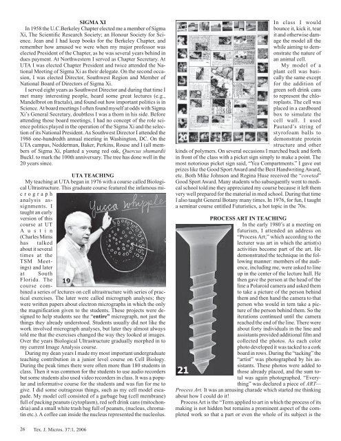
give. I did some outrageous things, such as my cell model escapade.<br />
My model cell consisted <strong>of</strong> a garbage bag (cell membrane)<br />
full <strong>of</strong> packing peanuts (cytoplasm), red s<strong>of</strong>t drink cans (mitochondria)<br />
and a small white trash bag full <strong>of</strong> peanuts, (nucleus, chromatin<br />
etc.). A c<strong>of</strong>fee can inside the nucleus represented the nucleolus.<br />
26 Tex. J. Micros. 37:1, 2006<br />
In class I would<br />
bounce it, kick it, tear<br />
it and otherwise damage<br />
the model all the<br />
while aiming to demonstrate<br />
the nature <strong>of</strong><br />
an animal cell.<br />
My model <strong>of</strong> a<br />
plant cell was basically<br />
the same except<br />
for the addition <strong>of</strong><br />
green s<strong>of</strong>t drink cans<br />
to represent the chloroplasts.<br />
The cell was<br />
placed in a cardboard<br />
box to simulate the<br />
cell wall. I used<br />
Pautard’s string <strong>of</strong><br />
styr<strong>of</strong>oam balls to<br />
demonstrate protein<br />
structure and other<br />
kinds <strong>of</strong> polymers. On several occasions I marched back and forth<br />
in front <strong>of</strong> the class with a picket sign simply to make a point. The<br />
most notorious picket sign said, “Yea Compartments.” I gave out<br />
prizes like the Good Sport Award and the Best Handwriting Award,<br />
etc. Both Mike Johnson and Regina Huse received the “coveted”<br />
Good Sport Award. Many students who subsequently went to medical<br />
school told me they appreciated my course because it left them<br />
very well prepared for the material in med school. During that time<br />
I also taught General Botany many times. In 1976, for fun, I taught<br />
a seminar course entitled Futuristics, a hot topic in the 70s.<br />
PROCESS ART IN TEACHING<br />
In the early 1980’s at a meeting on<br />
futurism, I attended an address on<br />
“Process Art;” which according to the<br />
lecturer was art in which the artist(s)<br />
activities become part <strong>of</strong> the art. He<br />
demonstrated the technique in the following<br />
manner: members <strong>of</strong> the audience,<br />
including me, were asked to line<br />
up in the center <strong>of</strong> the lecture hall. He<br />
then gave the person at the head <strong>of</strong> the<br />
line a Polaroid camera and asked them<br />
to take a picture <strong>of</strong> the person behind<br />
them and then hand the camera to that<br />
person who would in tern take a picture<br />
<strong>of</strong> the person behind them. So the<br />
iterations continued until the camera<br />
reached the end <strong>of</strong> the line. There were<br />
about forty individuals in the line and<br />
assistants provided additional film and<br />
collected the photos. As each color<br />
photo developed it was tacked to a cork<br />
board in rows. During the “tacking” the<br />
“artist” was photographed by his assistants.<br />
These photos were added to<br />
those already placed, and the sum total<br />
was again photographed. “Everything”<br />
was declared a piece <strong>of</strong> ART—<br />
Process Art. It was an amusing charade which started me thinking<br />
about how I could do it!<br />
Process Art is the “Term applied to art in which the process <strong>of</strong> its<br />
making is not hidden but remains a prominent aspect <strong>of</strong> the completed<br />
work so that a part or even the whole <strong>of</strong> its subject is the


