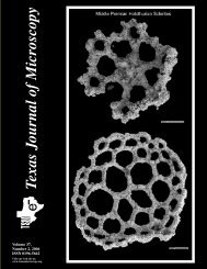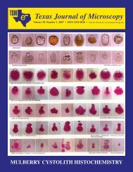
Texas Journal of Microscopy Texas Journal of Microscopy
Texas Journal of Microscopy Texas Journal of Microscopy
Texas Journal of Microscopy Texas Journal of Microscopy

Create successful ePaper yourself
Turn your PDF publications into a flip-book with our unique Google optimized e-Paper software.
Abstracts<br />
BIOLOGICAL SCIENCES<br />
SPRING 2006<br />
VISUALIZATION OF NS5A ACTIVITY VIA<br />
FLUORESCENCE MICROSCOPY. ALEJANDRO D’BROT,<br />
GIRIDHAR AKKARAJU. Department <strong>of</strong> Biology, <strong>Texas</strong> Christian<br />
University, Fort Worth, <strong>Texas</strong> 76129.<br />
Our research goal is to identify potential inhibitors <strong>of</strong> NS5A, a<br />
Hepatitis C virus (HCV) protein that inhibits the antiviral response<br />
in cells. If NS5A can be inhibited, the antiviral response can be<br />
restored in infected patients, and with the use <strong>of</strong> HCV specific<br />
drugs, HCV can be eliminated from the patients system. An IFNbeGFP<br />
+ NS5A reporter cell was constructed to assay NS5A<br />
inhibition. IFNb is one <strong>of</strong> the many genes inhibited by NS5A, so<br />
by using an IFNb promoter attached to an eGFP gene we can test<br />
whether or not the IFNb promoter is being turned on during viral<br />
infection, and consequently, whether or not NS5A is inhibiting it.<br />
If NS5A is active, it will inhibit the IFNb promoter and prevent the<br />
cells from expressing eGFP (and consequently from glowing green<br />
under a fluorescent microscope). If NS5A is inhibited, the cells<br />
will express eGFP and glow green. A stable cell line that exhibits<br />
correct gene expression has been created, and the cells were treated<br />
with plant extracts (provided by Dr. Manfred Reinecke, TCU), which<br />
had been shown to possess antiviral properties. Therefore, there is a<br />
chance that the compounds in plant extracts might also inhibit NS5A.<br />
CYPSELAR EPIDERMAL MORPHOLOGY OF<br />
PSEUDOGNAPHALIUM AND RELATED GENERA<br />
(ASTERACEAE). ANDREW WALTKE and GUY NESOM, Department<br />
<strong>of</strong> Biology, <strong>Texas</strong> Christian University, Fort Worth, TX<br />
76109; Botanical Research Institute <strong>of</strong> <strong>Texas</strong>, Fort Worth, TX 76102.<br />
Several primary types <strong>of</strong> cypselar (fruit) epidermal morphology<br />
are found among six putatively closely related genera, as seen under<br />
SEM <strong>of</strong> 25 Asteraceae species. The primarily New World genera<br />
Pseudognaphalium, Achyrocline, and Stenophalium have elongate,<br />
imbricate epidermal cells (distal end overlapping the base <strong>of</strong><br />
the adjacent cell). Variation occurs among species in the degree<br />
that the distal ends are raised, and the surface may appear (1) smooth<br />
(distal ends appressed), (2) roughened by imbricate papillae (distal<br />
ends slightly raised above the base <strong>of</strong> the adjacent cell), or (3)<br />
distinctly imbricate-papillate (distal ends greatly raised above the<br />
base <strong>of</strong> the adjacent cell). Among the Old World genera, species <strong>of</strong><br />
Laphangium and Homognaphalium have cypselar surfaces with<br />
rectangular epidermal cells producing irregularly scattered, 3-celled,<br />
elongate, glandular papillae. The large genus Helichrysum (ca. 500+<br />
species, restricted to the Old World) is polymorphic, with some<br />
species (including the type <strong>of</strong> the genus) producing glandular papillate<br />
surfaces, other species producing smooth or imbricate-papillate<br />
surfaces. Laphangium luteoalbum, which has glandular<br />
achenes, occurs in North America but probably is native to Eurasia,<br />
where its closest relatives are found. The taxonomic implication<br />
<strong>of</strong> these morphologies is that continued recognition <strong>of</strong> the New<br />
World genera may necessitate recognition <strong>of</strong> segregate genera<br />
among the species <strong>of</strong> Helichrysum. Alternatively, the New World<br />
genera might be included within Helichrysum.<br />
8 Tex. J. Micros. 37:1, 2006<br />
SEED VARIATION IN GAILLARDIA PULCHELLA FOUG.<br />
OF CENTRAL TEXAS. BRITT LEIGH BENEDICT*, ANN E.<br />
RUSHING, and DARRELL S. VODOPICH, Department <strong>of</strong> Biology,<br />
Baylor University, Waco, TX 76798-7388.<br />
Seeds <strong>of</strong> 20 field-collected Gaillardia pulchella populations were<br />
evaluated to determine if significant variation <strong>of</strong> seed characteristics<br />
existed either among or within populations and to detect relationships<br />
between those characteristics. Seeds were examined using<br />
both light and scanning electron microscopy. Seed characteristics<br />
studied were: total length, base length, base width, hair length,<br />
wing length, wing width, and wing number. The characteristics<br />
with the most within-populations variation were base length, base<br />
width, wing length, and hair length. Among populations, no evident<br />
north-south or east-west trends were found in the traits that<br />
showed the most variation within populations. No significant correlations<br />
were found between seed characteristics.<br />
TECHNIQUE FOR RETAINING THE MORPHOLOGY OF<br />
CELLS GROWN ON A POLYCAPROLACTONE SCAF-<br />
FOLD. DONGMEI FAN, GIRIDHAR AKKARAJU, JEFFERY L<br />
COFFER, ERNEST F. COUCH, Dept. <strong>of</strong> Chemistry and Dept. <strong>of</strong><br />
Biology, <strong>Texas</strong> Christian University, Fort Worth, TX 76129.<br />
There is great interest in tissue engineering for the production <strong>of</strong><br />
bone and other products. To this end cells can be grown on scaffolds<br />
in order to create a matrix similar to that found in nature. The<br />
goal is to induce cells to not only grow, but to differentiate; in the<br />
case <strong>of</strong> bone, into osteocytes that and produce an extracellular<br />
matrix. In this study mouse stromal cell were seeded on micr<strong>of</strong>ibers<br />
made in our laboratory from polycaprolactone, a nontoxic biodegradable<br />
polymer. After allowing the cells to proliferate they were<br />
fixed in a mixture <strong>of</strong> glutaraldehyde and paraformaldehyde in a<br />
phosphate buffer. They were then washed in buffer and post-fixed<br />
in OsO 4 . The scaffold and cells were then dehydrated through increasing<br />
concentrations <strong>of</strong> ethanol. This was followed by three<br />
changes <strong>of</strong> propylene oxide. This was a critical step because the<br />
propylene oxide removed the scaffold but left the cells in their original<br />
elongated shape. Also, the cells clung together forming a thin<br />
mat. A portion <strong>of</strong> the cells were flat embedded in Araldite 502 for<br />
TEM and some <strong>of</strong> the cells were returned to ethanol and dried with<br />
HMDS for SEM. A whole mount was made from some <strong>of</strong> the remaining<br />
cells. The whole mount was then observed and photographed<br />
with phase-contrast optics. Both light and electron microscopy<br />
showed well-preserved cells, which retained their original<br />
shape. This technique will be very useful in following cellular<br />
changes over time and their response to growth factors.<br />
DETERMINATION OF EGGSHELL MICROSTRUCTURAL<br />
CHARACTERSICTS AND ASSOCIATED PHYSIOLOGI-<br />
CAL PROFILES IN MG-VACCINATED EGG-LAYING<br />
CHICKENS. SARAH B. MAY and SANDRA L. WEST-<br />
MORELAND. The Center for Electron <strong>Microscopy</strong>, University <strong>of</strong><br />
<strong>Texas</strong> at Arlington, Arlington, <strong>Texas</strong> 76019.<br />
This experiment determined the effect <strong>of</strong> vaccination <strong>of</strong> commercial<br />
layers with F-strain Mycoplasma gallisepticum on eggshell<br />
thickness. The experiment involved multiple variables in addition<br />
to the vaccination, including three different diets and two<br />
ages <strong>of</strong> lay. Cross-section micrographs were taken on each eggshell<br />
and thickness measurements were made. These data were sta-