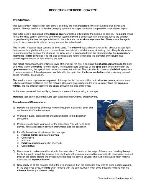
DISSECTION EXERCISE: COW EYE Introduction:
DISSECTION EXERCISE: COW EYE Introduction:
DISSECTION EXERCISE: COW EYE Introduction:

You also want an ePaper? Increase the reach of your titles
YUMPU automatically turns print PDFs into web optimized ePapers that Google loves.
<strong>Introduction</strong>:<br />
<strong>DISSECTION</strong> <strong>EXERCISE</strong>: <strong>COW</strong> <strong>EYE</strong><br />
The eyes contain receptors for light stimuli, and they are well protected by the surrounding skull bones and<br />
eyelids. The eye itself is a hollow ball, roughly spherical in shape. Its wall is composed of three distinct layers.<br />
The outer layer is composed of the fibrous layer consisting of two parts: the sclera and cornea. The sclera which<br />
forms the white portion of the eye and the transparent cornea is continuous with the sclera forms the anterior<br />
bulge where light enters the eye. Attached to the sclera are the extrinsic eye muscles. These move the eye in<br />
order to focus on objects without having to move the entire head.<br />
The (middle) Vascular layer consists of three parts. The choroid coat, a black layer, which absorbs excess light<br />
that passes through the retina and contains blood vessels tat nourish the eye. Anteriorly, the ciliary body forms a<br />
ring of muscle that controls the shape of the lens, which is suspended from the ciliary body by the suspensory<br />
ligaments (ciliary zonules). The iris also contracts and relaxes changing the diameter of the pupil thus<br />
controlling the amount of light entering the eye.<br />
The retina composes the inner Neural layer of the wall of the eye. It contains the photoreceptors: rods for blackand-white<br />
vision and cones for color vision. The neuron fibers coalesce at the optic disc, where they form the<br />
optic nerve exiting the eyeball and carries impulses to the brain. The optic disc is known as the blind spot since<br />
it has no receptors. A tiny depression just lateral to the optic disc, the fovea centralis contains densely packed<br />
cones for sharp, direct vision.<br />
The interior space or posterior segment of the eye behind the lens is filled with vitreous humor, a transparent<br />
jellylike substance that helps hold the retina in place and gives shape to the eye. A watery fluid, the aqueous<br />
humor, fills the anterior segment, the space between the lens and cornea.<br />
In this exercise we will be identifying these structures of the eye using a cow eye.<br />
Materials (per pair of students): Cow eye, dissection instruments, dissection tray<br />
Procedure and Observations:<br />
1) Review the structures of the eye from the diagram in your text book and<br />
on the model of the human eye.<br />
2) Working in pairs, each partner should participate in the dissection<br />
procedure.<br />
3) Prepare yourself and your area for the dissection. You will need to be<br />
gloved, have a dissection tray with instruments and the specimen.<br />
4) Identify the exterior structures of the cow eye:<br />
a. Fibrous Tunic: Sclera and cornea<br />
b. Conjunctiva<br />
c. adipose<br />
d. Extrinsic muscles (may be attached)<br />
e. Optic nerve<br />
5) Use a razor to make a small incision in the clear, about 5 mm from the edge of the cornea. Holding the eye<br />
firmly, but gently insert the scissors (the blunt side of the scissors should be inserted) into this incision and cut<br />
through the sclera around the eyeball while holding the cornea upward. The fluid that exudes when making<br />
this cut is the aqueous humor.<br />
6) Now gently lift off the anterior part of the eye and place it on the dissecting tray with its inner surface upward.<br />
In the preserved eyes, the lens often remains with the cornea, but in fresh eyes it usually remains with the<br />
vitreous humor (or vitreous body).<br />
Dissection Exercise: Cow Eye, page 135
7) Examine the anterior portion. Locate the lens and ciliary body (thickened black ring). Remove the lens (as<br />
applicable) from the ciliary body by gently cutting the tiny suspensory ligaments. Once removed, the iris<br />
and pupil can be seen clearly. There may be a little aqueous humor remaining next to the inner surface of the<br />
cornea.<br />
Observe the color of the iris and the shape of the pupil in your specimen and your neighbors specimen, what<br />
do you notice about the color of a cow’s eyes? How does this compare with human eye color and the shape<br />
of our pupil?<br />
8) There are two spaces within the eye. The anterior segment is the space between the cornea and lens. It is<br />
filled with aqueous humor. The posterior segment is the space between the lens and the retina which is<br />
filled with vitreous humor.<br />
Observe the vitreous humor within the posterior segment of the eyeball. Looking through the vitreous humor,<br />
the beige retina held in place against the posterior wall of the eyeball. Identify the blind spot or the optic<br />
disc. The fovea centralis is a region of high acuity densely populated with cones for color vision. The cow<br />
has only black and white vision so this is not found as part of the cow eye retina.<br />
9) Pour the vitreous humor onto the dissecting pan and note its jellylike consistency. The delicate retina easily<br />
separates from the underlying choroid but it is attached at the blind spot. (The junction of the optic nerve with<br />
the retina).<br />
10) Note the iridescent portion of the choroid layer; it is a modified choroid called the tapetum lucidum which<br />
aids dim-light vision by reflecting light that has passed through the retina back through the retina. This<br />
iridescence causes animals’ eyes to ‘shine’ at night by reflecting the light back to its light source.<br />
11) Complete your lab notebook dissection steps and answer the analysis questions.<br />
Once you have completed all of the identifications, dispose of the eye as directed by your instructor.<br />
Dissection Exercise: Cow Eye, page 136