JERZY TRZCINSKI, MAREK WRÓBEL AND LESZEK KIESZCZYNSKI
JERZY TRZCINSKI, MAREK WRÓBEL AND LESZEK KIESZCZYNSKI
JERZY TRZCINSKI, MAREK WRÓBEL AND LESZEK KIESZCZYNSKI

Create successful ePaper yourself
Turn your PDF publications into a flip-book with our unique Google optimized e-Paper software.
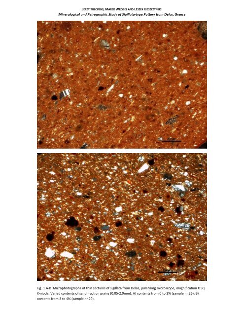
<strong>JERZY</strong> TRZCIŃSKI, <strong>MAREK</strong> <strong>WRÓBEL</strong> <strong>AND</strong> <strong>LESZEK</strong> KIESZCZYŃSKI<br />
Mineralogical and Petrographic Study of Sigillata-type Pottery from Delos, Greece<br />
Fig. 1.A-B Microphotographs of thin sections of sigillata from Delos, polarizing microscope, magnification X 50,<br />
X-nicols. Varied contents of sand fraction grains (0.05-2.0mm): A) contents from 0 to 2% (sample nr 26); B)<br />
contents from 3 to 4% (sample nr 29).
<strong>JERZY</strong> TRZCIŃSKI, <strong>MAREK</strong> <strong>WRÓBEL</strong> <strong>AND</strong> <strong>LESZEK</strong> KIESZCZYŃSKI<br />
Mineralogical and Petrographic Study of Sigillata-type Pottery from Delos, Greece<br />
Fig. 1.C-D Microphotographs of thin sections of sigillata from Delos, polarizing microscope, magnification X 50,<br />
X-nicols. Varied contents of sand fraction grains (0.05-2.0mm): C) contents from 5 to 7% (sample nr 40); D)<br />
contents from 8 to 15% (sample nr 24).
A<br />
B<br />
<strong>JERZY</strong> TRZCIŃSKI, <strong>MAREK</strong> <strong>WRÓBEL</strong> <strong>AND</strong> <strong>LESZEK</strong> KIESZCZYŃSKI<br />
Mineralogical and Petrographic Study of Sigillata-type Pottery from Delos, Greece<br />
Fig. 2. Microphotographs of thin sections of sigillata from Delos, polarizing microscope, X-nicols. Fragments of<br />
polycrystalline quartz (indicated by arrows): A) sample nr 24, magnification X 100; B) sample nr 25,<br />
magnification X 200.
A<br />
B<br />
<strong>JERZY</strong> TRZCIŃSKI, <strong>MAREK</strong> <strong>WRÓBEL</strong> <strong>AND</strong> <strong>LESZEK</strong> KIESZCZYŃSKI<br />
Mineralogical and Petrographic Study of Sigillata-type Pottery from Delos, Greece<br />
Fig. 3. Microphotographs of thin sections of sigillata from Delos, polarizing microscope, X-nicols. Grains of Kfeldspars<br />
and plagioclases (indicated by arrows): A) sample nr 10, magnification X 80; B) sample nr 35,<br />
magnification X 200.
A<br />
B<br />
<strong>JERZY</strong> TRZCIŃSKI, <strong>MAREK</strong> <strong>WRÓBEL</strong> <strong>AND</strong> <strong>LESZEK</strong> KIESZCZYŃSKI<br />
Mineralogical and Petrographic Study of Sigillata-type Pottery from Delos, Greece<br />
Fig. 4 Microphotographs of thin sections of sigillata from Delos, polarizing microscope, X-nicols. Pyroclastic<br />
fragments (indicated by arrows): A) sample nr 18, magnification X 160; B) sample nr 35, magnification X 500.
A<br />
B<br />
<strong>JERZY</strong> TRZCIŃSKI, <strong>MAREK</strong> <strong>WRÓBEL</strong> <strong>AND</strong> <strong>LESZEK</strong> KIESZCZYŃSKI<br />
Mineralogical and Petrographic Study of Sigillata-type Pottery from Delos, Greece<br />
Fig. 5 Microphotographs of thin sections of sigillata from Delos, polarizing microscope, X-nicols, magnification<br />
X 200. Amphibole grains (indicated by arrows): A) sample 38; B) sample 39.
A<br />
B<br />
<strong>JERZY</strong> TRZCIŃSKI, <strong>MAREK</strong> <strong>WRÓBEL</strong> <strong>AND</strong> <strong>LESZEK</strong> KIESZCZYŃSKI<br />
Mineralogical and Petrographic Study of Sigillata-type Pottery from Delos, Greece<br />
Fig. 6 Microphotographs of thin sections of sigillata from Delos, polarizing microscope, X-nicols. Limestone<br />
fragments (indicated by arrows): A) sample nr 38, magnification X 100; B) sample nr 39, magnification X 100.
A<br />
B<br />
<strong>JERZY</strong> TRZCIŃSKI, <strong>MAREK</strong> <strong>WRÓBEL</strong> <strong>AND</strong> <strong>LESZEK</strong> KIESZCZYŃSKI<br />
Mineralogical and Petrographic Study of Sigillata-type Pottery from Delos, Greece<br />
20 µm<br />
20 µm<br />
Fig. 7. Microphotographs of thin sections of sigillata from Delos, scanning electron microscope, BSE (back-scattered<br />
electrons) mode, magnification X 600: A) sample nr 2, B) sample nr 9. Large and medium angular and poorly rounded, often<br />
cracked grey quartz grains are visible (marked by arrows). Fine dark aggregates with lighter rim are embedded in the matrix<br />
consisting of elongated and platelet grains of clay minerals. The body is topped by a layer of fine-grained slip.
A<br />
B<br />
<strong>JERZY</strong> TRZCIŃSKI, <strong>MAREK</strong> <strong>WRÓBEL</strong> <strong>AND</strong> <strong>LESZEK</strong> KIESZCZYŃSKI<br />
Mineralogical and Petrographic Study of Sigillata-type Pottery from Delos, Greece<br />
10 µm<br />
10 µm<br />
Fig. 8. Microphotographs of thin sections of sigillata from Delos, scanning electron microscope, BSE mode,<br />
magnification X 1800: A) sample nr 2; B) sample nr 11. Around quartz grains clearly visible lighter and dissolved<br />
halo (marked by arrows), forming together with clay grains the fixed structure of the body. Both in the slip and<br />
in the matrix of the body (B) numerous fine light grains enriched in Fe.
A<br />
B<br />
<strong>JERZY</strong> TRZCIŃSKI, <strong>MAREK</strong> <strong>WRÓBEL</strong> <strong>AND</strong> <strong>LESZEK</strong> KIESZCZYŃSKI<br />
Mineralogical and Petrographic Study of Sigillata-type Pottery from Delos, Greece<br />
50 µm<br />
0,1 mm<br />
Fig. 9. Microphotographs of thin sections of sigillata from Delos, scanning electron microscope, BSE mode: A) sample nr 9, magnification X 330; B) sample<br />
nr 5, magnification X 160. Fragments of clayey rock, marked by arrows in photograph B, consist mainly of a mass of aggregated clayey crystals in form of<br />
platelets several to below twenty micrometers long. In this mass grains of quartz and other minerals from several to some tens of micrometers in size are<br />
embedded, marked by arrows in photograph A. The rock fragments are surrounded by empty rims resulting from differential thermal expansion and water<br />
loss at firing synæresis. In the clayey matrix of the body quartz grains, often cracked are embedded (marked by arrow in photograph A) and dark, oval<br />
aggregates (marked by arrows in photograph B). Clayey matrix forms a lattice structure.
A<br />
B<br />
<strong>JERZY</strong> TRZCIŃSKI, <strong>MAREK</strong> <strong>WRÓBEL</strong> <strong>AND</strong> <strong>LESZEK</strong> KIESZCZYŃSKI<br />
Mineralogical and Petrographic Study of Sigillata-type Pottery from Delos, Greece<br />
Fig. 10 Microphotographs of thin sections of sigillata from Delos, polarizing microscope, X-nicols. Fragments of<br />
clayey rocks, indicated by arrows: A) sample nr 21, magnification X 500; B) sample nr 37, magnification X 100.
A<br />
B<br />
<strong>JERZY</strong> TRZCIŃSKI, <strong>MAREK</strong> <strong>WRÓBEL</strong> <strong>AND</strong> <strong>LESZEK</strong> KIESZCZYŃSKI<br />
Mineralogical and Petrographic Study of Sigillata-type Pottery from Delos, Greece<br />
200 µm<br />
20 µm<br />
Fig. 11. Microphotographs of thin sections of sigillata from Delos, scanning electron microscope, BSE mode: A) sample nr 13, magnification X 80; B) sample nr<br />
40, magnification X 500. In the clayey mass large grains of feldspars (marked by arrows in photograph A), grey grains of quartz of various size, secondary<br />
aggregates and large isometric and anisometric pores – black spaces are visible. In photograph B in the clayey matrix of the body with high contents of large<br />
aggregates of mica platelets (marked by arrows) a single grain of feldspar is visible (marked by arrow).
A B A<br />
B<br />
<strong>JERZY</strong> TRZCIŃSKI, <strong>MAREK</strong> <strong>WRÓBEL</strong> <strong>AND</strong> <strong>LESZEK</strong> KIESZCZYŃSKI<br />
Mineralogical and Petrographic Study of Sigillata-type Pottery from Delos, Greece<br />
20 20 µm<br />
µm<br />
20 µm<br />
Fig. 12. Microphotographs of thin sections of sigillata from Delos, scanning electron microscope, BSE mode, magnification X<br />
330: A) sample nr 2; B) sample nr 5. In lower part of photograph A, large horizontal platelet grains of kaolinite, marked by<br />
arrows are visible. In the matrix grains of quartz (grey) and in the right top corner a group of dark aggregates are<br />
embedded. In photograph B a long aggregate of mica (marked by arrow) and oval dark aggregates are visible. An isolated<br />
grain of quartz is set in slip (marked by arrow).
A<br />
B<br />
<strong>JERZY</strong> TRZCIŃSKI, <strong>MAREK</strong> <strong>WRÓBEL</strong> <strong>AND</strong> <strong>LESZEK</strong> KIESZCZYŃSKI<br />
Mineralogical and Petrographic Study of Sigillata-type Pottery from Delos, Greece<br />
Fig. 13. Microphotographs of thin sections of sigillata from Delos, polarizing microscope, X-nicols. Flakes of<br />
micas (elongated platelet grains): A) sample nr 20, magnification X 80; B) sample nr 38, magnification X 200.
A<br />
B<br />
<strong>JERZY</strong> TRZCIŃSKI, <strong>MAREK</strong> <strong>WRÓBEL</strong> <strong>AND</strong> <strong>LESZEK</strong> KIESZCZYŃSKI<br />
Mineralogical and Petrographic Study of Sigillata-type Pottery from Delos, Greece<br />
200 µm<br />
200 µm<br />
Fig. 14. Microphotographs of thin sections of sigillata from Delos, scanning electron microscope, BSE mode,<br />
magnification X 80: A) sample nr 5; B) sample nr 9. Dark secondary aggregates 20-50 μm in size are marked by<br />
arrows. They are evenly distributed in poorly rotation oriented clay mass In the matrix embedded large grains<br />
of quartz (grey) and large anisometric pores (black) are visible. In photograph B, a large fragment of clayey rock<br />
(marked by arrow) is visible in upper central part.
A<br />
B<br />
<strong>JERZY</strong> TRZCIŃSKI, <strong>MAREK</strong> <strong>WRÓBEL</strong> <strong>AND</strong> <strong>LESZEK</strong> KIESZCZYŃSKI<br />
Mineralogical and Petrographic Study of Sigillata-type Pottery from Delos, Greece<br />
10 µm<br />
20 µm<br />
Fig. 15. Microphotographs of thin sections of from Delos, scanning electron microcope, BSE mode: A) sample nr 5, magnification X 600; B) sample nr 9,<br />
magnification X 1800. In photograph A in the centre a secondary dark aggregate, 40 μm in size is visible. Its centre is sintered (upper arrow), surrounded<br />
by void due to thermal differential expension (central arrow) and a rim with fluidal structure (lower arrow). Around the aggregate lattice structure of<br />
clayey matrix is well visible, with large rotation conformingly oriented platelet crystals of kaolinite. In photograph B a single dark aggregate consisting of a<br />
rim (marked by upper arrow) and a lighter core, with more Fe (lower arrow). Space between the core and the rim is very porous (black areas).
A<br />
B<br />
<strong>JERZY</strong> TRZCIŃSKI, <strong>MAREK</strong> <strong>WRÓBEL</strong> <strong>AND</strong> <strong>LESZEK</strong> KIESZCZYŃSKI<br />
Mineralogical and Petrographic Study of Sigillata-type Pottery from Delos, Greece<br />
Fig. 16 Microphotographs of thin sections of sigillata from Delos, polarizing microscope, X-nicols. Carbonateiron-clayey<br />
aggregates, indicated by arrows: A) sample nr 27, magnification X 50; B) sample nr 23,<br />
magnification X 200.
A<br />
B<br />
<strong>JERZY</strong> TRZCIŃSKI, <strong>MAREK</strong> <strong>WRÓBEL</strong> <strong>AND</strong> <strong>LESZEK</strong> KIESZCZYŃSKI<br />
Mineralogical and Petrographic Study of Sigillata-type Pottery from Delos, Greece<br />
Fig. 17. Microphotographs of thin sections of sigillata from Delos, polarizing microscope, X-nicols. Zircon grains<br />
(indicated by arrows): sample nr 23, magnification X 200; B) sample nr 34, magnification X 500.
A<br />
B<br />
<strong>JERZY</strong> TRZCIŃSKI, <strong>MAREK</strong> <strong>WRÓBEL</strong> <strong>AND</strong> <strong>LESZEK</strong> KIESZCZYŃSKI<br />
Mineralogical and Petrographic Study of Sigillata-type Pottery from Delos, Greece<br />
Fig. 18 Microphotographs of thin sections of sigillata from Delos, polarizing microscope, X-nicols. Aggregate<br />
rims (indicated by arrows): sample nr 26, magnification X 100; B) sample nr 36, magnification X 200.
A<br />
B<br />
<strong>JERZY</strong> TRZCIŃSKI, <strong>MAREK</strong> <strong>WRÓBEL</strong> <strong>AND</strong> <strong>LESZEK</strong> KIESZCZYŃSKI<br />
Mineralogical and Petrographic Study of Sigillata-type Pottery from Delos, Greece<br />
10 µm<br />
10 µm<br />
Fig. 19 Crophotographs of thin sections of sigillata from Delos, scanning electron microscope, BSE mode,<br />
magnification X 1800: A) sample nr 5; B) sample nr 1. The body is topped by unstructured and finely grained slip<br />
layer up to 20 μm thick with isolated quartz grains 2-3 μm in size, marked by arrows and dispersed very fine<br />
light grains, rich in Fe. The slip shows cracks parallel to surface due to shrinkage stresses.