Tropical phytopathogens 1 - Plant Pathology & Quarantine
Tropical phytopathogens 1 - Plant Pathology & Quarantine
Tropical phytopathogens 1 - Plant Pathology & Quarantine

Create successful ePaper yourself
Turn your PDF publications into a flip-book with our unique Google optimized e-Paper software.
<strong>Tropical</strong> <strong>phytopathogens</strong> 1: Pseudocercospora punicae<br />
<strong>Plant</strong> <strong>Pathology</strong> & <strong>Quarantine</strong><br />
Phengsintham P 1,2 , Chukeatirote E 1 , McKenzie EHC 3 , Hyde KD 1* and Braun U 4<br />
1School<br />
of Science, Mae Fah Luang University, Chiang Rai 57100. Thailand<br />
2<br />
Biology Department, Faculty of Sciences, National University of Laos<br />
3<br />
Landcare Research, Private Bag 92170, Auckland, New Zealand<br />
4<br />
Martin–Luther–Universität, Institut für Biologie, Bereich Geobotanik und Botanischer Garten, Herbarium, Neuwerk 21<br />
D–06099 Halle/S. Germany<br />
Phengsintham P, Chukeatirote E, McKenzie EHC, Hyde KD, Braun U. 2011 – <strong>Tropical</strong><br />
phythopathogens 1: Pseudocercospora punicae. <strong>Plant</strong> <strong>Pathology</strong> & <strong>Quarantine</strong> 1(1), 1–6.<br />
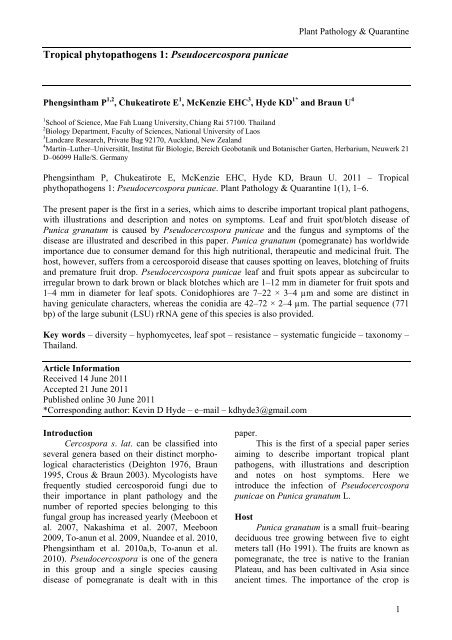
The present paper is the first in a series, which aims to describe important tropical plant pathogens,<br />
with illustrations and description and notes on symptoms. Leaf and fruit spot/blotch disease of<br />
Punica granatum is caused by Pseudocercospora punicae and the fungus and symptoms of the<br />
disease are illustrated and described in this paper. Punica granatum (pomegranate) has worldwide<br />
importance due to consumer demand for this high nutritional, therapeutic and medicinal fruit. The<br />
host, however, suffers from a cercosporoid disease that causes spotting on leaves, blotching of fruits<br />
and premature fruit drop. Pseudocercospora punicae leaf and fruit spots appear as subcircular to<br />
irregular brown to dark brown or black blotches which are 1–12 mm in diameter for fruit spots and<br />
1–4 mm in diameter for leaf spots. Conidophiores are 7–22 × 3–4 µm and some are distinct in<br />
having geniculate characters, whereas the conidia are 42–72 × 2–4 µm. The partial sequence (771<br />
bp) of the large subunit (LSU) rRNA gene of this species is also provided.<br />
Key words – diversity – hyphomycetes, leaf spot – resistance – systematic fungicide – taxonomy –<br />
Thailand.<br />
Article Information<br />
Received 14 June 2011<br />
Accepted 21 June 2011<br />
Published online 30 June 2011<br />
*Corresponding author: Kevin D Hyde – e–mail – kdhyde3@gmail.com<br />
Introduction<br />
Cercospora s. lat. can be classified into<br />
several genera based on their distinct morphological<br />
characteristics (Deighton 1976, Braun<br />
1995, Crous & Braun 2003). Mycologists have<br />
frequently studied cercosporoid fungi due to<br />
their importance in plant pathology and the<br />
number of reported species belonging to this<br />
fungal group has increased yearly (Meeboon et<br />
al. 2007, Nakashima et al. 2007, Meeboon<br />
2009, To-anun et al. 2009, Nuandee et al. 2010,<br />
Phengsintham et al. 2010a,b, To-anun et al.<br />
2010). Pseudocercospora is one of the genera<br />
in this group and a single species causing<br />
disease of pomegranate is dealt with in this<br />
paper.<br />
This is the first of a special paper series<br />
aiming to describe important tropical plant<br />
pathogens, with illustrations and description<br />
and notes on host symptoms. Here we<br />
introduce the infection of Pseudocercospora<br />
punicae on Punica granatum L.<br />
Host<br />
Punica granatum is a small fruit–bearing<br />
deciduous tree growing between five to eight<br />
meters tall (Ho 1991). The fruits are known as<br />
pomegranate, the tree is native to the Iranian<br />
Plateau, and has been cultivated in Asia since<br />
ancient times. The importance of the crop is<br />
1
Fig. 1 – Leaf and fruit spots caused by Pseudocercospora punicae on Punica granatum.<br />
rising due to worldwide increasing consumer<br />
demand for its high nutrition and use as<br />
therapeutic foods (Palou et al. 2009). It is also<br />
used in traditional medicine (http://en.wikipe<br />
dia.org/wiki/Pomegranate).<br />
Pala et al. (2009) studied fungal pathogens<br />
causing postharvest decay of pomegranate<br />
and concluded that Alternaria alternata, Coniella<br />
granati and Aspergillus niger were the<br />
major biotic diseases. Fruit cracks, sunburn and<br />
hail damage were the most commonly detected<br />
abiotic diseases. Pomegranate’s leaf and fruit<br />
spots can be caused by various organisms such<br />
as: Colletotrichum gloeosporioides, Sphaceloma<br />
punicae, Cercospora punicae, Drechslera<br />
sp. and Phomopsis sp., which take a heavy toll<br />
on the crop yield and quality (Jamadar & Patil<br />
2007).<br />
2<br />
Cercospora punicae was first recorded in<br />
Japan by Hennings in 1906 (Chupp, 1954),<br />
while Petcharat and Kanjanamaneesathian (1989)<br />
recorded Cercospora punicae in southern<br />
Thailand, but without any description. Deighton<br />
(1976) redescribed this species and transferred<br />
it to the genus Pseudocercospora.<br />
Symptoms<br />
These are rather variable (Fig. 1). The<br />
affected fruits show small irregular black spots,<br />
which later coalesce into large spots measuring<br />
1–12 mm diam. Leaf spots/lesions are<br />
subcircular to irregular, 1–4 mm diam., at first<br />
brown, dingy gray to pale tan and eventually<br />
brown to dark brown at the margin.
Fig. 2 – Pseudocercospora punicae on Punica<br />
granatum: 1. Stroma with attached conidiophores.<br />
2. Conidiophore. 3. External hypha<br />
with attached conidiophores. 4–8. Conidia.<br />
Bars: = 10 µm.<br />
Taxonomy<br />
Pseudocercospora punicae (Henn.) Deighton,<br />
Mycol. Pap. 140: 151 (1976). Figs 2–3<br />
≡ Cercospora punicae Henn., Bot. Jahrb.<br />
genous Syst. 37: 165. 1906.<br />
Description – Caespituli/colonies amphigenous,<br />
conspicuous. Mycelium internal and<br />
external; internal hyphae branched, 2–3 µm<br />
wide (x = 2.2 µm), septate, constricted at the<br />
septa, distance between septa 6–10 µm ( x =<br />
8.8 µm), brownish or subhyaline, wall 0.3–0.5<br />
µm wide ( x = 0.34 µm), smooth, forming<br />
plate–like plectenchymatous stromatic hyphal<br />
aggregations; external hyphae branched, 2–4<br />
µm wide (x = 3 µm), septate, constricted at the<br />
septa, distance between septa 4–8 µm (x = 4.7<br />
µm), brownish or subhyaline, wall 0.3–0.5 µm<br />
wide (x = 0.43 µm), smooth. Stromata oval to<br />
ellipsoidal, 10–35 µm diam. ( x = 22.5 µm),<br />
brown to dark brown, stroma cells oval,<br />
ellipsoidal to angular, 3–9 µm wide ( x = 6.3<br />
µm), dark brown, wall 0.3–0.5 µm wide (x =<br />
0.45 µm), smooth. Conidiophores fasciculate or<br />
<strong>Plant</strong> <strong>Pathology</strong> & <strong>Quarantine</strong><br />
solitary, arising from stromata (4–40 per<br />
fascicle) and external mycelium, geniculate,<br />
branched, 7–22 × 3–4 µm ( x = 13.8 × 3.28<br />
µm), 0–1–septate, slightly constricted at the<br />
septa, distance between septa 5–17 µm long (x<br />
= 8.33 µm), uniformly pale to medium brown,<br />
paler and narrower towards the tip, wall 0.3–<br />
0.5 µm (x = 0.48 µm), smooth. Conidiogenous<br />
cells integrated, terminal, 7–17 × 3–4 µm (x =<br />
10 × 3.11 µm), apex obtuse; conidiogenous loci<br />
inconspicuous, unthickened, not darkened.<br />
Conidia solitary, obclavate, straight to slightly<br />
curved, 42–72 × 2–4 µm ( x = 54.23 × 2.46<br />
µm), 2–6–septate, pale olivaceous–brown, wall<br />
0.3–0.5 µm wide ( x = 0.36 µm), smooth, tip<br />
subacute, base truncate, hila 1.5–2 µm wide (x<br />
= 1.55 µm).<br />
Known hosts – Punica granatum L.<br />
(Lythraceae).<br />
Known distribution – Africa: Egypt,<br />
Ethiopia, Kenya, Mauritius, Mozambique,<br />
Sudan, Tanzania, Uganda, Zambia; Asia:<br />
Afghanistan; Cambodia, China, Hong Kong,<br />
India, Indonesia, Iran, Japan, Laos, Malaysia,<br />
Maldives, Nepal, Pakistan, Philippines, Saudi<br />
Arabia, Singapore, Taiwan, Thailand; Europe<br />
(Caucasus): Azerbaijan, Georgia; North<br />
America: Cuba, Dominican Rep., Guatemala,<br />
Jamaica, Panama, Puerto Rico, USA (FL, HI,<br />
TX), Virgin islands; South America: Brazil,<br />
Colombia, Venezuela (Crous & Braun 2003).<br />
Cultural characteristics – Mycelial colonies<br />
on PDA after three weeks at 25°C gray to<br />
dark gray, reaching 16–18 mm diam., hyphae<br />
2–9 µm wide (x = 5 µm), septate, constricted<br />
at the septa, distance between septa 5–26 µm<br />
(x = 11.37 µm), brownish or subhyaline, wall<br />
0.3–1 µm wide ( x = 0.61 µm), smooth.<br />
Conidiophores and conidia not formed in<br />
culture.<br />
Material examined – Thailand, Chiang<br />
Rai Province, Muang District, Ban Du Village,<br />
on leaf of Punica granatum (Lythraceae), 1<br />
July 2010, P. Phengsintham (MFLU10-0323).<br />
Culture (MFLUCC 11-0284), GenBank accession<br />
no JN107998; Laos, Xiangkhouang<br />
Province, Phonsavan Village, 26 April 2011, P.<br />
Phengsintham (NUOL P623).<br />
Remarks – The collection from Thailand<br />
differs from Pseudocercospora punicae as<br />
described by Hsieh & Goh (1990) in having<br />
geniculate conidiophores.<br />
3
Fig. 3 – Pseudocercospora punicae on Punica granatum from leaf spots: 1–2. Lesions on host<br />
leaves (1. upper surface and 2. lower surface). 3. Caespituli. 4. Stromata with attached<br />
conidiophores. 5–8. Conidia. 9. Culture. Bars: 1, 2, 9 = 10 mm. 4–8 = 10 µm.<br />
Table 1 Conidial size and septation of Pseudocercospora punicae isolates/strains.<br />
Species<br />
Conidia References<br />
Septa Size (µm)<br />
Cercospora punicae = Pseudocercospora punicae 2–8-septate 25–85 × 2.5–5 Chupp (1954)<br />
Pseudocercospora punicae 2–8-septate 18–85 × 2.5–5 Hsieh & Goh (1990)<br />
Pseudocercospora punicae 2–6-septate 42–72 × 2–4 Present study<br />
The leaf spots and fruits caused by this<br />
fungal species lead to reduced harvest and<br />
quality. The conidial septation and size of<br />
Pseudocercospora punicae are shown in Table<br />
1.<br />
Additionally, we describe here a partial<br />
sequence of the large subunit LSU (rRNA gene<br />
of P. punicae. For this, the genomic DNA from<br />
fungal mycelium was isolated using the pro-<br />
4<br />
tocol based on Cai et al. (1999). The incomplete<br />
sequence of the LSU rRNA operon was<br />
then amplified using the primers LROR and<br />
LR5 and sequenced using the same primers by<br />
SinoGenoMax Company limited, China for<br />
sequencing. The sequence obtained was 771 bp<br />
and is deposited in GenBank (JN107998). This<br />
sequence information can be useful for phylogenetic<br />
study in future work.
Management of the Disease<br />
Mancozeb 0.25 % sprayed at 15 days<br />
interval gives good control of the disease on<br />
leaves (http://agritech.tnau.ac.in/crop_protec<br />
tion/crop_prot_crop%20diseases_fruits_pomeg<br />
ranate.html).<br />
Two to three sprayings at 15 days interval<br />
of Dithane M-45 or Captan @ 2.5 g in one<br />
litre of water after fruit formation gives good<br />
control of the disease on fruit. (http://nhb.<br />
gov.in/bulletin_files/fruits/pomegranate/pom00<br />
2.pdf). Diseased fruits should be collected and<br />
destroyed.<br />
Acknowledgements<br />
The authors would like to thank the<br />
Mushroom Research Foundation (MRF) for<br />
financial support. Special thanks also go to the<br />
MRF organizers and members of Prof. KD<br />
Hyde’s laboratory, Mae Fah Luang University<br />
(MFLU) for their assistance. Cai Lei is<br />
sincerely thanked for laboratory facilities for<br />
molecular work. The Global Research Network<br />
for Fungal Biology and King Saud University<br />
are thanked for support.<br />
References<br />
Braun W. 1995 – A monograph of<br />
Cercosporella, Ramularia and allied<br />
genera (phytopathogenic hyphomycetes)<br />
Vol. I. IHW–Verling, Eching.<br />
Cai L, Hyde KD, Taylor PWJ, Weir B, Waller<br />
J, Abang MM, Zhang JZ, Yang YL,<br />
Phoulivong S, Liu ZY, Prihastuti H,<br />
Shivas RG, McKenzie EHC, Johnston,<br />
PR. 2009 – A polyphasic approach for<br />
studying Colletotrichum. Fungal Diversity<br />
39 185–206.<br />
Chupp C. 1954 – A monograph of the fungus<br />
genus Cercospora. Ithaca, New York.<br />
Crous PW, Braun U. 2003 – Mycosphaerella<br />
and its anamorphs: 1. Names published in<br />
Cercospora and Passalora. CBS Biodiversity<br />
Series 1, 1–569.<br />
Deighton FC. 1976 – Studies on Cercospora<br />
and allied genera. VI. Pseudocercospora<br />
Speg., Pantospora Cif. and Cercoseptoria<br />
Petr. Mycological Papers 140, 1–168.<br />
Hsieh WH, Goh TK. 1990 – Cercospora and<br />
similar fungi from Taiwan. Maw Chang<br />
Book Company, Taipei, Taiwan.<br />
<strong>Plant</strong> <strong>Pathology</strong> & <strong>Quarantine</strong><br />
Jamadar MM, Patil DR. 2007 – Bio–efficacy of<br />
new formulations against leaf /fruit spot<br />
on pomegranate. Karnataka Journal of<br />
Agricultural Sciences 20, 865–866.<br />
Meeboon J. 2009 – Diversity and phylogeny of<br />
true cercosporoid fungi from Northern<br />
Thailand. Ph.D. Thesis, Chiang Mai<br />
University, Thailand (Unpublished).<br />
Meeboon J, Hidayat I, To-anun C. 2007 –<br />
Annotated list of cercosporoid fungi in<br />
Northern Thailand. Journal of Agricultural<br />
Technology 3, 51–63.<br />
Nakashima C, Motohashi K, Meeboon J, To–<br />
anun C. 2007 – Studies on Cercospora<br />
and allied genera in Northern Thailand.<br />
Fungal Diversity 26, 257–270.<br />
Nuandee N, Kongtragoul P, Boonying W,<br />
Hidayat I, Miyamoto Y, Izumi Y,<br />
Akimitsu K, Nalumpang S. 2010 –<br />
Characterization of Carbendazim resistant<br />
Cercospora causal agent of lettuce<br />
leaf spots in Chiang Mai Province. KKU<br />
Research Journal 15, 1053–1060.<br />
Pala H, Tatli A, Yilmaz C, Özgüven AI. 2009 –<br />
Important diseases of pomegranate fruit<br />
and control possibilities in Turkey. Acta<br />
Horticultura (ISHS) 818, 285–290.<br />
Palou L, del Río MA. 2009 – Assessment of<br />
fungal pathogens causing postharvest<br />
decay of pomegranate in southeast Spain.<br />
Acta Hortuktura (ISHS) 818, 305–312.<br />
Petcharat V, Kajanamaneesathian M. 1989 –<br />
Species of plant pathogenic Cercospora<br />
in Southern Thailand. Thai Journal of<br />
Phytopathology 9, 23–27.<br />
Ho PH, 1991 – Flora of Vietnam V 2, 74.<br />
Phengsintham P, Chukeatirote E, Abd-Elsalam<br />
KA, Hyde KD, Braun U. 2010a –<br />
Cercospora and allied genera from Laos<br />
3. Cryptogamie, Mycologie 31, 305–322.<br />
Phengsintham P, Chukeatirote E, McKenzie<br />
EHC, Moslem MA, Hyde KD, Braun U.<br />
2010b – Two new species and a new<br />
record of cercosporoids from Thailand.<br />
Mycosphere 1, 205–212.<br />
To-anun C, Nguenhom J, Meeboon J, Hidayat I.<br />
2009 – Two fungi associated with<br />
necrotic leaflets of areca palm (Areca<br />
catechu). Mycological Progress 8, 115–<br />
121.<br />
To-anun C, Hidayat I, Meeboon J. 2010 –<br />
Cercospora cristellae, a new cerco-<br />
5
6<br />
sporoid fungus associated with weed<br />
Cristella parasitica from Northern<br />
Thailand. Journal of Agricultural<br />
Technology 6, 331–339.