Intranasal Tooth
Intranasal Tooth
Intranasal Tooth

Create successful ePaper yourself
Turn your PDF publications into a flip-book with our unique Google optimized e-Paper software.
<strong>Intranasal</strong> tooth<br />
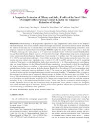
Fig. 2 A gross view of the extracted tooth revealing some<br />
attached mucosa and dental caries.<br />
DISCUSSION<br />
<strong>Intranasal</strong> teeth are rare. In 1979, Smith et al. reported<br />
2 cases and found 27 well-documented cases in the literature<br />
since the original description of intranasal teeth in<br />
1897 1 . In 2001, Gupta et al. reported one case of an<br />
intranasal tooth in a 4-year-old boy with a repair for cleft<br />
lip and alveolus, and identified another 20 cases 2 . Since<br />
then, an additional 28 cases have been reported 3-11 , with<br />
approximately 78 cases of intranasal teeth reported. Most<br />
of the articles are case reports and only two larger series of<br />
consecutive cases have been reported 3,12 .<br />
This literature survey revealed that the age of detection<br />
of intranasal teeth ranged from 3 to 71 years; approximately<br />
half of these cases were discovered before adulthood<br />
3 . Slightly more males than females were affected<br />
and in most cases, only one tooth was found in the nasal<br />
cavity. However, multiple ectopic teeth have also been<br />
reported 1,4,12 . Almost all of these cases were unilateral with<br />
no dominance of either side of the nasal cavity; three cases<br />
were bilateral 5,12,13 . The types of ectopic intranasal teeth<br />
were supernumerary, deciduous, and permanent.<br />
<strong>Intranasal</strong> teeth present a variety of symptoms that<br />
include the sensation of a foreign body in the nose, unilateral<br />
nasal obstruction, purulent nasal discharge, bloodstained<br />
rhinorrhea, repeated epistaxis, nasal or facial pain,<br />
headache, and epiphora 1,3,4 . <strong>Intranasal</strong> teeth may also be<br />
asymptomatic and noticed incidentally on a routine clinical<br />
or radiographic examination. Clinically, intranasal teeth<br />
occur most commonly on the floor of the nose, approxi-<br />
256<br />
mately halfway between the nostril and choana 3 . Eruption<br />
of ectopic intranasal teeth may present as hard white<br />
masses without a covering of nasal mucosa, for which<br />
diagnosis is usually straightforward. However, the tooth<br />
may be embedded in the nasal mucosa surrounded with<br />
debris, granulation, and ulcerative materials, for which a<br />
differential diagnosis should be formulated.<br />
The differential diagnoses that could indicate an intranasal<br />
tooth include foreign body; rhinolith; inflammatory<br />
lesions due to syphilis, tuberculosis, or fungal infection<br />
with calcification; benign tumors, including hemangioma,<br />
osteoma, calcified polyps, enchondroma, and dermoid<br />
cysts; and malignant tumors, such as chondrosarcoma and<br />
osteosarcoma 6 .<br />
Radiographic examinations, including Caldwell’s view,<br />
Waters’ view, and a lateral view of skull, occlusal radiography,<br />
panoramic radiography and computed tomography<br />
scans, are helpful in confirming the diagnosis of intranasal<br />
teeth. Panoramic radiography also reveals a detailed<br />
condition of the dentition. Computed tomography can<br />
indicate tooth-equivalent attenuation, identify the lesion<br />
centrally, and evaluate the depth of the eruption site, which<br />
are all highly discriminating features that not only delineate<br />
but also confirm the diagnosis 6,7 . Additionally, careful<br />
inspection of dentition and consultation with a dentist is<br />
necessary to find out about possible missing teeth and to<br />
differentiate the diagnosis of supernumerary, deciduous,<br />
or permanent teeth 3 .<br />
The etiology of intranasal teeth remains unclear. However,<br />
some of the causes proposed include displacement<br />
due to trauma; odontogenic or rhinogenic infection; genetic<br />
factors; developmental disturbances, such as cleft lip,<br />
alveolus or palate; and obstruction at the time of tooth<br />
eruption secondary to crowding of the dentition, retained<br />
primary teeth, or dense bone 1,2 . Another possibility is that<br />
the tooth may be pushed further up into the nasal cavity<br />
during surgical repair of an alveolus cleft 2 .<br />
Treatment of intranasal teeth is early surgical removal<br />
because of potential morbidity. These morbidities include<br />
rhinosinusitis, osteomyelitis, dacryocystitis due to nasolacrimal<br />
duct obstruction, nasal septal abscess or perforation,<br />
oronasal or intraoral fistula, aspergillosis, and nasal<br />
deformity 3-6 . A very rare association of ectopic tooth with<br />
squamous cell carcinoma of the palate in a 12-year-old<br />
Nigerian boy has been reported 14 . Surgical removal of an<br />
intranasal tooth in asymptomatic patients is recommended<br />
to prevent complications, or it should be observed with<br />
close clinical follow-up 1 . The surgical removal includes<br />
transnasal and transpalatal approaches and may combine<br />
ancillary procedures 3 . Usually, an intranasal tooth can be