Intranasal Tooth
Intranasal Tooth
Intranasal Tooth

You also want an ePaper? Increase the reach of your titles
YUMPU automatically turns print PDFs into web optimized ePapers that Google loves.
J Med Sci 2005;25(5):255-258<br />
http://jms.ndmctsgh.edu.tw/2505255.pdf<br />
Copyright © 2005 JMS<br />
Received: December 24, 2004; Revised: January 17, 2005;<br />
Accepted: February 3, 2005.<br />
* Corresponding author: Jin-Chin Lee, Department of Otolaryngology-Head<br />
and Neck Surgery, Tri-Service General<br />
Hospital, 325, Cheng-Gong Road Section 2, Taipei 114,<br />
Taiwan, Republic of China. Tel: +886-2-8792-7192; Fax:<br />
+886-2-8792-7193.<br />
<strong>Intranasal</strong> <strong>Tooth</strong><br />
Hsin-Chien Chen, and Jin-Chin Lee *<br />
Department of Otolaryngology-Head and Neck Surgery,<br />
Tri-Service General Hospital, National Defense Medical Center,<br />
Taipei, Taiwan, Republic of China<br />
Hsin-Chien Chen, et al.<br />
Ectopic or supernumerary teeth are common, but a tooth presenting in the nasal cavity is rare. Teeth in the nasal cavity may<br />
be asymptomatic or associated with different symptoms. We report a 22-year-old man with a 2-week history of suffering from<br />
posterior nasal dripping and the sensation of a foreign body in the nasal cavity. A supernumerary tooth was extracted from<br />
the nose by endoscopy under general anesthesia. The identification of intranasal teeth by clinical and radiographic<br />
examinations is not difficult. The management of such teeth is important since they have the potential to cause considerable<br />
morbidity. The purpose of this article is to report one case of an intranasal tooth and to review the literature.<br />
Key words: ectopic tooth, intranasal tooth, nasal cavity, supernumerary tooth<br />
INTRODUCTION<br />
Ectopic eruption of teeth can occur in a variety of locations<br />
in the head region and in other regions of the body.<br />
Teeth have been reported to erupt into the maxillary sinus,<br />
mandibular condyle, coronoid process, orbit, palate, chin<br />
and skin, and have also been found in the ovaries, testes,<br />
anterior mediastinum, retroperitoneal area, and the presacral<br />
and coccygeal regions 1,2 . However, teeth erupting into<br />
the nasal cavity are rare. A review of the literature reveals<br />
that approximately 78 cases of intranasal teeth have been<br />
reported 1-11 . <strong>Intranasal</strong> teeth may present with a variety of<br />
symptoms, or they may be asymptomatic. Clinical examination<br />
and radiographic imaging are extremely helpful in<br />
making the diagnosis. Treatment by surgical removal of<br />
the symptomatic intranasal tooth alleviates symptoms and<br />
prevents complications. Here, we present a case of an<br />
intranasal tooth and discuss the symptoms, diagnosis and<br />
management.<br />
CASE REPORT<br />
A 22-year-old man complained of posterior nasal dripping<br />
and the sensation of a foreign body in the right nasal<br />
cavity that had been present for 2 weeks. Physical exami-<br />
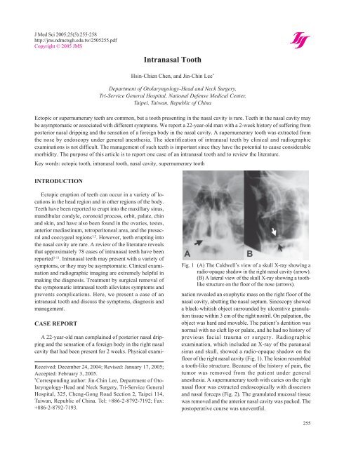
Fig. 1 (A) The Caldwell’s view of a skull X-ray showing a<br />
radio-opaque shadow in the right nasal cavity (arrow).<br />
(B) A lateral view of the skull X-ray showing a toothlike<br />
structure on the floor of the nose (arrows).<br />
nation revealed an exophytic mass on the right floor of the<br />
nasal cavity, abutting the nasal septum. Sinoscopy showed<br />
a black-whitish object surrounded by ulcerative granulation<br />
tissue within 3 cm of the right nostril. On palpation, the<br />
object was hard and movable. The patient’s dentition was<br />
normal with no cleft lip or palate, and he had no history of<br />
previous facial trauma or surgery. Radiographic<br />
examination, which included an X-ray of the paranasal<br />
sinus and skull, showed a radio-opaque shadow on the<br />
floor of the right nasal cavity (Fig. 1). The lesion resembled<br />
a tooth-like structure. Because of the history of pain, the<br />
tumor was removed from the patient under general<br />
anesthesia. A supernumerary tooth with caries on the right<br />
nasal floor was extracted endoscopically with dissectors<br />
and nasal forceps (Fig. 2). The granulated mucosal tissue<br />
was removed and the anterior nasal cavity was packed. The<br />
postoperative course was uneventful.<br />
255
<strong>Intranasal</strong> tooth<br />
Fig. 2 A gross view of the extracted tooth revealing some<br />
attached mucosa and dental caries.<br />
DISCUSSION<br />
<strong>Intranasal</strong> teeth are rare. In 1979, Smith et al. reported<br />
2 cases and found 27 well-documented cases in the literature<br />
since the original description of intranasal teeth in<br />
1897 1 . In 2001, Gupta et al. reported one case of an<br />
intranasal tooth in a 4-year-old boy with a repair for cleft<br />
lip and alveolus, and identified another 20 cases 2 . Since<br />
then, an additional 28 cases have been reported 3-11 , with<br />
approximately 78 cases of intranasal teeth reported. Most<br />
of the articles are case reports and only two larger series of<br />
consecutive cases have been reported 3,12 .<br />
This literature survey revealed that the age of detection<br />
of intranasal teeth ranged from 3 to 71 years; approximately<br />
half of these cases were discovered before adulthood<br />
3 . Slightly more males than females were affected<br />
and in most cases, only one tooth was found in the nasal<br />
cavity. However, multiple ectopic teeth have also been<br />
reported 1,4,12 . Almost all of these cases were unilateral with<br />
no dominance of either side of the nasal cavity; three cases<br />
were bilateral 5,12,13 . The types of ectopic intranasal teeth<br />
were supernumerary, deciduous, and permanent.<br />
<strong>Intranasal</strong> teeth present a variety of symptoms that<br />
include the sensation of a foreign body in the nose, unilateral<br />
nasal obstruction, purulent nasal discharge, bloodstained<br />
rhinorrhea, repeated epistaxis, nasal or facial pain,<br />
headache, and epiphora 1,3,4 . <strong>Intranasal</strong> teeth may also be<br />
asymptomatic and noticed incidentally on a routine clinical<br />
or radiographic examination. Clinically, intranasal teeth<br />
occur most commonly on the floor of the nose, approxi-<br />
256<br />
mately halfway between the nostril and choana 3 . Eruption<br />
of ectopic intranasal teeth may present as hard white<br />
masses without a covering of nasal mucosa, for which<br />
diagnosis is usually straightforward. However, the tooth<br />
may be embedded in the nasal mucosa surrounded with<br />
debris, granulation, and ulcerative materials, for which a<br />
differential diagnosis should be formulated.<br />
The differential diagnoses that could indicate an intranasal<br />
tooth include foreign body; rhinolith; inflammatory<br />
lesions due to syphilis, tuberculosis, or fungal infection<br />
with calcification; benign tumors, including hemangioma,<br />
osteoma, calcified polyps, enchondroma, and dermoid<br />
cysts; and malignant tumors, such as chondrosarcoma and<br />
osteosarcoma 6 .<br />
Radiographic examinations, including Caldwell’s view,<br />
Waters’ view, and a lateral view of skull, occlusal radiography,<br />
panoramic radiography and computed tomography<br />
scans, are helpful in confirming the diagnosis of intranasal<br />
teeth. Panoramic radiography also reveals a detailed<br />
condition of the dentition. Computed tomography can<br />
indicate tooth-equivalent attenuation, identify the lesion<br />
centrally, and evaluate the depth of the eruption site, which<br />
are all highly discriminating features that not only delineate<br />
but also confirm the diagnosis 6,7 . Additionally, careful<br />
inspection of dentition and consultation with a dentist is<br />
necessary to find out about possible missing teeth and to<br />
differentiate the diagnosis of supernumerary, deciduous,<br />
or permanent teeth 3 .<br />
The etiology of intranasal teeth remains unclear. However,<br />
some of the causes proposed include displacement<br />
due to trauma; odontogenic or rhinogenic infection; genetic<br />
factors; developmental disturbances, such as cleft lip,<br />
alveolus or palate; and obstruction at the time of tooth<br />
eruption secondary to crowding of the dentition, retained<br />
primary teeth, or dense bone 1,2 . Another possibility is that<br />
the tooth may be pushed further up into the nasal cavity<br />
during surgical repair of an alveolus cleft 2 .<br />
Treatment of intranasal teeth is early surgical removal<br />
because of potential morbidity. These morbidities include<br />
rhinosinusitis, osteomyelitis, dacryocystitis due to nasolacrimal<br />
duct obstruction, nasal septal abscess or perforation,<br />
oronasal or intraoral fistula, aspergillosis, and nasal<br />
deformity 3-6 . A very rare association of ectopic tooth with<br />
squamous cell carcinoma of the palate in a 12-year-old<br />
Nigerian boy has been reported 14 . Surgical removal of an<br />
intranasal tooth in asymptomatic patients is recommended<br />
to prevent complications, or it should be observed with<br />
close clinical follow-up 1 . The surgical removal includes<br />
transnasal and transpalatal approaches and may combine<br />
ancillary procedures 3 . Usually, an intranasal tooth can be
emoved under direct vision with a head-light or mirror. In<br />
recent years, endoscopic extraction of intranasal teeth has<br />
been reported with great advantage and less morbidity 3-5,7-9 .<br />
This report presents a case of a symptomatic ectopic<br />
supernumerary tooth arising in the nasal cavity. Endoscopic<br />
removal of the intranasal tooth was performed due<br />
to local infection of intranasal dental caries. Once an<br />
intranasal tooth is symptomatic, surgical removal is indicated.<br />
REFERENCES<br />
1. Smith RA, Gordon NC, De Luchi SF. <strong>Intranasal</strong> Teeth.<br />
Report of two cases and review of the literature. Oral<br />
Surg Oral Med Oral Path 1979;47:120-122.<br />
2. Gupta YK, Shah N. <strong>Intranasal</strong> tooth as a complication<br />
of cleft lip and alveolus in a four year old child: case<br />
report and literature review. Int J Paediatr Dent 2001;<br />
11:221-224.<br />
3. Lee FP. Endoscopic extraction of an intranasal tooth:<br />
a review of 13 Cases. Laryngoscope 2001;111:1027-<br />
1031.<br />
4. Alexandrakis G, Hubbell RN, Aitken PA. Nasolacrimal<br />
duct obstruction secondary to ectopic teeth. Ophthalmology<br />
2000;107:189-192.<br />
5. Sokolov M, Jecker P, Roth Y. Nasal teeth associated<br />
with rhinosinusitis. Rhinology 2004;42:107-110.<br />
Hsin-Chien Chen, et al.<br />
6. Chen A, Huang JK, Cheng SJ, Sheu CY. Nasal teeth:<br />
report of three cases. Am J Neuroradiol 2002;23:671-<br />
673.<br />
7. Lin IH, Hwang CF, Su CY, Kao YF, Peng JP. <strong>Intranasal</strong><br />
tooth: report of three cases. Chang Gung Med J<br />
2004;27:385-389.<br />
8. Kim DH, Kim JM, Chae SW, Hwang SJ, Lee SH, Lee<br />
HM. Endoscopic removal of an intranasal ectopic<br />
tooth. Int J Pediatr Otorhinolaryngol 2003;67:79-81.<br />
9. Meijer BA, van der Wal KG. Unilateral nasal bleeding<br />
due to an intranasal tooth. Ned Tijdschr Tandheelkd<br />
2003;110:362-364.<br />
10. Ondzotto G. Ectopic tooth in the nasal cavity. Rev<br />
Stomatol Chir Maxillofac 2003;104:352-354.<br />
11. Kuroda H, Tsutsumi K, Tomisawa H, Koizuka I. A<br />
case of an inverted tooth in the nasal cavity. Auris<br />
Nasus Larynx 2003;30(Suppl):S127-129.<br />
12. Martinson FD, Cockshott WP. Ectopic nasal dentition.<br />
Clin Radiol 1972;23:451-454.<br />
13. Srivastava RP, Pradhan AC, Yadav VNS. <strong>Tooth</strong> in<br />
nasal cavity: a case report of cleft lip and palate. J Ind<br />
Dent Assoc 1977;49:145-146.<br />
14. Ogisi FO, Odita JC. Ectopic nasal dentition associated<br />
with squamous cell carcinoma of palate in a 12-yearold<br />
boy. Br J Oral Maxillofacial Surg 1988;26:58-61.<br />
257
<strong>Intranasal</strong> tooth<br />
258