CE 421 - Dental Anatomy: A Review - DentalCare.com
CE 421 - Dental Anatomy: A Review - DentalCare.com
CE 421 - Dental Anatomy: A Review - DentalCare.com

Create successful ePaper yourself
Turn your PDF publications into a flip-book with our unique Google optimized e-Paper software.
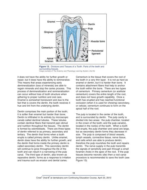
Figure 13. Divisions and Tissues of a Tooth. Parts of the teeth and<br />
surrounding tissues.<br />
Image courtesy of The <strong>Anatomy</strong> and Physiology Learning System, 2nd Ed.<br />
it does not have the ability for further growth or<br />
repair, but it does have the ability to remineralize.<br />
This means that areas experiencing early<br />
demineralization (loss of minerals) are able to<br />
regain minerals and stop the caries process. This<br />
process of demineralization and remineralization<br />
can occur without loss of tooth structure when<br />
adhering to proper nutrition and oral care.<br />
Enamel is somewhat translucent and due to the<br />
fact that is covers the dentin, the tooth receives it<br />
hue and tint from the underlying dentin.<br />
Dentin <strong>com</strong>prises the main portion of the tooth;<br />
it is softer than enamel but harder than bone.<br />
Dentin is infiltrated in its entirely by microscopic<br />
canals called dentinal tubules. These tubules<br />
contain dentinal fibers that transmit pain stimuli<br />
and nutrition throughout the tissues. The dentin<br />
is formed by odontoblasts. There are three types<br />
of dentin referred to as primary, secondary and<br />
tertiary. The dentin that forms when a tooth<br />
erupts is called primary dentin. Unlike enamel,<br />
dentin does have the ability for further growth, and<br />
the dentin that forms inside the primary dentin is<br />
called secondary dentin. This secondary dentin<br />
will continue to grow throughout the life of the<br />
tooth and can result in a narrowing of the pulp<br />
canal. The third type of dentin, also known as<br />
reparative dentin, forms as a response to irritation<br />
and trauma such as erosion and dental caries.<br />
11<br />
Cementum is the tissue that covers the root of<br />
the tooth in a very thin layer. It is not as hard as<br />
enamel or dentin, but it is harder than bone. It<br />
contains attachment fibers that help to anchor<br />
the tooth within the bone. There are two types<br />
of cementum. Primary cementum (or acellular<br />
cementum) covers the entire length of the root,<br />
and does not have growth capability. Once a<br />
tooth has erupted and has reached functional<br />
occlusion (when it is used for chewing) secondary,<br />
or cellular, cementum continues to form on the<br />
apical half of the root.<br />
The pulp is located in the center of the tooth,<br />
and is surrounded by dentin. The pulp cavity is<br />
divided into two areas: the pulp chamber, located<br />
in the crown of the tooth; and the pulp canal(s),<br />
located in the root(s) of the tooth. When a tooth<br />
first erupts, the pulp chamber and canal are large,<br />
but as secondary dentin forms they decrease in<br />
size. The pulp is <strong>com</strong>posed of blood vessels,<br />
lymph vessels, connective tissue, nerve tissue,<br />
and cells which are able to produce dentin;<br />
therefore the pulp nourishes the tooth and repairs<br />
dentin. The nerve supply in the pulp transmits<br />
the signals of sensitivity and pain through a small<br />
foramen (hole) in the apex of the root. If the pulp<br />
tissues be<strong>com</strong>e necrotic (die) then a root canal<br />
procedure is re<strong>com</strong>mended to save the tooth (see<br />
Figure 13).<br />
Crest ® Oral-B ®<br />
at dentalcare.<strong>com</strong> Continuing Education Course, April 23, 2013