CE 421 - Dental Anatomy: A Review - DentalCare.com
CE 421 - Dental Anatomy: A Review - DentalCare.com
CE 421 - Dental Anatomy: A Review - DentalCare.com

You also want an ePaper? Increase the reach of your titles
YUMPU automatically turns print PDFs into web optimized ePapers that Google loves.
• Palatine Rugae: Irregular ridges or folds of<br />
masticatory mucosa that extend horizontally<br />
from either side of the palatine raphe.<br />
• Soft Palate: Forms the posterior section of<br />
the palate and is not supported by underlying<br />
bone. It can be lifted to meet the posterior<br />
pharyngeal wall to seal the nasopharynx<br />
during swallowing and speech.<br />
• Uvula: Small conical mass of tissue that<br />
hangs from the palatine velum (free edge of<br />
the soft palate).<br />
• Fauces: The passageway from the oral cavity<br />
to the pharynx (throat).<br />
• Pillars of Fauces: Two arches of muscle<br />
tissue surrounding posterior oral cavity.<br />
The Anterior Pillar of Fauces is also called<br />
palatoglossal arch, the Posterior Pillar of<br />
Fauces is also called the palatopharyngeal<br />
arch. They contract and narrow the fauces<br />
during deglutition (swallowing).<br />
• Isthmus of Fauces: The space between the<br />
left and right anterior and posterior pillars of<br />
fauces. It contains the palatine tonsils.<br />
(These landmarks are identified in Figure 4)<br />
Landmarks of the Tongue<br />
The following are landmarks of the tongue.<br />
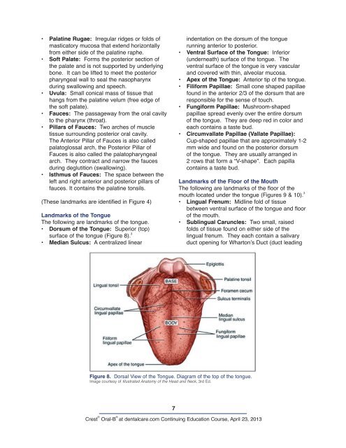
• Dorsum of the Tongue: Superior (top)<br />
surface of the tongue (Figure 8). 3<br />
• Median Sulcus: A centralized linear<br />
7<br />
indentation on the dorsum of the tongue<br />
running anterior to posterior.<br />
• Ventral Surface of the Tongue: Inferior<br />
(underneath) surface of the tongue. The<br />
ventral surface of the tongue is very vascular<br />
and covered with thin, alveolar mucosa.<br />
• Apex of the Tongue: Anterior tip of the tongue.<br />
• Filiform Papillae: Small cone shaped papillae<br />
found in the anterior 2/3 of the dorsum that are<br />
responsible for the sense of touch.<br />
• Fungiform Papillae: Mushroom-shaped<br />
papillae spread evenly over the entire dorsum<br />
of the tongue. They are deep red in color and<br />
each contains a taste bud.<br />
• Circumvallate Papillae (Vallate Papillae):<br />
Cup-shaped papillae that are approximately 1-2<br />
mm wide and found on the posterior dorsum<br />
of the tongue. They are usually arranged in<br />
2 rows that form a “V-shape”. Each papilla<br />
contains a taste bud.<br />
Landmarks of the Floor of the Mouth<br />
The following are landmarks of the floor of the<br />
mouth located under the tongue (Figures 9 & 10). 3<br />
• Lingual Frenum: Midline fold of tissue<br />
between ventral surface of the tongue and floor<br />
of the mouth.<br />
• Sublingual Caruncles: Two small, raised<br />
folds of tissue found on either side of the<br />
lingual frenum. They each contain a salivary<br />
duct opening for Wharton’s Duct (duct leading<br />
Figure 8. Dorsal View of the Tongue. Diagram of the top of the tongue.<br />
Image courtesy of Illustrated <strong>Anatomy</strong> of the Head and Neck, 3rd Ed.<br />
Crest ® Oral-B ®<br />
at dentalcare.<strong>com</strong> Continuing Education Course, April 23, 2013