Scara b s - University of Nebraska State Museum
Scara b s - University of Nebraska State Museum
Scara b s - University of Nebraska State Museum

Create successful ePaper yourself
Turn your PDF publications into a flip-book with our unique Google optimized e-Paper software.
WITHIN THIS ISSUE<br />
Breeding Dynastes ........... 1<br />
Dark Protaetia ................ 10<br />
SOLA 2011 ...................... 13<br />
The Complete Guide to<br />
Rearing the Rainbow<br />
<strong>Scara</strong>b ............................... 16<br />
BACK ISSUES<br />
Available At These Sites:<br />
Coleopterists Society<br />
www.coleopsoc.org/default.asp?Action=Show_<br />
Resources&ID=<strong>Scara</strong>bs<br />
<strong>University</strong> <strong>of</strong> <strong>Nebraska</strong><br />
www-museum.unl.edu/<br />
research/entomology/<br />
<strong>Scara</strong>bs-Newsletter.htm<br />
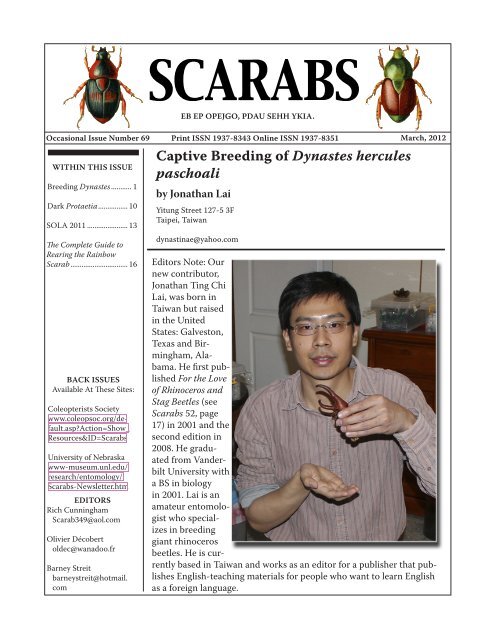
SCARABS<br />
Occasional Issue Number 69 Print ISSN 1937-8343 Online ISSN 1937-8351<br />
March, 2012<br />
EDITORS<br />
Rich Cunningham<br />
<strong>Scara</strong>b349@aol.com<br />
Olivier Décobert<br />
oldec@wanadoo.fr<br />
Barney Streit<br />
barneystreit@hotmail.<br />
com<br />
EB EP OPEJGO, PDAU SEHH YKIA.<br />
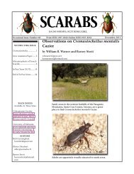
Captive Breeding <strong>of</strong> Dynastes hercules<br />
paschoali<br />
by Jonathan Lai<br />
Yitung Street 127-5 3F<br />
Taipei, Taiwan<br />
dynastinae@yahoo.com<br />
Editors Note: Our<br />
new contributor,<br />
Jonathan Ting Chi<br />
Lai, was born in<br />
Taiwan but raised<br />
in the United<br />
<strong>State</strong>s: Galveston,<br />
Texas and Birmingham,Alabama.<br />
He first published<br />
For the Love<br />
<strong>of</strong> Rhinoceros and<br />
Stag Beetles (see<br />
<strong>Scara</strong>bs 52, page<br />
17) in 2001 and the<br />
second edition in<br />
2008. He graduated<br />
from Vanderbilt<br />
<strong>University</strong> with<br />
a BS in biology<br />
in 2001. Lai is an<br />
amateur entomologist<br />
who specializes<br />
in breeding<br />
giant rhinoceros<br />
beetles. He is currently<br />
based in Taiwan and works as an editor for a publisher that publishes<br />
English-teaching materials for people who want to learn English<br />
as a foreign language.
Photo 1: Dynastes hercules paschoali is known to lack<br />
cephalic denticles.<br />
Photo 2: Male larvae are capable <strong>of</strong> growing to over 130<br />
grams.<br />
Photo 3: Larvae are very docile unless provoked. I have<br />
yet to be bitten by one.<br />
Page 2<br />
Over the ages, giant rhinoceros<br />
beetles have fascinated man, to an<br />
extent that they are named after<br />
Greek gods and heroes. The most<br />
famous <strong>of</strong> them all is perhaps the<br />
Hercules Beetle Dynastes hercules,<br />
which is capable <strong>of</strong> growing<br />
to over 17 cm. This mega-titan <strong>of</strong><br />
the insect world was described as<br />
early as 1753 by Carl Linnaeus. To<br />
date, a total <strong>of</strong> 12 subspecies are<br />
known. One <strong>of</strong> the rarest <strong>of</strong> them<br />
is D. h. paschoali, found only on<br />
the Atlantic coast <strong>of</strong> southeastern<br />
Brazil. In fact, it was listed as<br />
an endangered species by Brazil<br />
in 2008. Although this subspecies<br />
was described by Grossi and<br />
Arnaud in 1993, even today, very<br />
few specimens are known and<br />
information regarding this subspecies<br />
is virtually nonexistent. As<br />
passionate as the author is about<br />
D. hercules, he was not able to<br />
see even a photograph <strong>of</strong> a specimen<br />
until 2007. In July <strong>of</strong> 2009,<br />
the author was ecstatic to be able<br />
to obtain 24 newly-hatched F2<br />
generation larvae that ultimately<br />
came from adult beetles collected<br />
in 2007 in Bahia, Brazil, and thus<br />
began an endeavor to unravel the<br />
mysteries <strong>of</strong> D. h. paschoali. The<br />
main objective was to determine<br />
the level <strong>of</strong> difficulty in captive-<br />
rearing this subspecies, and to give<br />
the public an opportunity to finally<br />
look at this elusive giant up close.<br />
(Photo 1)<br />
The 24 first instar larvae were<br />
divided into three groups <strong>of</strong> 8<br />
larvae. Each group was kept in a<br />
plastic container measuring 30<br />
x 20 x 15 cm. The lid <strong>of</strong> the container<br />
was drilled with 6 ventila-
tion holes, each about 0.5 cm in<br />
diameter. A thin cloth was used<br />
to cover the holes to prevent unwanted<br />
pests such as fungus gnats<br />
and cockroaches from entering. The<br />
larvae were fed hardwood sawdust<br />
that had been composted for 4-6<br />
months to rid it <strong>of</strong> indigestible or<br />
toxic substances such as lignin<br />
and plant alkaloids. Three pellets<br />
<strong>of</strong> dried dog food each weighing<br />
roughly 1 gram were distributed<br />
on the bottom <strong>of</strong> each container<br />
before the containers were filled<br />
with substrate to a depth <strong>of</strong> 6 cm.<br />
The humidity <strong>of</strong> the substrate was<br />
adjusted to be roughly 50%. No<br />
substrate change was performed<br />
until the larvae molted to second<br />
instar. The first instar stage lasted<br />
approximately 30 days.<br />
Upon reaching the second instar<br />
stage, the larvae were sexed and<br />
there were 15 males and 9 females.<br />
Unfortunately, one female larva<br />
died <strong>of</strong> molting failure caused by a<br />
jammed head capsule. The remaining<br />
larvae were divided into three<br />
groups <strong>of</strong> 5 larvae and two groups<br />
<strong>of</strong> 4 larvae with mixed sexes. The<br />
dimensions <strong>of</strong> the five rearing<br />
containers remained unchanged.<br />
However, the depth <strong>of</strong> the substrate<br />
was increased to 9 cm. No substrate<br />
change was performed until the<br />
larvae molted to third instar. The<br />
second instar stage lasted approximately<br />
45 days.<br />
Upon reaching third instar, it was<br />
obvious that the male larvae had<br />
bigger heads, with head capsule<br />
diameters ranging from 16.0-17.1<br />
mm (a breeder in Taiwan raised a<br />
larva with 18.0 mm head capsule).<br />
Photo 4: Pre-pupa.<br />
Photo 5: Split <strong>of</strong> dorsal skin marks beginning <strong>of</strong> pupation.<br />
Notice the extremely compressed horns.<br />
Photo 6: Four minutes after the onset <strong>of</strong> pupation.<br />
Page 3
Page 4<br />
Photo 7: Eight minutes after the onset <strong>of</strong> pupation.<br />
Photo 8: 47 minutes after the onset <strong>of</strong> pupation.<br />
Photo 9: Four hours and nine minutes after the onset <strong>of</strong><br />
pupation.<br />
Females ranged from 14.5-15.5 mm<br />
(later experience showed female<br />
larvae were able to attain 16.2<br />
mm). From now on, all larvae were<br />
kept individually. Each female was<br />
kept in a cylindrical container with<br />
13 cm diameter and 19 cm height<br />
(approximately 2.5 liters). The<br />
depth <strong>of</strong> the substrate was kept at<br />
16-17 cm. Each male was kept in a<br />
cylindrical container with 15.5 cm<br />
diameter and 22 cm height (approximately<br />
4.1 liters). The depth<br />
<strong>of</strong> the substrate was kept at 19-20<br />
cm. Three ventilation holes were<br />
drilled in the lids <strong>of</strong> each container.<br />
A substrate change was performed<br />
every six weeks. The entire content<br />
<strong>of</strong> a container would be carefully<br />
dumped out (as digging for the<br />
larva with a spoon could harm the<br />
larva). The container would be<br />
thoroughly washed and the larva<br />
gently rinsed with tap water to<br />
prevent pathogen buildup. In order<br />
to retain beneficial endosymbionts<br />
that help larvae to digest cellulose,<br />
6-8 fecal pellets would be crushed<br />
and returned to the container. The<br />
containers for females would receive<br />
4 pellets <strong>of</strong> dog food and the<br />
containers for males would receive<br />
6 pellets <strong>of</strong> dog food. The larva<br />
would be put back and the last step<br />
would be refilling the container<br />
with clean substrate. Larvae were<br />
kept in a climate-controlled environment<br />
<strong>of</strong> 25º C in the summer<br />
and 20º C in the winter.<br />
Eight months after hatching,<br />
female larvae were beginning to<br />
reach their maximum weight,<br />
ranging from 65-80 grams. Two<br />
months later, females began to<br />
construct pupal cells with 10-30
degree inclination. They rested<br />
inside these oval chambers and became<br />
pupae approximately 30 days<br />
later. The pupal stage lasted about<br />
60 days at 25º C. However, underground<br />
life was not over. New adults<br />
rested in pupal cells for yet another<br />
60 days or more before emerging to<br />
the surface to look for food. They<br />
were fed a diet <strong>of</strong> sliced apples for<br />
20-30 days until fully mature and<br />
ready to mate. Females measured<br />
66-71 mm. (However, a Taiwanese<br />
breeder raised a 78 mm female.)<br />
Male larvae took much longer to<br />
develop because they grew so much<br />
larger, almost twice the weight <strong>of</strong><br />
females. They didn’t begin to reach<br />
maximum weight until 14 months<br />
after hatching. Some extra-large<br />
male larvae were still gaining weight<br />
16 months after hatching. The<br />
maximum weight <strong>of</strong> male larvae<br />
ranged from 107-126 grams (Photos<br />
2 & 3). Once a male larva reached<br />
100 grams, it was relocated to a 30<br />
x 20 x 24 cm container so it would<br />
have enough horizontal space to<br />
construct a pupal cell large enough<br />
to accommodate its long thoracic<br />
horn. (Incidentally, male larvae kept<br />
in 4.1 liter containers also made<br />
pupal cells, but the front and the<br />
end were obstructed by the walls<br />
<strong>of</strong> the containers, resulting in very<br />
short pupal cells. Such larvae were<br />
relocated during the pre-pupal<br />
stage to artificial pupal cells made<br />
from floral foams (otherwise, the<br />
horns would be bent). Male larvae<br />
began to construct pupal cells 16-18<br />
months after hatching. The pupal<br />
cells were inclined, with angles<br />
ranging from 10-30 degrees. The<br />
pre-pupal and pupal stages lasted<br />
Photo 10: 48 hours after the onset <strong>of</strong> pupation.<br />
Photo 11: Pupa very close to eclosion<br />
Photo 12: Breaking pupal skin marks the beginning <strong>of</strong><br />
eclosion.<br />
Photo 13: Ten minutes after the beginning <strong>of</strong> eclosion.<br />
Page 5
Photo 14: 34 minutes after the beginning <strong>of</strong> eclosion.<br />
Photo 15: One hour and 39 minutes after the beginning<br />
<strong>of</strong> eclosion.<br />
Photo 16: Five hours and three minutes after the beginning<br />
<strong>of</strong> eclosion.<br />
Photo 17: Eight hours and 25 minutes after the beginning<br />
<strong>of</strong> eclosion.<br />
Page 6<br />
approximately 45 and 80 days,<br />
respectively, at 20º C (calculation<br />
taken during winter months)<br />
(Photos 4-18). Unfortunately, one<br />
male was unable to successfully<br />
shed his upper larval skin during<br />
pupation and became a severely<br />
deformed pupa that died a few<br />
days later. The remaining 14 males<br />
ranged from 130-145 mm as measured<br />
from the tip <strong>of</strong> the thoracic<br />
horn to the end <strong>of</strong> the elytra (Photo<br />
19). New adults stayed inactive<br />
in their pupal cells for 45-55 days.<br />
(The author is confident that D. h.<br />
paschoali is a subspecies capable<br />
<strong>of</strong> producing male larvae <strong>of</strong> over<br />
140 grams, and subsequently male<br />
adults <strong>of</strong> over 150 mm. He hopes<br />
to reach this goal in the F3 generation.)<br />
The weight <strong>of</strong> a larva did not determine<br />
the absolute length <strong>of</strong> the<br />
adult beetle. It was possible for a<br />
heavier larva to become a shorter<br />
beetle. For example, a heavier larva<br />
might have developed a thicker,<br />
but not necessarily longer horn,<br />
thus becoming shorter than its<br />
lighter counterpart. But generally<br />
speaking, heavier larvae became<br />
longer adults.<br />
Although the 14 males were<br />
brothers, they exhibited a wide<br />
range <strong>of</strong> appearance. In regard to<br />
elytra color (one color per beetle),<br />
various shades <strong>of</strong> green, brown,<br />
and yellow could be found among<br />
the siblings. As for the elytral<br />
spotting, spotless, moderately<br />
spotted, intensely spotted, small<br />
spots, large spots, striped, and<br />
both striped and spotted were<br />
observed (Photo 20). The thoracic
horn ranged from thin (stenoceras)<br />
to thick (pachyceras). The apex <strong>of</strong><br />
the cephalic horn ranged from unhooked<br />
to hooked. The protrusion<br />
right next to the apex ranged from<br />
minute to conspicuous. All <strong>of</strong> the<br />
beetles lacked cephalic denticles.<br />
(Photo 21)<br />
The continual propagation <strong>of</strong> this<br />
captive culture was presented<br />
with a problem. All <strong>of</strong> the females<br />
emerged 4-6 months ahead <strong>of</strong> the<br />
males. Experience with other subspecies<br />
<strong>of</strong> D. hercules has revealed<br />
that females need to be mated<br />
within two months <strong>of</strong> emergence<br />
from the pupal cell, as after this<br />
period, their vitality quickly declines.<br />
It would not be possible for<br />
males and females with an emergence<br />
discrepancy <strong>of</strong> 4-6 months to<br />
mate. Obviously, adult males from<br />
elsewhere that had emerged in sync<br />
with the author’s females had to be<br />
acquired if these females were to<br />
reproduce. Luckily, the author was<br />
able to obtain two mature males<br />
from other breeders. Both <strong>of</strong> the<br />
males were 129 mm in length.<br />
All 8 females were successfully<br />
mated. One male mated with 6<br />
females, while the other mated with<br />
2 females. Interestingly, cold temperature<br />
prolonged mating time.<br />
The first male mated in a temperature<br />
range <strong>of</strong> 18-20º C. Mating<br />
times were 35, 40, 40, 40, 45, and 30<br />
minutes. The second male mated<br />
in a temperature range <strong>of</strong> 15-16º C.<br />
Mating times were 85 and 80 minutes.<br />
Each female mated only once.<br />
(Photo 22)<br />
Photo 18: 18 hours and 45 minutes after the beginning<br />
<strong>of</strong> eclosion.<br />
Photo 19: D. h. paschoali over 144 mm.<br />
Photo 20: Left to right: spotless, small spots, large<br />
spots, stripes.<br />
Page 7
Page 8<br />
Photo 21: Some variations in the cephalic horn.<br />
Photo 22: D. h. paschoali mating.<br />
After mating, each female was<br />
individually placed in a 30 x 25 x<br />
30 cm breeding container filled<br />
to a depth <strong>of</strong> 20 cm with the same<br />
substrate used to rear larvae. The<br />
bottom 3-4 cm was thoroughly<br />
compressed by hand to aid the<br />
females’ oviposition (as females<br />
oviposit, they pack the substrate<br />
into hard clumps in which they<br />
lay their eggs. It is speculated that<br />
the hard clumps protect eggs from<br />
predators). The females were continuously<br />
provided with slices <strong>of</strong><br />
apple placed on a plate located on<br />
the top <strong>of</strong> the substrate. The slices<br />
were changed every 3 days to prevent<br />
spoilage. The females would<br />
alternate between spending a few<br />
days on the apples and remaining<br />
for several days underground. The<br />
entire content <strong>of</strong> each container<br />
was dumped out to check for eggs<br />
every 3-4 weeks. Substrate clumps<br />
had to be taken apart to harvest<br />
the eggs. The eggs were then<br />
placed on the bottom <strong>of</strong> a clear<br />
plastic container and covered very<br />
gently with 4 cm <strong>of</strong> substrate. The<br />
development <strong>of</strong> the eggs could be<br />
observed by looking at the bottom<br />
<strong>of</strong> the container. Eggs absorbed<br />
moisture and gradually expanded<br />
to about twice their original size<br />
before hatching. Incubation lasted<br />
approximately 30 days (Photo 23).<br />
Below is a breeding summary <strong>of</strong><br />
the 8 females:<br />
The adult females lived up to<br />
200 days, and the adult males<br />
up to 300 days. Despite feeding<br />
throughout their lives, the<br />
adults progressively lost weight<br />
until death. The life cycle <strong>of</strong> D. h.<br />
paschoali was successfully docu-
mented. Given the high survival<br />
rate <strong>of</strong> the F2 generation larvae<br />
(over 90%) and the large imagoes<br />
produced, it was concluded that<br />
captive raising D. h. paschoali was<br />
not only possible, but not difficult<br />
(given the larvae were fed proper<br />
substrate).<br />
Photo 23: Eggs in various stages <strong>of</strong> development with<br />
newly-hatched larva on far right.<br />
Female # 1 2 3 4 5 6 7 8<br />
Size 67 mm 68 mm 66 mm 68 mm 68 mm 70 mm 68 mm 68 mm<br />
Eggs<br />
Laid<br />
103 76 67 157 89 63 101 54<br />
Larvae<br />
Hatched<br />
Weight<br />
92 42 45 90 67 42 89 40<br />
at Mating<br />
20 g 22 g 21 g 22 g 22 g 23 g 21 g 19 g<br />
Weight<br />
at Death<br />
Time in<br />
14 g 16 g 14 g 14 g 17 g 16 g 16 g 14 g<br />
BreedingContainer<br />
119 days 141 days 172 days 177 days 142 days 171 days 142 days 97 days<br />
It is worth pointing out that although<br />
the F2 generation individuals<br />
exhibited sex-based discrepancies<br />
in eclosion, subsequent<br />
experience with another, unrelated<br />
group <strong>of</strong> 11 F2 generation larvae<br />
showed no such discrepancy<br />
(both groups kept under identical<br />
conditions); all males and females<br />
eclosed at about the same time.<br />
(Photo 24).<br />
At the time <strong>of</strong> this writing, many<br />
F3 generation larvae are healthy<br />
third instar larvae.<br />
Photo 24: Power <strong>of</strong> captive propagation.<br />
Page 9
Page 10<br />
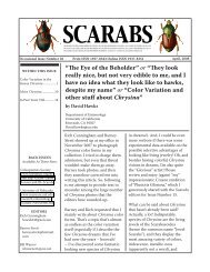
Some Dark French Protaetia<br />
by Olivier Décobert<br />
oldec@wanadoo.fr<br />
After reading some <strong>of</strong> the past issues <strong>of</strong> <strong>Scara</strong>bs, the reader could think that French Protaetia<br />
are always full colored and shiny. I must admit that I forgot to write about dark species <strong>of</strong> this<br />
genus in my country.<br />
I had intended to tell more about this subject after reading the“Black is beautiful, Baby” that<br />
Brett Ratcliffe recalled on the first page <strong>of</strong> <strong>Scara</strong>bs 46 (December, 2009). But I had many other<br />
articles in my mind at that time and again, I forgot...<br />
In April, 2011, I was in the Oriental Pyrenees mountains when I found a larva under a rock<br />
(Photo 1). My first thought was “melolonthid” but it was indeed a cetonid larva, which is more<br />
habitually found in decaying wood. Here, this larva was in a little cavity dug in the soil. I don’t<br />
know what it exactly ate, probably roots <strong>of</strong> grasses or small surrounding plants.<br />
Photo 1: Larva.
As usual, the question was “What is the species?” It is impossible to know before rearing and<br />
obtaining the adult. This event happened two months later, after a transition by a pupa in a shell<br />
(Photos 2, 3 & 4). The adult was clearly Protaetia morio (Fabricius), a black species, whose size<br />
can vary between 12 and 20 mm. It was the time to show some dark Protaetia in <strong>Scara</strong>bs!<br />
Photo 2: Shell Photo 3: Pupa in shell. Photo 4: Pupa.<br />
As one can see on Photo 5, the emerging adult is not yet black, and this will take two or three<br />
days to be definitive (Photo 6).<br />
Photos 5 and 6: Teneral adult <strong>of</strong> Protaetia morio (left) and mature adult (right).<br />
Page 11
Photo 7: Protaetia morio form.<br />
Page 12<br />
White marks exist on the pronotum and elytra, and can sometimes be<br />
numerous (Photo 7). This form is called “quadripunctata” (Fabricius). I<br />
found this one on the southwestern coast <strong>of</strong> France.<br />
A close species is Protaetia oblonga (Gory & Percheron),<br />
smaller (12-16 mm) and rather brown than black. I found the<br />
specimen shown in Photo 8 near the French Alps.<br />
Photo 9: Protaetia opaca.<br />
Protaetia opaca (Fabricius) (Photo 9) is a bigger species (up to 27<br />
mm). This cetonid likes to frequent beehives to eat the honey! I<br />
found this specimen in the South <strong>of</strong> France.<br />
It is impossible to forget Corsica Island, where a dark blue<br />
endemic cetonid lives: Protaetia sardea (Gory & Percheron),<br />
about 25 mm in size (Photo 10).<br />
Photo 8: Protaetia oblonga.<br />
Photo 10: Protaetia sardea.
SOLA - 2011<br />
by Andrew Smith<br />
Canadian <strong>Museum</strong> <strong>of</strong> Nature<br />
asmith@unlserve.unl.edu<br />
Photo 1: The 14th annual SOLA (Sacred Order <strong>of</strong><br />
Lamellate Antennae) <strong>Scara</strong>b Workers Symposium<br />
was held in Reno, Nevada on November 13th.<br />
Yours truly was again the host and organizer <strong>of</strong><br />
this event. As always, SOLA was part <strong>of</strong> the larger<br />
Entomological Society <strong>of</strong> America (ESA) meeting.<br />
Photo 2: The customary SOLA salute was given<br />
at the beginning <strong>of</strong> the symposium. Those who<br />
abstained or demonstrated poor form were<br />
invited to attend the carabid symposium down<br />
the hall.<br />
Photo 3: Bob Anderson gave a sobering<br />
presentation “<strong>Scara</strong>b beetles as bycatch”<br />
reminding everyone that scarab beetles are not<br />
the primary focus <strong>of</strong> all field work.<br />
Photo 4: Paul Skelley and M.J. Paulsen, both<br />
sporting semi-impressive facial hair, gave<br />
separate presentations on “Bagging big trophies”<br />
and “Fighting for a meaningful classification <strong>of</strong><br />
Lucanidae.” SOLA presentations apparently get<br />
bonus marks if the title begins with an action<br />
verb in the present continuous tense.<br />
Page 13
Photo 5: Sergey Tarasov wowed the crowd<br />
with his detailed phylogenetic work on dung<br />
beetles during his presentation “Clarifying<br />
systematics <strong>of</strong> <strong>Scara</strong>baeinae: cybertaxonomy,<br />
revision and phylogeny <strong>of</strong> Oriental deltochiline<br />
genera Cassolus and Parachorius (Coleoptera:<br />
<strong>Scara</strong>baeidae: <strong>Scara</strong>baeinae).”<br />
Photo 6: Bruce Gill finally gets his chance to tell<br />
everyone about his field work in Africa with his<br />
presentation “A second try - searching for scarabs<br />
in Katanga, D.R. Congo.” Bruce had to cancel this<br />
presentation last year, which lead to an awkward<br />
20 minutes <strong>of</strong> listening to crickets chirping (see<br />
<strong>Scara</strong>bs 60, page 10).<br />
Page 14<br />
Photo 7: Alan Mudge gave more interesting<br />
insights into African field work during his<br />
presentation “Cetoniine diversity <strong>of</strong> the Upper<br />
Guinean forests and savannas <strong>of</strong> Ghana.”<br />
Photo 8: Mike Klein gave out lots <strong>of</strong> door prizes<br />
after the SOLA meeting. Many were featured<br />
in his presentation “<strong>Scara</strong>b collecting: trash or<br />
treasure?”
Photo 9: Bill Warner chastising a hapless<br />
audience member on his pronunciation <strong>of</strong><br />
Chnaunanthus during his presentation “Tiny,<br />
obscure, and floral, a preliminary look at the<br />
hard-to-pronounce genus Chnaunanthus.”<br />
Photo 10: Jorge Leon-Cortes ponders a question<br />
during his presentation on “The ecology<br />
and distribution <strong>of</strong> two exotic scarab beetles<br />
in Chiapas: Euoniticellus intermedius and<br />
Digitonthophagus gazella.”<br />
Photo 11: M.J. Paulsen proudly displays his<br />
award for the 2011 Outstanding SOLA Paper.<br />
M.J. demonstrated outstanding perseverance<br />
and dedication in battling through the sorry<br />
state <strong>of</strong> lucanid classification to provide us with<br />
a new meaningful classification. M.J. promised<br />
to hand out reprints <strong>of</strong> his lucanid classification<br />
paper at the 2012 SOLA meeting.<br />
Photo 12: The SOLA meeting was again<br />
inundated by Tenebrionidae workers, who in<br />
recent years have been organizing their own<br />
ESA symposia. Imitation is the sincerest form<br />
<strong>of</strong> flattery we suppose! Shown here are Kojan<br />
Kanda, Ron Somerby, Chuck Triplehorn,<br />
Aaron Smith, Don Thomas, Rolf Aalbu, Warren<br />
Steiner, and Pat Bouchard. A special thanks<br />
to Jil Swearington for providing all <strong>of</strong> the<br />
photographs from the 2011 SOLA meeting!<br />
Page 15
Page 16<br />
Literature Notice: The Complete Guide to<br />
Rearing the Rainbow <strong>Scara</strong>b and Other<br />
Dung Beetles<br />
by Barney Streit<br />
Our new layout editor Tiffany displaying our copy <strong>of</strong><br />
The Complete Guide to Rearing the Rainbow <strong>Scara</strong>b and<br />
Other Dung Beetles.<br />
Tiffany points out that interested readers should also<br />
download <strong>Scara</strong>baeus Number 6 (available at the two<br />
web sites listed on page one) and read Dave Edmonds’<br />
article “Facilities for Raising Dung Beetles.” Dave also<br />
wrote a nice summary <strong>of</strong> scarabaeine nesting behavior<br />
in <strong>Scara</strong>baeus Number 2.<br />
The Complete Guide to Rearing the<br />
Rainbow <strong>Scara</strong>b and Other Dung<br />
Beetles, by Steven Barney and<br />
Orin McMonigle, is now available<br />
through Amazon (http://www.<br />
amazon.com/).<br />
Although perhaps a bit expensive<br />
($14) for a 40-page s<strong>of</strong>tcover<br />
guide, this is a nifty little book. It<br />
contains everything you would<br />
need to know about rearing dung<br />
beetles. There are sections about<br />
the background <strong>of</strong> scarabs, dung<br />
beetle behavior, captive breeding<br />
<strong>of</strong> Phanaeus, trapping and<br />
collecting, food and bait, other<br />
U.S. dung beetles, a glossary and<br />
bibliography. There are both black<br />
and white and color plates. The<br />
featured dung beetle, Phanaeus<br />
vindex (the “rainbow scarab”), is<br />
thoroughly discussed.<br />
Other guides in this series include<br />
The Complete Guide to Rearing the<br />
Eastern Hercules Beetle and Other<br />
Rhinocerus Beetles, The Complete<br />
Guide to Rearing the Elephant Stag<br />
Beetle and Other Stags, and The<br />
Complete Guide to Rearing Flower<br />
and Jewel <strong>Scara</strong>bs.