Scara b s - University of Nebraska State Museum
Scara b s - University of Nebraska State Museum
Scara b s - University of Nebraska State Museum

You also want an ePaper? Increase the reach of your titles
YUMPU automatically turns print PDFs into web optimized ePapers that Google loves.
WITHIN THIS ISSUE<br />
Breeding Dynastes ........... 1<br />
Dark Protaetia ................ 10<br />
SOLA 2011 ...................... 13<br />
The Complete Guide to<br />
Rearing the Rainbow<br />
<strong>Scara</strong>b ............................... 16<br />
BACK ISSUES<br />
Available At These Sites:<br />
Coleopterists Society<br />
www.coleopsoc.org/default.asp?Action=Show_<br />
Resources&ID=<strong>Scara</strong>bs<br />
<strong>University</strong> <strong>of</strong> <strong>Nebraska</strong><br />
www-museum.unl.edu/<br />
research/entomology/<br />
<strong>Scara</strong>bs-Newsletter.htm<br />
SCARABS<br />
Occasional Issue Number 69 Print ISSN 1937-8343 Online ISSN 1937-8351<br />
March, 2012<br />
EDITORS<br />
Rich Cunningham<br />
<strong>Scara</strong>b349@aol.com<br />
Olivier Décobert<br />
oldec@wanadoo.fr<br />
Barney Streit<br />
barneystreit@hotmail.<br />
com<br />
EB EP OPEJGO, PDAU SEHH YKIA.<br />
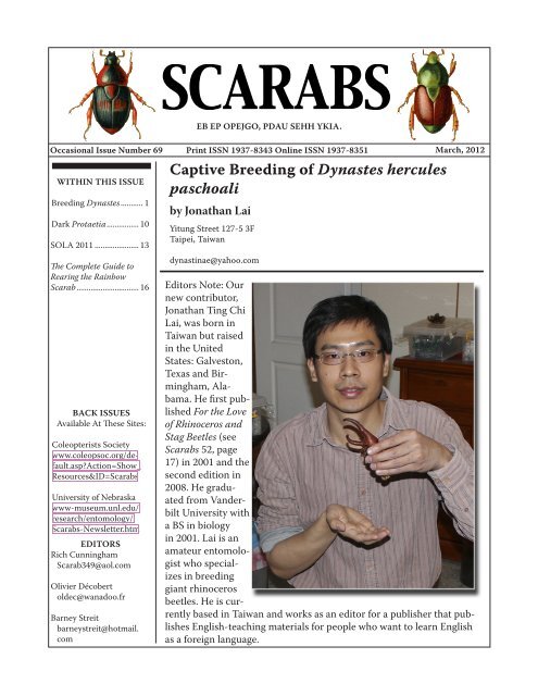
Captive Breeding <strong>of</strong> Dynastes hercules<br />
paschoali<br />
by Jonathan Lai<br />
Yitung Street 127-5 3F<br />
Taipei, Taiwan<br />
dynastinae@yahoo.com<br />
Editors Note: Our<br />
new contributor,<br />
Jonathan Ting Chi<br />
Lai, was born in<br />
Taiwan but raised<br />
in the United<br />
<strong>State</strong>s: Galveston,<br />
Texas and Birmingham,Alabama.<br />
He first published<br />
For the Love<br />
<strong>of</strong> Rhinoceros and<br />
Stag Beetles (see<br />
<strong>Scara</strong>bs 52, page<br />
17) in 2001 and the<br />
second edition in<br />
2008. He graduated<br />
from Vanderbilt<br />
<strong>University</strong> with<br />
a BS in biology<br />
in 2001. Lai is an<br />
amateur entomologist<br />
who specializes<br />
in breeding<br />
giant rhinoceros<br />
beetles. He is currently<br />
based in Taiwan and works as an editor for a publisher that publishes<br />
English-teaching materials for people who want to learn English<br />
as a foreign language.
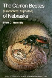
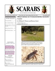
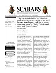
Photo 1: Dynastes hercules paschoali is known to lack<br />
cephalic denticles.<br />
Photo 2: Male larvae are capable <strong>of</strong> growing to over 130<br />
grams.<br />
Photo 3: Larvae are very docile unless provoked. I have<br />
yet to be bitten by one.<br />
Page 2<br />
Over the ages, giant rhinoceros<br />
beetles have fascinated man, to an<br />
extent that they are named after<br />
Greek gods and heroes. The most<br />
famous <strong>of</strong> them all is perhaps the<br />
Hercules Beetle Dynastes hercules,<br />
which is capable <strong>of</strong> growing<br />
to over 17 cm. This mega-titan <strong>of</strong><br />
the insect world was described as<br />
early as 1753 by Carl Linnaeus. To<br />
date, a total <strong>of</strong> 12 subspecies are<br />
known. One <strong>of</strong> the rarest <strong>of</strong> them<br />
is D. h. paschoali, found only on<br />
the Atlantic coast <strong>of</strong> southeastern<br />
Brazil. In fact, it was listed as<br />
an endangered species by Brazil<br />
in 2008. Although this subspecies<br />
was described by Grossi and<br />
Arnaud in 1993, even today, very<br />
few specimens are known and<br />
information regarding this subspecies<br />
is virtually nonexistent. As<br />
passionate as the author is about<br />
D. hercules, he was not able to<br />
see even a photograph <strong>of</strong> a specimen<br />
until 2007. In July <strong>of</strong> 2009,<br />
the author was ecstatic to be able<br />
to obtain 24 newly-hatched F2<br />
generation larvae that ultimately<br />
came from adult beetles collected<br />
in 2007 in Bahia, Brazil, and thus<br />
began an endeavor to unravel the<br />
mysteries <strong>of</strong> D. h. paschoali. The<br />
main objective was to determine<br />
the level <strong>of</strong> difficulty in captive-<br />
rearing this subspecies, and to give<br />
the public an opportunity to finally<br />
look at this elusive giant up close.<br />
(Photo 1)<br />
The 24 first instar larvae were<br />
divided into three groups <strong>of</strong> 8<br />
larvae. Each group was kept in a<br />
plastic container measuring 30<br />
x 20 x 15 cm. The lid <strong>of</strong> the container<br />
was drilled with 6 ventila-
tion holes, each about 0.5 cm in<br />
diameter. A thin cloth was used<br />
to cover the holes to prevent unwanted<br />
pests such as fungus gnats<br />
and cockroaches from entering. The<br />
larvae were fed hardwood sawdust<br />
that had been composted for 4-6<br />
months to rid it <strong>of</strong> indigestible or<br />
toxic substances such as lignin<br />
and plant alkaloids. Three pellets<br />
<strong>of</strong> dried dog food each weighing<br />
roughly 1 gram were distributed<br />
on the bottom <strong>of</strong> each container<br />
before the containers were filled<br />
with substrate to a depth <strong>of</strong> 6 cm.<br />
The humidity <strong>of</strong> the substrate was<br />
adjusted to be roughly 50%. No<br />
substrate change was performed<br />
until the larvae molted to second<br />
instar. The first instar stage lasted<br />
approximately 30 days.<br />
Upon reaching the second instar<br />
stage, the larvae were sexed and<br />
there were 15 males and 9 females.<br />
Unfortunately, one female larva<br />
died <strong>of</strong> molting failure caused by a<br />
jammed head capsule. The remaining<br />
larvae were divided into three<br />
groups <strong>of</strong> 5 larvae and two groups<br />
<strong>of</strong> 4 larvae with mixed sexes. The<br />
dimensions <strong>of</strong> the five rearing<br />
containers remained unchanged.<br />
However, the depth <strong>of</strong> the substrate<br />
was increased to 9 cm. No substrate<br />
change was performed until the<br />
larvae molted to third instar. The<br />
second instar stage lasted approximately<br />
45 days.<br />
Upon reaching third instar, it was<br />
obvious that the male larvae had<br />
bigger heads, with head capsule<br />
diameters ranging from 16.0-17.1<br />
mm (a breeder in Taiwan raised a<br />
larva with 18.0 mm head capsule).<br />
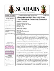
Photo 4: Pre-pupa.<br />
Photo 5: Split <strong>of</strong> dorsal skin marks beginning <strong>of</strong> pupation.<br />
Notice the extremely compressed horns.<br />
Photo 6: Four minutes after the onset <strong>of</strong> pupation.<br />
Page 3
Page 4<br />
Photo 7: Eight minutes after the onset <strong>of</strong> pupation.<br />
Photo 8: 47 minutes after the onset <strong>of</strong> pupation.<br />
Photo 9: Four hours and nine minutes after the onset <strong>of</strong><br />
pupation.<br />
Females ranged from 14.5-15.5 mm<br />
(later experience showed female<br />
larvae were able to attain 16.2<br />
mm). From now on, all larvae were<br />
kept individually. Each female was<br />
kept in a cylindrical container with<br />
13 cm diameter and 19 cm height<br />
(approximately 2.5 liters). The<br />
depth <strong>of</strong> the substrate was kept at<br />
16-17 cm. Each male was kept in a<br />
cylindrical container with 15.5 cm<br />
diameter and 22 cm height (approximately<br />
4.1 liters). The depth<br />
<strong>of</strong> the substrate was kept at 19-20<br />
cm. Three ventilation holes were<br />
drilled in the lids <strong>of</strong> each container.<br />
A substrate change was performed<br />
every six weeks. The entire content<br />
<strong>of</strong> a container would be carefully<br />
dumped out (as digging for the<br />
larva with a spoon could harm the<br />
larva). The container would be<br />
thoroughly washed and the larva<br />
gently rinsed with tap water to<br />
prevent pathogen buildup. In order<br />
to retain beneficial endosymbionts<br />
that help larvae to digest cellulose,<br />
6-8 fecal pellets would be crushed<br />
and returned to the container. The<br />
containers for females would receive<br />
4 pellets <strong>of</strong> dog food and the<br />
containers for males would receive<br />
6 pellets <strong>of</strong> dog food. The larva<br />
would be put back and the last step<br />
would be refilling the container<br />
with clean substrate. Larvae were<br />
kept in a climate-controlled environment<br />
<strong>of</strong> 25º C in the summer<br />
and 20º C in the winter.<br />
Eight months after hatching,<br />
female larvae were beginning to<br />
reach their maximum weight,<br />
ranging from 65-80 grams. Two<br />
months later, females began to<br />
construct pupal cells with 10-30
degree inclination. They rested<br />
inside these oval chambers and became<br />
pupae approximately 30 days<br />
later. The pupal stage lasted about<br />
60 days at 25º C. However, underground<br />
life was not over. New adults<br />
rested in pupal cells for yet another<br />
60 days or more before emerging to<br />
the surface to look for food. They<br />
were fed a diet <strong>of</strong> sliced apples for<br />
20-30 days until fully mature and<br />
ready to mate. Females measured<br />
66-71 mm. (However, a Taiwanese<br />
breeder raised a 78 mm female.)<br />
Male larvae took much longer to<br />
develop because they grew so much<br />
larger, almost twice the weight <strong>of</strong><br />
females. They didn’t begin to reach<br />
maximum weight until 14 months<br />
after hatching. Some extra-large<br />
male larvae were still gaining weight<br />
16 months after hatching. The<br />
maximum weight <strong>of</strong> male larvae<br />
ranged from 107-126 grams (Photos<br />
2 & 3). Once a male larva reached<br />
100 grams, it was relocated to a 30<br />
x 20 x 24 cm container so it would<br />
have enough horizontal space to<br />
construct a pupal cell large enough<br />
to accommodate its long thoracic<br />
horn. (Incidentally, male larvae kept<br />
in 4.1 liter containers also made<br />
pupal cells, but the front and the<br />
end were obstructed by the walls<br />
<strong>of</strong> the containers, resulting in very<br />
short pupal cells. Such larvae were<br />
relocated during the pre-pupal<br />
stage to artificial pupal cells made<br />
from floral foams (otherwise, the<br />
horns would be bent). Male larvae<br />
began to construct pupal cells 16-18<br />
months after hatching. The pupal<br />
cells were inclined, with angles<br />
ranging from 10-30 degrees. The<br />
pre-pupal and pupal stages lasted<br />
Photo 10: 48 hours after the onset <strong>of</strong> pupation.<br />
Photo 11: Pupa very close to eclosion<br />
Photo 12: Breaking pupal skin marks the beginning <strong>of</strong><br />
eclosion.<br />
Photo 13: Ten minutes after the beginning <strong>of</strong> eclosion.<br />
Page 5
Photo 14: 34 minutes after the beginning <strong>of</strong> eclosion.<br />
Photo 15: One hour and 39 minutes after the beginning<br />
<strong>of</strong> eclosion.<br />
Photo 16: Five hours and three minutes after the beginning<br />
<strong>of</strong> eclosion.<br />
Photo 17: Eight hours and 25 minutes after the beginning<br />
<strong>of</strong> eclosion.<br />
Page 6<br />
approximately 45 and 80 days,<br />
respectively, at 20º C (calculation<br />
taken during winter months)<br />
(Photos 4-18). Unfortunately, one<br />
male was unable to successfully<br />
shed his upper larval skin during<br />
pupation and became a severely<br />
deformed pupa that died a few<br />
days later. The remaining 14 males<br />
ranged from 130-145 mm as measured<br />
from the tip <strong>of</strong> the thoracic<br />
horn to the end <strong>of</strong> the elytra (Photo<br />
19). New adults stayed inactive<br />
in their pupal cells for 45-55 days.<br />
(The author is confident that D. h.<br />
paschoali is a subspecies capable<br />
<strong>of</strong> producing male larvae <strong>of</strong> over<br />
140 grams, and subsequently male<br />
adults <strong>of</strong> over 150 mm. He hopes<br />
to reach this goal in the F3 generation.)<br />
The weight <strong>of</strong> a larva did not determine<br />
the absolute length <strong>of</strong> the<br />
adult beetle. It was possible for a<br />
heavier larva to become a shorter<br />
beetle. For example, a heavier larva<br />
might have developed a thicker,<br />
but not necessarily longer horn,<br />
thus becoming shorter than its<br />
lighter counterpart. But generally<br />
speaking, heavier larvae became<br />
longer adults.<br />
Although the 14 males were<br />
brothers, they exhibited a wide<br />
range <strong>of</strong> appearance. In regard to<br />
elytra color (one color per beetle),<br />
various shades <strong>of</strong> green, brown,<br />
and yellow could be found among<br />
the siblings. As for the elytral<br />
spotting, spotless, moderately<br />
spotted, intensely spotted, small<br />
spots, large spots, striped, and<br />
both striped and spotted were<br />
observed (Photo 20). The thoracic
horn ranged from thin (stenoceras)<br />
to thick (pachyceras). The apex <strong>of</strong><br />
the cephalic horn ranged from unhooked<br />
to hooked. The protrusion<br />
right next to the apex ranged from<br />
minute to conspicuous. All <strong>of</strong> the<br />
beetles lacked cephalic denticles.<br />
(Photo 21)<br />
The continual propagation <strong>of</strong> this<br />
captive culture was presented<br />
with a problem. All <strong>of</strong> the females<br />
emerged 4-6 months ahead <strong>of</strong> the<br />
males. Experience with other subspecies<br />
<strong>of</strong> D. hercules has revealed<br />
that females need to be mated<br />
within two months <strong>of</strong> emergence<br />
from the pupal cell, as after this<br />
period, their vitality quickly declines.<br />
It would not be possible for<br />
males and females with an emergence<br />
discrepancy <strong>of</strong> 4-6 months to<br />
mate. Obviously, adult males from<br />
elsewhere that had emerged in sync<br />
with the author’s females had to be<br />
acquired if these females were to<br />
reproduce. Luckily, the author was<br />
able to obtain two mature males<br />
from other breeders. Both <strong>of</strong> the<br />
males were 129 mm in length.<br />
All 8 females were successfully<br />
mated. One male mated with 6<br />
females, while the other mated with<br />
2 females. Interestingly, cold temperature<br />
prolonged mating time.<br />
The first male mated in a temperature<br />
range <strong>of</strong> 18-20º C. Mating<br />
times were 35, 40, 40, 40, 45, and 30<br />
minutes. The second male mated<br />
in a temperature range <strong>of</strong> 15-16º C.<br />
Mating times were 85 and 80 minutes.<br />
Each female mated only once.<br />
(Photo 22)<br />
Photo 18: 18 hours and 45 minutes after the beginning<br />
<strong>of</strong> eclosion.<br />
Photo 19: D. h. paschoali over 144 mm.<br />
Photo 20: Left to right: spotless, small spots, large<br />
spots, stripes.<br />
Page 7
Page 8<br />
Photo 21: Some variations in the cephalic horn.<br />
Photo 22: D. h. paschoali mating.<br />
After mating, each female was<br />
individually placed in a 30 x 25 x<br />
30 cm breeding container filled<br />
to a depth <strong>of</strong> 20 cm with the same<br />
substrate used to rear larvae. The<br />
bottom 3-4 cm was thoroughly<br />
compressed by hand to aid the<br />
females’ oviposition (as females<br />
oviposit, they pack the substrate<br />
into hard clumps in which they<br />
lay their eggs. It is speculated that<br />
the hard clumps protect eggs from<br />
predators). The females were continuously<br />
provided with slices <strong>of</strong><br />
apple placed on a plate located on<br />
the top <strong>of</strong> the substrate. The slices<br />
were changed every 3 days to prevent<br />
spoilage. The females would<br />
alternate between spending a few<br />
days on the apples and remaining<br />
for several days underground. The<br />
entire content <strong>of</strong> each container<br />
was dumped out to check for eggs<br />
every 3-4 weeks. Substrate clumps<br />
had to be taken apart to harvest<br />
the eggs. The eggs were then<br />
placed on the bottom <strong>of</strong> a clear<br />
plastic container and covered very<br />
gently with 4 cm <strong>of</strong> substrate. The<br />
development <strong>of</strong> the eggs could be<br />
observed by looking at the bottom<br />
<strong>of</strong> the container. Eggs absorbed<br />
moisture and gradually expanded<br />
to about twice their original size<br />
before hatching. Incubation lasted<br />
approximately 30 days (Photo 23).<br />
Below is a breeding summary <strong>of</strong><br />
the 8 females:<br />
The adult females lived up to<br />
200 days, and the adult males<br />
up to 300 days. Despite feeding<br />
throughout their lives, the<br />
adults progressively lost weight<br />
until death. The life cycle <strong>of</strong> D. h.<br />
paschoali was successfully docu-
mented. Given the high survival<br />
rate <strong>of</strong> the F2 generation larvae<br />
(over 90%) and the large imagoes<br />
produced, it was concluded that<br />
captive raising D. h. paschoali was<br />
not only possible, but not difficult<br />
(given the larvae were fed proper<br />
substrate).<br />
Photo 23: Eggs in various stages <strong>of</strong> development with<br />
newly-hatched larva on far right.<br />
Female # 1 2 3 4 5 6 7 8<br />
Size 67 mm 68 mm 66 mm 68 mm 68 mm 70 mm 68 mm 68 mm<br />
Eggs<br />
Laid<br />
103 76 67 157 89 63 101 54<br />
Larvae<br />
Hatched<br />
Weight<br />
92 42 45 90 67 42 89 40<br />
at Mating<br />
20 g 22 g 21 g 22 g 22 g 23 g 21 g 19 g<br />
Weight<br />
at Death<br />
Time in<br />
14 g 16 g 14 g 14 g 17 g 16 g 16 g 14 g<br />
BreedingContainer<br />
119 days 141 days 172 days 177 days 142 days 171 days 142 days 97 days<br />
It is worth pointing out that although<br />
the F2 generation individuals<br />
exhibited sex-based discrepancies<br />
in eclosion, subsequent<br />
experience with another, unrelated<br />
group <strong>of</strong> 11 F2 generation larvae<br />
showed no such discrepancy<br />
(both groups kept under identical<br />
conditions); all males and females<br />
eclosed at about the same time.<br />
(Photo 24).<br />
At the time <strong>of</strong> this writing, many<br />
F3 generation larvae are healthy<br />
third instar larvae.<br />
Photo 24: Power <strong>of</strong> captive propagation.<br />
Page 9
Page 10<br />
Some Dark French Protaetia<br />
by Olivier Décobert<br />
oldec@wanadoo.fr<br />
After reading some <strong>of</strong> the past issues <strong>of</strong> <strong>Scara</strong>bs, the reader could think that French Protaetia<br />
are always full colored and shiny. I must admit that I forgot to write about dark species <strong>of</strong> this<br />
genus in my country.<br />
I had intended to tell more about this subject after reading the“Black is beautiful, Baby” that<br />
Brett Ratcliffe recalled on the first page <strong>of</strong> <strong>Scara</strong>bs 46 (December, 2009). But I had many other<br />
articles in my mind at that time and again, I forgot...<br />
In April, 2011, I was in the Oriental Pyrenees mountains when I found a larva under a rock<br />
(Photo 1). My first thought was “melolonthid” but it was indeed a cetonid larva, which is more<br />
habitually found in decaying wood. Here, this larva was in a little cavity dug in the soil. I don’t<br />
know what it exactly ate, probably roots <strong>of</strong> grasses or small surrounding plants.<br />
Photo 1: Larva.
As usual, the question was “What is the species?” It is impossible to know before rearing and<br />
obtaining the adult. This event happened two months later, after a transition by a pupa in a shell<br />
(Photos 2, 3 & 4). The adult was clearly Protaetia morio (Fabricius), a black species, whose size<br />
can vary between 12 and 20 mm. It was the time to show some dark Protaetia in <strong>Scara</strong>bs!<br />
Photo 2: Shell Photo 3: Pupa in shell. Photo 4: Pupa.<br />
As one can see on Photo 5, the emerging adult is not yet black, and this will take two or three<br />
days to be definitive (Photo 6).<br />
Photos 5 and 6: Teneral adult <strong>of</strong> Protaetia morio (left) and mature adult (right).<br />
Page 11
Photo 7: Protaetia morio form.<br />
Page 12<br />
White marks exist on the pronotum and elytra, and can sometimes be<br />
numerous (Photo 7). This form is called “quadripunctata” (Fabricius). I<br />
found this one on the southwestern coast <strong>of</strong> France.<br />
A close species is Protaetia oblonga (Gory & Percheron),<br />
smaller (12-16 mm) and rather brown than black. I found the<br />
specimen shown in Photo 8 near the French Alps.<br />
Photo 9: Protaetia opaca.<br />
Protaetia opaca (Fabricius) (Photo 9) is a bigger species (up to 27<br />
mm). This cetonid likes to frequent beehives to eat the honey! I<br />
found this specimen in the South <strong>of</strong> France.<br />
It is impossible to forget Corsica Island, where a dark blue<br />
endemic cetonid lives: Protaetia sardea (Gory & Percheron),<br />
about 25 mm in size (Photo 10).<br />
Photo 8: Protaetia oblonga.<br />
Photo 10: Protaetia sardea.
SOLA - 2011<br />
by Andrew Smith<br />
Canadian <strong>Museum</strong> <strong>of</strong> Nature<br />
asmith@unlserve.unl.edu<br />
Photo 1: The 14th annual SOLA (Sacred Order <strong>of</strong><br />
Lamellate Antennae) <strong>Scara</strong>b Workers Symposium<br />
was held in Reno, Nevada on November 13th.<br />
Yours truly was again the host and organizer <strong>of</strong><br />
this event. As always, SOLA was part <strong>of</strong> the larger<br />
Entomological Society <strong>of</strong> America (ESA) meeting.<br />
Photo 2: The customary SOLA salute was given<br />
at the beginning <strong>of</strong> the symposium. Those who<br />
abstained or demonstrated poor form were<br />
invited to attend the carabid symposium down<br />
the hall.<br />
Photo 3: Bob Anderson gave a sobering<br />
presentation “<strong>Scara</strong>b beetles as bycatch”<br />
reminding everyone that scarab beetles are not<br />
the primary focus <strong>of</strong> all field work.<br />
Photo 4: Paul Skelley and M.J. Paulsen, both<br />
sporting semi-impressive facial hair, gave<br />
separate presentations on “Bagging big trophies”<br />
and “Fighting for a meaningful classification <strong>of</strong><br />
Lucanidae.” SOLA presentations apparently get<br />
bonus marks if the title begins with an action<br />
verb in the present continuous tense.<br />
Page 13
Photo 5: Sergey Tarasov wowed the crowd<br />
with his detailed phylogenetic work on dung<br />
beetles during his presentation “Clarifying<br />
systematics <strong>of</strong> <strong>Scara</strong>baeinae: cybertaxonomy,<br />
revision and phylogeny <strong>of</strong> Oriental deltochiline<br />
genera Cassolus and Parachorius (Coleoptera:<br />
<strong>Scara</strong>baeidae: <strong>Scara</strong>baeinae).”<br />
Photo 6: Bruce Gill finally gets his chance to tell<br />
everyone about his field work in Africa with his<br />
presentation “A second try - searching for scarabs<br />
in Katanga, D.R. Congo.” Bruce had to cancel this<br />
presentation last year, which lead to an awkward<br />
20 minutes <strong>of</strong> listening to crickets chirping (see<br />
<strong>Scara</strong>bs 60, page 10).<br />
Page 14<br />
Photo 7: Alan Mudge gave more interesting<br />
insights into African field work during his<br />
presentation “Cetoniine diversity <strong>of</strong> the Upper<br />
Guinean forests and savannas <strong>of</strong> Ghana.”<br />
Photo 8: Mike Klein gave out lots <strong>of</strong> door prizes<br />
after the SOLA meeting. Many were featured<br />
in his presentation “<strong>Scara</strong>b collecting: trash or<br />
treasure?”
Photo 9: Bill Warner chastising a hapless<br />
audience member on his pronunciation <strong>of</strong><br />
Chnaunanthus during his presentation “Tiny,<br />
obscure, and floral, a preliminary look at the<br />
hard-to-pronounce genus Chnaunanthus.”<br />
Photo 10: Jorge Leon-Cortes ponders a question<br />
during his presentation on “The ecology<br />
and distribution <strong>of</strong> two exotic scarab beetles<br />
in Chiapas: Euoniticellus intermedius and<br />
Digitonthophagus gazella.”<br />
Photo 11: M.J. Paulsen proudly displays his<br />
award for the 2011 Outstanding SOLA Paper.<br />
M.J. demonstrated outstanding perseverance<br />
and dedication in battling through the sorry<br />
state <strong>of</strong> lucanid classification to provide us with<br />
a new meaningful classification. M.J. promised<br />
to hand out reprints <strong>of</strong> his lucanid classification<br />
paper at the 2012 SOLA meeting.<br />
Photo 12: The SOLA meeting was again<br />
inundated by Tenebrionidae workers, who in<br />
recent years have been organizing their own<br />
ESA symposia. Imitation is the sincerest form<br />
<strong>of</strong> flattery we suppose! Shown here are Kojan<br />
Kanda, Ron Somerby, Chuck Triplehorn,<br />
Aaron Smith, Don Thomas, Rolf Aalbu, Warren<br />
Steiner, and Pat Bouchard. A special thanks<br />
to Jil Swearington for providing all <strong>of</strong> the<br />
photographs from the 2011 SOLA meeting!<br />
Page 15
Page 16<br />
Literature Notice: The Complete Guide to<br />
Rearing the Rainbow <strong>Scara</strong>b and Other<br />
Dung Beetles<br />
by Barney Streit<br />
Our new layout editor Tiffany displaying our copy <strong>of</strong><br />
The Complete Guide to Rearing the Rainbow <strong>Scara</strong>b and<br />
Other Dung Beetles.<br />
Tiffany points out that interested readers should also<br />
download <strong>Scara</strong>baeus Number 6 (available at the two<br />
web sites listed on page one) and read Dave Edmonds’<br />
article “Facilities for Raising Dung Beetles.” Dave also<br />
wrote a nice summary <strong>of</strong> scarabaeine nesting behavior<br />
in <strong>Scara</strong>baeus Number 2.<br />
The Complete Guide to Rearing the<br />
Rainbow <strong>Scara</strong>b and Other Dung<br />
Beetles, by Steven Barney and<br />
Orin McMonigle, is now available<br />
through Amazon (http://www.<br />
amazon.com/).<br />
Although perhaps a bit expensive<br />
($14) for a 40-page s<strong>of</strong>tcover<br />
guide, this is a nifty little book. It<br />
contains everything you would<br />
need to know about rearing dung<br />
beetles. There are sections about<br />
the background <strong>of</strong> scarabs, dung<br />
beetle behavior, captive breeding<br />
<strong>of</strong> Phanaeus, trapping and<br />
collecting, food and bait, other<br />
U.S. dung beetles, a glossary and<br />
bibliography. There are both black<br />
and white and color plates. The<br />
featured dung beetle, Phanaeus<br />
vindex (the “rainbow scarab”), is<br />
thoroughly discussed.<br />
Other guides in this series include<br />
The Complete Guide to Rearing the<br />
Eastern Hercules Beetle and Other<br />
Rhinocerus Beetles, The Complete<br />
Guide to Rearing the Elephant Stag<br />
Beetle and Other Stags, and The<br />
Complete Guide to Rearing Flower<br />
and Jewel <strong>Scara</strong>bs.