Bivalvia: Protobranchia: Nuculidae - Western Australian Museum
Bivalvia: Protobranchia: Nuculidae - Western Australian Museum
Bivalvia: Protobranchia: Nuculidae - Western Australian Museum

Create successful ePaper yourself
Turn your PDF publications into a flip-book with our unique Google optimized e-Paper software.
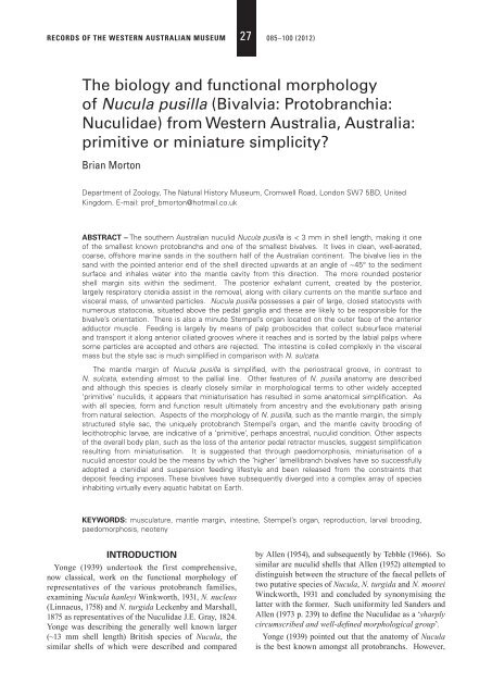
RECORDS OF THE WESTERN AUSTRALIAN MUSEUM 27 085–100 (2012)<br />
The biology and functional morphology<br />
of Nucula pusilla (<strong>Bivalvia</strong>: <strong>Protobranchia</strong>:<br />
<strong>Nuculidae</strong>) from <strong>Western</strong> Australia, Australia:<br />
primitive or miniature simplicity?<br />
Brian Morton<br />
Department of Zoology, The Natural History <strong>Museum</strong>, Cromwell Road, London SW7 5BD, United<br />
Kingdom. E-mail: prof_bmorton@hotmail.co.uk<br />
ABSTRACT – The southern <strong>Australian</strong> nuculid Nucula pusilla is < 3 mm in shell length, making it one<br />
of the smallest known protobranchs and one of the smallest bivalves. It lives in clean, well-aerated,<br />
coarse, offshore marine sands in the southern half of the <strong>Australian</strong> continent. The bivalve lies in the<br />
sand with the pointed anterior end of the shell directed upwards at an angle of ~45° to the sediment<br />
surface and inhales water into the mantle cavity from this direction. The more rounded posterior<br />
shell margin sits within the sediment. The posterior exhalant current, created by the posterior,<br />
largely respiratory ctenidia assist in the removal, along with ciliary currents on the mantle surface and<br />
visceral mass, of unwanted particles. Nucula pusilla possesses a pair of large, closed statocysts with<br />
numerous statoconia, situated above the pedal ganglia and these are likely to be responsible for the<br />
bivalve’s orientation. There is also a minute Stempel’s organ located on the outer face of the anterior<br />
adductor muscle. Feeding is largely by means of palp proboscides that collect subsurface material<br />
and transport it along anterior ciliated grooves where it reaches and is sorted by the labial palps where<br />
some particles are accepted and others are rejected. The intestine is coiled complexly in the visceral<br />
mass but the style sac is much simplifi ed in comparison with N. sulcata.<br />
The mantle margin of Nucula pusilla is simplifi ed, with the periostracal groove, in contrast to<br />
N. sulcata, extending almost to the pallial line. Other features of N. pusilla anatomy are described<br />
and although this species is clearly closely similar in morphological terms to other widely accepted<br />
‘primitive’ nuculids, it appears that miniaturisation has resulted in some anatomical simplifi cation. As<br />
with all species, form and function result ultimately from ancestry and the evolutionary path arising<br />
from natural selection. Aspects of the morphology of N. pusilla, such as the mantle margin, the simply<br />
structured style sac, the uniquely protobranch Stempel’s organ, and the mantle cavity brooding of<br />
lecithotrophic larvae, are indicative of a ‘primitive’, perhaps ancestral, nuculid condition. Other aspects<br />
of the overall body plan, such as the loss of the anterior pedal retractor muscles, suggest simplifi cation<br />
resulting from miniaturisation. It is suggested that through paedomorphosis, miniaturisation of a<br />
nuculid ancestor could be the means by which the ‘higher’ lamellibranch bivalves have so successfully<br />
adopted a ctenidial and suspension feeding lifestyle and been released from the constraints that<br />
deposit feeding imposes. These bivalves have subsequently diverged into a complex array of species<br />
inhabiting virtually every aquatic habitat on Earth.<br />
KEYWORDS: musculature, mantle margin, intestine, Stempel’s organ, reproduction, larval brooding,<br />
paedomorphosis, neoteny<br />
INTRODUCTION<br />
Yonge (1939) undertook the first comprehensive,<br />
now classical, work on the functional morphology of<br />
representatives of the various protobranch families,<br />
examining Nucula hanleyi Winkworth, 1931, N. nucleus<br />
(Linnaeus, 1758) and N. turgida Leckenby and Marshall,<br />
1875 as representatives of the <strong>Nuculidae</strong> J.E. Gray, 1824.<br />
Yonge was describing the generally well known larger<br />
(~13 mm shell length) British species of Nucula, the<br />
similar shells of which were described and compared<br />
by Allen (1954), and subsequently by Tebble (1966). So<br />
similar are nuculid shells that Allen (1952) attempted to<br />
distinguish between the structure of the faecal pellets of<br />
two putative species of Nucula, N. turgida and N. moorei<br />
Winckworth, 1931 and concluded by synonymising the<br />
latter with the former. Such uniformity led Sanders and<br />
Allen (1973 p. 239) to defi ne the <strong>Nuculidae</strong> as a ‘sharply<br />
circumscribed and well-defi ned morphological group’.<br />
Yonge (1939) pointed out that the anatomy of Nucula<br />
is the best known amongst all protobranchs. However,
86 B. MORTON<br />
there has never been (i) any examination of the anatomy<br />
of an <strong>Australian</strong> species nor (ii) of such a minute taxa<br />
as N. pusilla Angas, 1877, the object of this study. This<br />
is surprising considering Lamprell and Healy (1998)<br />
redescribed 21 species of nuculids belonging to three<br />
genera (Nucula Gray, 1824, Rumptunucula Bergmans,<br />
1978 and Leionucula Quenstedt, 1930) from <strong>Australian</strong><br />
waters, but is possibly less so when the small size of N.<br />
pusilla is considered. The research reported here thus<br />
complements that of Yonge (1939) and draws on the<br />
anatomical researches of earlier workers such as Drew<br />
(1899; 1901), Pelseneer (1911) and Heath (1937) as well<br />
as more contemporary authors who have researched the<br />
fi ner details of different nuculid species. These latter<br />
authors will be identifi ed in the relevant sections of this<br />
text. This study also seeks to provide better illustrations<br />
of a nuculid taxon, congeners of which have not<br />
previously been illustrated as living entities but as either<br />
dried or preserved specimens, for example Hampson<br />
(1971), Allen (1978), and Yonge (1939).<br />
Finally, because Nucula pusilla is so small,<br />
this study may give some insights into what the<br />
primitive, ancestral bivalve was like as they were<br />
also tiny (Runnegar 1978). Yonge (1939) considered<br />
the <strong>Nuculidae</strong> to be the least specialised of the<br />
<strong>Protobranchia</strong> and to be representative of the most<br />
primitive living lamellibranch bivalves. Conversely,<br />
Graham (1949) studied the stomach of N. hanleyi and<br />
noted (page 742): ‘the structure of the stomach of Nucula<br />
does not in some respects suggest primitiveness, but a<br />
certain degree of specialisation’. Following a study of<br />
N. nucleus, Purchon (1956) linked the protobranchs with<br />
the anomalodesmatan septibranchs because of stomach<br />
simplicity, although such a similarity is derived from the<br />
fact that representatives of both ingest large particles of<br />
food resulting in a high degree of extracellular digestion.<br />
In the case of the latter, this is because of the ingestion<br />
of preyed-upon small crustaceans, for example, in<br />
species of Cuspidaria (Reid and Reid 1974), not detritus<br />
as in protobranchs.<br />
Modern biologists now view animals under a<br />
somewhat different light suggesting that in order<br />
to survive, all species must be specialised to meet<br />
the demands of their ecology in its broadest sense.<br />
Hence, although the <strong>Nuculidae</strong> may in some ways be<br />
representative of the primitive, ancestral bivalve form<br />
and lifestyle, suggested by Yonge (1939), arguably the<br />
greatest 20th century authority on the <strong>Bivalvia</strong>, the<br />
minuteness of N. pusilla may illuminate aspects of the<br />
ancestral nuculid and/or bivalve condition, as well as the<br />
role that minuteness plays in determining the complexity<br />
(or not) of functional anatomy. Whether N. pusilla<br />
would reveal any insights into how its anatomy refl ected<br />
either primitive or miniature simplicity became the<br />
overall aim of this study.<br />
MATERIALS AND METHODS<br />
Individuals of Nucula pusilla were obtained by sieving<br />
coarse shallow subtidal sands from off Cape Vlamingh,<br />
Rottnest Island, off Perth, <strong>Western</strong> Australia, during<br />
the course of the Fifth International Marine Biological<br />
Workshop, convened on Rottnest Island from 1–24<br />
January 1991. The antero-posterior shell lengths of each<br />
individual plus empty right valves were measured to the<br />
nearest 0.1 mm using vernier calipers. Subsequently,<br />
some of the largest individuals were dissected and<br />
the ciliary currents of the mantle cavity elucidated<br />
by the application of drops of carmine suspended in<br />
seawater. Two other individuals of ~1 mm and ~2 mm<br />
shell length were fi xed in Bouin’s fl uid and following<br />
routine histological procedures, sectioned transversely<br />
at 4 μm, and resulting slides stained in either Ehrlich’s<br />
haematoxylin and eosin or Masson’s trichrome.<br />
For comparison, two individuals of Nucula sulcata<br />
(Bronn, 1831) were obtained from the Milport Marine<br />
Station courtesy of the then Director J.A. Allen. These<br />
were dissected and sectioned using the same procedures<br />
as described above for N. pusilla.<br />
RESULTS<br />
TAXONOMY AND FOSSIL HISTORY<br />
Keen (1969) considered that representatives of<br />
the <strong>Nuculidae</strong> have a fossil history dating from the<br />
Ordovician and that Recent species occur almost<br />
worldwide. Angas (1877, page 177, plate XXVI, fi gure<br />
26) described and recorded Nucula pusilla from shellysand<br />
from Port Jackson, South Australia. Bergmans<br />
(1978) described the taxonomy and distribution of<br />
fourteen species of Recent <strong>Australian</strong> Nucula and<br />
selected a lectotype for N. pusilla Angas, 1877, which is<br />
held in the collections of the Natural History <strong>Museum</strong>,<br />
London (BMNH 1877.5.12.61), and considered N.<br />
hedleyi Pritchard and Gatliff, 1904, N. micans Angas,<br />
1879 and Pronucula concentrica Cotton, 1930 to be<br />
junior synonyms. According to Huber (2010), another<br />
synonym is N. minuta Tenison-Woods, 1876 but this<br />
species is restricted to the Canadian Arctic and is likely<br />
to be a valid taxon. The shell of the lectotype of N.<br />
pusilla is illustrated in Bergmans (1978, fi gures 2–4).<br />
In the collection of fossil nuculids contained in the<br />
<strong>Western</strong> <strong>Australian</strong> <strong>Museum</strong> (WAM), N. pusilla has<br />
been obtained from Pliocene, Pleistocene and Recent<br />
strata at Jandikot and Redcliffe in Perth and from West<br />
Gingin, north of Perth, and Rottnest Island.<br />
BIOLOGY<br />
Nucula pusilla can be considered to have a southern<br />
<strong>Australian</strong>, cold temperate distribution, and to live in<br />
clean, nearshore, oceanic sediments. With a recorded<br />
maximum shell length of 2.92 mm (Bergmans 1978), N.<br />
pusilla is a minute occupant of sediments comprising<br />
medium to coarse well sorted sands (0.25–1.00 mm)<br />
(Glover and Taylor 1999). Adult N. pusilla individuals<br />
are only a little bigger than the largest grains in the<br />
sediments in which they live.
BIOLOGY AND MORPHOLOGY OF NUCULA PUSILLA 87<br />
FIGURE 1 Nucula pusilla. Shell length frequency<br />
histogram of a sample of (i) living individuals<br />
and (ii) empty right shell valves collected<br />
from offshore sands at Cape Vlamingh,<br />
Rottnest Island, <strong>Western</strong> Australia.<br />
FIGURE 2 Nucula pusilla. An illustration of the living<br />
animal buried in a natural position in the<br />
sand and showing the anterior inhalant and<br />
posterior exhalant streams. Also shown<br />
are the tips of the palp proboscides in the<br />
sediment and the posterior ends of the<br />
ctenidia.<br />
Figure 1 is a shell length frequency histogram of a<br />
sample of (i) living individuals and (ii) empty right shell<br />
valves of Nucula pusilla collected from offshore sands<br />
at Cape Vlamingh, Rottnest Island, <strong>Western</strong> Australia.<br />
It comprises two peaks of living individuals of ~1.4<br />
mm and ~2.4 mm shell length and empty (right) valves<br />
of approximately matching sizes (1.4 mm and 2.2 mm<br />
shell lengths). The smallest living individual had a<br />
shell length of 0.8 mm. I have no data on the population<br />
density of N. pusilla at Cape Vlamingh but this may not<br />
be signifi cant as the species is particularly susceptible<br />
to changes in water quality. Macleod and Forbes (2004)<br />
found numbers of N. pusilla declined rapidly in the<br />
sediments beneath fi nfi sh farms in Tasmania subjected<br />
to eutrophication from artifi cial feeds, but recovered<br />
once impacts ceased.<br />
In laboratory aquaria with a bed of sand from their<br />
natural habitat, living individuals of Nucula pusilla<br />
reburied themselves quickly and adopted positions<br />
generally similar to that illustrated in Figure 2. In<br />
FIGURE 3 Nucula pusilla. The method of locomotion<br />
with the foot extended forward and the shell<br />
pulled towards it by means of the contraction<br />
of the posterior pedal retractor muscle. (For<br />
abbreviations see Appendix).<br />
this natural position, they buried themselves with the<br />
posterior ends downwards and the pointed anterior<br />
ends of their shells oriented somewhat upwards out of<br />
the sediment. The application of suspended carmine to<br />
living individuals identifi ed an anterior inhalant stream<br />
and some of this was eventually passed out of the animal<br />
postero-ventrally as a posterior exhalant stream. Also<br />
illustrated are the tips of the palp proboscides extended<br />
into the sediment and the posterior ends of the ctenidia<br />
that extend beyond the posterior margin of the shell<br />
valves (Figure 2).<br />
The foot of various species of Nucula has been<br />
described as a ‘creeping’ organ (Forbes and Hanley<br />
1853, Pelseneer 1891, Verrill and Bush 1897, 1898).<br />
Yonge (1939) rectifi ed this error and described, but<br />
did not illustrate, how locomotion was achieved.<br />
The method of locomotion exhibited by N. pusilla<br />
is illustrated in Figure 3. From a resting position<br />
illustrated in Figure 3A, the marginally frilled foot is<br />
extended forwards in an anteriorly oblique direction,
88 B. MORTON<br />
expanded, and flared outwards (Figure 3B). The<br />
shell is then pulled forwards by contraction of the<br />
paired posterior pedal retractor muscles (Figure 3C).<br />
Associated with this action the parted shell valves are<br />
brought together by adduction of the adductor muscles<br />
and this provides the lift and momentum for the whole<br />
animal to be pulled forward. Further foot extensions<br />
may occur subsequently, such that an individual may<br />
either continue forward motion or return to and adopt a<br />
position of repose (Figure 3D). The frilled ploughshare<br />
structure of the foot of N. pusilla is similar to those of<br />
the preserved nuculids illustrated by various authors, for<br />
example Pelseneer (1911, plate I, fi gure 1) and the highly<br />
active, and deep burrowing, Solemya parkinsoni Smith,<br />
1874 (Owen 1961, fi gure 2D) and S. velesiana Iredale,<br />
1931 (Taylor et al. 2008, fi gure 12A and B).<br />
ANATOMY<br />
The shell<br />
The shell of Nucula pusilla is minute, the largest<br />
individual here reported having a length of 2.8 mm.<br />
Bergmans (1978, table 1) recorded that the holotype<br />
of Pronucula concentrica Cotton, 1930 from Gulf St<br />
Vincent, which he considered to be a junior synonym<br />
of N. pusilla, had a shell length of 3.75 mm. All other<br />
specimens reported by Bergmans had shell lengths<br />
50 mm. At 20<br />
mm, N. sulcata is the largest British species, all four<br />
other species having maximum shell lengths of ~13 mm<br />
(Tebble 1966).<br />
The aragonitic shells of all nuculids, both Recent and<br />
fossil, examined by Taylor et al. (1969, table 1) comprise<br />
three layers, an outer layer of composite prisms, and<br />
middle and inner layers of lenticular and sheet nacre,<br />
respectively. The equivalve shell of N. pusilla (Figure<br />
4) is obliquely and triangularly ovate, or wedge-shaped,<br />
and distinctly inequilateral, the antero-dorsal margin<br />
sloping more gently than the postero-dorsal resulting<br />
in truncate anterior and more rounded posterior valve<br />
margins. Externally (Figure 4A), the shell is relatively<br />
thin, pale greenish-white, fi nely and commarginally<br />
striated externally, and crossed with extremely delicate<br />
radiating striae. The ventral margin is smoothly<br />
arcuate, and the postero-dorsal umbones swollen with<br />
opisthogyrate umbones. Internally (Figure 4B) the shell<br />
of N. pusilla is nacreous, as in all nuculids, and slightly<br />
translucent.<br />
When the shell of Nucula pusilla is angled slightly<br />
(Figure 5A) the scars of the anterior (AA) and posterior<br />
(PA) adductor muscles can be clearly seen. The scar of<br />
the posterior pedal retractor (PPR) is visible beneath the<br />
posterior region of the hinge plate, and the extremely<br />
thin, obscure and deeply recessed pallial line (PL) is<br />
also more obvious. The prodissoconch of N. pusilla is<br />
large (~260 μm in length) and corroded (Figure 5B).<br />
Also apparent are tiny larval hinge teeth (LT) (Figure<br />
5B). Another view of the interior of the right shell<br />
valve shows the structure of the hinge in greater detail<br />
FIGURE 4 Nucula pusilla. The shell. A. Exterior view of<br />
the left shell valve. B. Interior view of the<br />
right shell valve.<br />
FIGURE 5 Nucula pusilla. A. Angled view of the interior<br />
of a 2 mm long left shell valve with the<br />
adductor muscles and pallial line identifi ed. B.<br />
Corroded prodissoconch, ~260 μm in length<br />
also showing the larval teeth. C. Interior view<br />
of the left shell valve of an individual of ~1.6<br />
mm shell length showing the structure of the<br />
hinge in greater detail. D. Detailed, oblique,<br />
view of the hinge teeth of the left shell valve.<br />
(For abbreviations see Appendix).<br />
(Figure 5C) and the central approximately amphidetic,<br />
opisthogyrate ligament with the prodissoconch situated<br />
above it, and the taxodont anterior (x6) and posterior<br />
(x3) hinge teeth. A more detailed view of the hinge<br />
teeth of the right shell valve (Figure 5D) shows that the<br />
anterior hinge teeth are pointedly curved towards the<br />
anterior, with a socket behind.<br />
The hinge plate is also illustrated in Figure 5C. The<br />
triangular ligament of the hinge plate is internal and<br />
situated on a resilifer, not a chondrophore, as suggested<br />
by Bergmans (1978). In an adult individual 2.5 mm<br />
in shell length, taxodont hinge teeth are present both<br />
anteriorly and posteriorly, the maximum numbers being<br />
eight and four, respectively, for example in the lectotype<br />
(Bergmans 1978, fi gure 3). The thin pallial line and<br />
the scars of the larger posterior and smaller anterior
BIOLOGY AND MORPHOLOGY OF NUCULA PUSILLA 89<br />
adductor muscles are visible, while the scar of the only<br />
pedal retractor muscle, the posterior, is hidden under the<br />
hinge plate.<br />
The musculature<br />
The musculature of Nucula pusilla comprises<br />
approximately equal anterior and posterior adductor<br />
muscles (Figure 3A, AA; PA), although the former<br />
is slightly larger than the latter, so the species<br />
is approximately isomyarian. There is a large<br />
posterior pedal retractor muscle (PRM), but no anterior<br />
equivalent. Neither Yonge (1939) nor Hampson (1971)<br />
describe any pedal retractor muscles for the nuculids<br />
they examined although Mikkelsen and Bieler (2008)<br />
illustrate and describe both a large posterior and a<br />
smaller anterior pedal retractor for an unidentifi ed,<br />
possibly generalised, species of Nucula. Haszprunar<br />
(1985, fi gure 1a) described a similar arrangement of<br />
muscles in N. sulcata.<br />
A Stempel’s organ is present on the anterior, outer face<br />
of the anterior adductor muscle of Nucula pusilla (Figure<br />
6, SO). It is a minute structure closely adherent to the<br />
anterior adductor muscle (AA) and embedded within<br />
the united inner folds of the mantle margin (IMF). The<br />
organ is ~6 μm in diameter and comprises a sphere of<br />
nucleolated cells enclosing a hollow central core with a<br />
statolith-like structure inside. The mantle margin above<br />
the anterior adductor muscle comprises outer (OMF) and<br />
middle (MMF) mantle folds with the periostracum (PE)<br />
arising between them. The outer mantle fold is here<br />
densely occupied by sub-epithelial secretory cells (SC).<br />
Comparison with Nucula sulcata<br />
The Stempel’s organ present in Nucula sulcata<br />
has been described in great detail by Haszprunar<br />
(1985). As in N. pusilla, the organ in N. sulcata is<br />
located on the anterior face of the anterior adductor<br />
muscle. It has a form reminiscent of the muscle of N.<br />
pusilla (Haszprunar 1985, fi gure 4), but is much more<br />
complex and comprises a closed tube that is innervated<br />
from the pleural ganglia. The lumen of the organ<br />
is fi lled with a ventral crest that comprises ciliated<br />
cells and it is believed that the structure functions<br />
as a mechanoreceptor, possibly associated with the<br />
coordination of the organs of feeding, the labial palps,<br />
palp proboscides, and possibly the ctenidia.<br />
The mantle margin<br />
The mantle margin of Nucula pusilla is divided<br />
throughout its extent, except dorsally beneath the hinge<br />
plate and between the adductor muscles. The mantle<br />
margin of N. pusilla is exceedingly simple (Figure 7)<br />
but does comprise the three folds typical of the majority<br />
of the bivalves except for some arcoids, e.g. Arca noae<br />
(Linnaeus, 1758) (Morton and Peharda 2008). The inner<br />
fold (Figure 7, IMF) is small and swollen and contains<br />
the pallial nerve (PN). The middle (MMF) and outer<br />
mantle folds (OMF) are exceedingly long and receive<br />
most of the components of the pallial retractor muscle<br />
(PRM), which is also long, refl ecting the distance of<br />
FIGURE 6 Nucula pusilla. Transverse section through<br />
Stempel’s organ located on the outer<br />
face of the anterior adductor muscle. (For<br />
abbreviations see Appendix).<br />
the pallial line inwards from each valve margin (Figure<br />
5A, PL). Between these two folds lies the periostracal<br />
groove (Figure 7, PEG) which basally secrets the<br />
periostracum that stains blue in Masson’s trichrome.<br />
Comparison with Nucula sulcata<br />
The mantle margin of this species of Nucula also<br />
comprises the three folds typical of the <strong>Bivalvia</strong> (Figure<br />
8) except that the folds are arranged differently from<br />
N. pusilla. The inner mantle fold (IMF) is much larger<br />
and the middle (MMF) and outer folds (OMF) are<br />
considerably shorter in N. sulcata. As a consequence,<br />
although the pedal retractor muscle (PRM) in N.<br />
sulcata is as long, relative to N. pusilla, the periostracal<br />
groove (PEG) is proportionally much shorter (Figure<br />
8). In the two much larger individuals of N. sulcata,<br />
the periostracum (PE) is seen to comprise two layers,<br />
an inner layer that stains blue in Masson’s trichrome<br />
suggesting a mucoid structure, and a thin outer chitinous<br />
layer.<br />
The organs of the mantle cavity<br />
The organs and ciliary currents of the mantle cavity<br />
of Nucula pusilla, seen from the left side, are illustrated<br />
in Figure 9. In living individuals (Figure 2), there is a<br />
strong anterior inhalant stream and an equally distinct<br />
posterior exhalant fl ow which are presumably almost<br />
completely a respiratory flow, as the water passes<br />
through and around the paired and curved ctenidia<br />
that comprise inner (ID) and outer demibranchs (OD),<br />
located in the posterior region of the mantle cavity. The<br />
ctenidia, or gills, of an unidentifi ed species of Nucula<br />
were studied by Orton (1912) and discussed by Atkins<br />
(1937) who also provided an illustration (Atkins 1938,<br />
fi gure 7A) of a transverse section through a gill fi lament.
90 B. MORTON<br />
FIGURE 7 Nucula pusilla. Transverse section through<br />
the right ventral mantle margin. (For<br />
abbreviations see Appendix).<br />
The ctenidia of N. pusilla possess few or no fi ltering or<br />
particle collecting facilities and any suspended material<br />
retained by these structures is rejected from their<br />
surfaces quickly, typically from the ventral extremities<br />
of the ctenidial fi laments, and from the mantle cavity<br />
with the posterior exhalant stream. Yonge (1939, fi gures<br />
9 and 10) illustrated similar ctenidia in N. hanleyi and N.<br />
turgida. Stasek (1961) showed, as illustrated here for N.<br />
pusilla, that the nuculid Acila castrensis (Hinds, 1843)<br />
possesses an intimate association between the ctenidia<br />
and the labial palp lamellae, suggesting that food<br />
collection in these species may also comprise an element<br />
of suspension feeding.<br />
The feeding structures of Nucula pusilla comprise<br />
the paired palp proboscides (Figure 9, LPP, RPP) that<br />
are situated in the mantle cavity between the anteriorly<br />
situated paired, inner and outer labial palps (ILP,<br />
OLP), and the posteriorly located, also paired ctenidia.<br />
The palp proboscides are long, truncated, extensible<br />
structures that probe into the habitat sediments from<br />
between the parted shell valves approximately mid<br />
ventrally. On their anterior faces are ciliary channels<br />
that collect particles of sediment and pass them upwards<br />
towards the labial palps.<br />
The position of the labial palps in the mantle cavity<br />
FIGURE 8 Nucula sulcata. Transverse section through<br />
the right ventral mantle margin. (For<br />
abbreviations see Appendix).<br />
FIGURE 9 Nucula pusilla. Organs and ciliary currents of<br />
the mantle cavity as seen from the left side<br />
and after removal of the left mantle lobe.<br />
(For abbreviations see Appendix).
BIOLOGY AND MORPHOLOGY OF NUCULA PUSILLA 91<br />
of Nucula pusilla is illustrated in Figure 9. Figure 10 is<br />
a more detailed representation of the labial palp – palp<br />
proboscide junction showing the ciliary currents of the<br />
former. In this fi gure only the left palp proboscides<br />
(LPP) and left labial palps (ILP, OLP) are illustrated.<br />
On the anterior face of the former is a particle collecting<br />
tract (PCT) that passes material collected from the<br />
sediment upwards where it fi nally accumulates in the<br />
distal oral groove (DOG), posterior to the labial palps.<br />
In this illustration, the outer labial palp (OLP) is lifted<br />
to expose the lamellar inner surfaces of both structures<br />
and the labial palp pouch (LPO). Between these is<br />
the proximal oral groove (POG) that extends from the<br />
anterior end of the distal oral grove and terminates at the<br />
mouth (not illustrated). In life, when the two palps hang<br />
down, collected material passing between them from the<br />
distal to the oral grooves would be subjected to ciliary<br />
currents on these palp surfaces. The nuculid ctenidial<br />
– palp proboscides – labial palp junction was defi ned<br />
by Stasek (1963) as Category I and illustrated for Acila<br />
castrensis (fi gures 1a and b).<br />
In broad terms, material potentially acceptable<br />
for ingestion passes over the apices of the palp<br />
lamellae towards the proximal oral groove (Figure<br />
10). Conversely, material potentially unacceptable<br />
for ingestion, either due to size or unpalatability, is<br />
passed into the depths of the grooves between adjacent<br />
palp ridges and towards the palp margins where it is<br />
transported in an aboral direction back to the palp<br />
proboscides and rejected.<br />
Details of the ciliary currents of two ridges that<br />
coordinate either the acceptance or rejection processes<br />
on the labial palps of Nucula pusilla are illustrated<br />
in Figure 11. As described above an orally directed<br />
acceptance tract is located on the crests of the palp<br />
ridges and transports material towards the proximal<br />
oral groove and mouth. In the bases of the grooves<br />
between the palp ridges are rejection tracts that<br />
transport unwanted particles towards the margins of<br />
the palps, where they are then passed aborally and<br />
rejected. On the oral and aboral surfaces of the palp<br />
ridges are resorting currents that are relatively simple<br />
in this ‘deposit’ feeder, compared to other nuculids and<br />
nuculanids, e.g. Acila castrensis and Yoldia ensifera<br />
Dall, 1897, respectively, which are able to (reportedly)<br />
also function as ‘suspension’ feeders and are much more<br />
complex (Stasek 1961, 1965).<br />
In Nucula pusilla, inwardly and outwardly directed<br />
tracts lead towards the tips of the ridges, and<br />
downwardly directed tracts towards both the oral and<br />
aboral faces of the palp ridges, meaning that the sorting<br />
of material for either acceptance or rejection is restricted<br />
to the apices of the palp ridges. This may be related to<br />
habitat, bearing in mind the coarse well sorted sand<br />
habitat of N. pusilla, most palp proboscid-collected<br />
material will be wholly unsuitable for acceptance and<br />
would need to be rejected. In this process, the functions<br />
of the mantle and visceral mass complement the ciliary<br />
currents of the labial palps.<br />
FIGURE 10 Nucula pusilla. Detail of the labial palp<br />
– palp proboscide junction showing the<br />
ciliary currents. (For abbreviations see<br />
Appendix).<br />
FIGURE 11 Nucula pusilla. Ciliary currents of two<br />
ridges and an intervening gutter of the<br />
labial palps.<br />
FIGURE 12 Nucula pusilla. Ciliary currents of the left<br />
mantle lobe.
92 B. MORTON<br />
FIGURE 13 Nucula pusilla. Ciliary currents of the<br />
visceral mass after removal of the right<br />
mantle lobe, ctenidia and palps.<br />
FIGURE 14 Nucula pusilla. A. Extended foot as<br />
seen from above. B. Transverse section<br />
through the foot with C., a detail of the<br />
tip of one of the pedal projections. (For<br />
abbreviations see Appendix).<br />
In summary, material rejected for ingestion by the<br />
outer labial palps of Nucula pusilla is collected by the<br />
ciliary currents of the mantle (Figure 12). These cilia<br />
pass material downwards and in a broadly posterior<br />
direction, contributing to a posteriorly directed current<br />
at the mantle margin in the groove between the general<br />
mantle surface and the inner mantle fold. At the<br />
posterior margin of the mantle, all unwanted material is<br />
rejected from the mantle cavity.<br />
Similarly, material rejected for ingestion by the<br />
inner labial palps of Nucula pusilla is collected by the<br />
ciliary currents of the visceral mass (Figure 13). On the<br />
dorsal region of the visceral mass, as with the mantle,<br />
unwanted material is passed downwards in a general<br />
posterior direction. Conversely, on the ventral region<br />
at the base of the foot, material is passed upwards and<br />
in a broadly posterior direction. This latter material<br />
has not been rejected by the inner labial palps but is<br />
unwanted material entering the mantle cavity, possibly<br />
in the inhalant stream, and has to be removed. Both<br />
ciliary areas contribute to a posteriorly directed rejection<br />
tract in the mid central region of the visceral mass and<br />
any material caught in this stream is rejected from the<br />
mantle cavity.<br />
The foot<br />
When seen from above, the extended and expanded<br />
foot of Nucula pusilla (Figure 14A, F) has the shape of<br />
a frilled, or stellate, ploughshare, that is, it is steeply<br />
sharp at the front (anteriorly) but flared outwards<br />
towards the rear (posteriorly). The marginal frill of the<br />
foot comprises pedal papillae (PP), the shell sits atop<br />
the foot to the rear and when the adductor muscles,<br />
anteriorly (AA) and posteriorly (PA) relax, the foot<br />
can be extended and, as described above (Figure 3), be<br />
withdrawn by contraction of the relaxed and extended<br />
posterior pedal retractor muscle (PPR).<br />
In transverse section (Figure 14B), the foot of Nucula<br />
pusilla (F) is laterally bilobed, so when contracted,<br />
the left and right sides fold downwards so it can be<br />
withdrawn into the mantle cavity between the shell<br />
valves. The paired posterior pedal retractors (PPR)<br />
extend into the visceral mass (VM) and between them,<br />
dorsally, is the rectum (R). The foot is abyssate (at<br />
least in the adult) and contains an extensive haemocoel<br />
(HA), which is expanded when blood is pumped into<br />
it. Numerous transverse muscle fi bres (TF) that link<br />
opposing epithelia prevent overfi lling.<br />
As noted above, the left and right margins of the foot<br />
are frilled by stumpy pedal papillae (PP). In transverse<br />
section (Figure 14C) the tips of these pedal projections<br />
are swollen terminally, and each swelling has an<br />
epithelium that comprises apical cells (AC) interspersed<br />
by distal darkly staining, triangular cells. As with the<br />
foot, the epithelia of each papilla are interlinked by<br />
transverse muscle fi bres (TF).<br />
Although no nerve cells have been identifi ed, the<br />
pedal papillae of Nucula pusilla have the general<br />
appearance of being sensory. The foot has the same<br />
frilled structure seen in Solemya parkinsoni illustrated<br />
in its infl ated form by Owen (1961, fi gure 2D). The<br />
foot of S. velesiana has the same structure and Taylor<br />
et al. (2008, fi gure 12) have shown that the swollen<br />
apical region of the pedal papillae of this species, as in<br />
N. pusilla, are sensory and possess numerous, possibly<br />
chemo- or mechano-receptive pores scattered over their<br />
surfaces.
BIOLOGY AND MORPHOLOGY OF NUCULA PUSILLA 93<br />
FIGURE 15 Nucula pusilla. Course of the intestine in<br />
the visceral mass, as seen from the right<br />
side. (For abbreviations see Appendix).<br />
The organs of the visceral mass<br />
No attempt has been made to identify the structure<br />
of the stomach of Nucula pusilla due to its minuteness.<br />
The nuculid stomach has been previously described by<br />
Graham (1949; N. hanleyi), Purchon (1956; N. nucleus)<br />
and Owen (1956; N. sulcata) in great detail. The long<br />
and convoluted intestine in the visceral mass of N.<br />
pusilla is illustrated from the right side in Figure 15.<br />
The mouth (M) is located on the postero-ventral base<br />
of the anterior adductor muscle (AA) and leads into a<br />
long oesophagus (O), which opens into the stomach<br />
(ST) anteriorly. The conjoined style sac and midgut<br />
(CSM) arise from the ventral base of the stomach and<br />
extend downwards into the visceral mass and, ventrally,<br />
the midgut (MG) separates from the style sac with an<br />
upward course, Adjacent to the dorsal region of the<br />
stomach, the midgut coils a number of times on the<br />
right side of the visceral mass and eventually becomes<br />
the hindgut (HG) which passes posteriorly and becomes<br />
the rectum (R), penetrating the ventricle of the heart<br />
(H) and looping over the posterior adductor (PA) to<br />
terminate on the posterior face of this muscle in an anus<br />
(A). A number of studies have been undertaken on the<br />
convoluted route of the intestine in other representatives<br />
of the <strong>Nuculidae</strong>, e.g. N. proxima Say, 1822 and N.<br />
annulata Hampson, 1971 by Hampson (1971, fi gures 2(a)<br />
and (b)), respectively, and for N. proxima Say, 1822 and<br />
N. cancellata Meek and Hayden, 1856 by Allen (1978,<br />
fi gures 10(a) and 10(b)) respectively. All reveal an<br />
underlying similarity, the midgut and hindgut are always<br />
convolute and loop on the right side of the visceral mass<br />
and refl ect the common deposit feeding mode of life<br />
and source of nutrition. In contrast, Pelseneer (1911)<br />
reported, that in N. (=Leionucula) cumingi (Hinds,<br />
1843), the rectum passes underneath the heart, not<br />
through it as in N. pusilla.<br />
Arising from the postero-ventral end of the stomachs<br />
of all nuculids (Hampson 1971) is a plump style sac that<br />
typically contains a protostyle. Histological transverse<br />
sections through the style sac of Nucula pusilla shows<br />
the midgut arises from the ventral terminus of the<br />
style sac and returns dorsally alongside it (Figure 16).<br />
Morton (1969) described the structure of the style sac of<br />
the lamellibranch Dreissena polymorpha (Pallas, 1771)<br />
and showed it to comprise four epithelial types, A, B, C<br />
and D identifi ed by Kato and Kubomura (1954). In N.<br />
pusilla, the major epithelial cells are type A (A) and are<br />
~15 μm tall with long (15 μm) cilia. Type B cells (B) are<br />
taller (~30 μm) and have similar sized cilia to the A type<br />
cells. Between these two cell types occur types C and D<br />
cells (~5 μm tall) as in D. polymorpha, but these cannot<br />
be differentiated in N. pusilla and there is, instead, a<br />
simple ciliated gutter (C/D). The contained style (S) is<br />
a simple amorphous mass of mucus and ingested debris.<br />
The midgut situated below the style sac is a simple<br />
ciliated tube (Figure 16).<br />
Originally, Owen (1956) considered that Nucula<br />
sulcata only digested ingested material extracellularly<br />
but subsequently demonstrated that digestion was<br />
both extra- and intracellular (Owen 1973), these being<br />
the functions of the style and digestive diverticula<br />
respectively. The function of the style is enhanced by<br />
its rotation against the gastric cuticle or shield that lines<br />
the left side of the stomach of N. sulcata (and N. pusilla<br />
[Figure 19, GS]) and which is enzymatically active<br />
(Halton and Owen 1968).<br />
Comparison with Nucula sulcata<br />
The style sac and midgut of Nucula sulcata are<br />
illustrated in transverse section for comparison (Figure<br />
17). The style sac of N. sulcata has the same A (A) and<br />
B (B) type cells comprising >90% of the epithelium. In<br />
this species, however, the C (C) and D (D) type cells are<br />
clearly differentiated with the former abutting the B type<br />
epithelium (B) and comprising short (~8 μm) ciliated<br />
cells and the latter abutting the A type cells (A) and<br />
comprising a longer row of taller (~12 μm) ciliated cells.<br />
There is also a simple amorphous style present with<br />
the style sac, unlike in N. pusilla, enclosed in a thick<br />
collagen sheath (CS), within which there is a defi nable<br />
blood vessel (BV). Unlike N. pusilla, the ciliated midgut<br />
epithelium of N. sulcata is pleated (PEP) and, as with the<br />
style sac, the midgut is surrounded by a collagen sheath<br />
(CS) with a distinctive blood vessel (BV) (Figure 17).<br />
The visceral mass of both Nucula sulcata and N.<br />
pusilla contain paired statocysts and are of the same<br />
structure (Figure 18). In transverse section, the<br />
statocysts are located above the paired pedal ganglia<br />
(PED). Each statocyst is ~25 μm in diameter, comprises<br />
a low, sparsely ciliated, epithelium, and within each<br />
statocyst is a cluster of tiny (
94 B. MORTON<br />
FIGURE 16 Nucula pusilla. Transverse sections<br />
through the style sac and midgut. (For<br />
abbreviations see Appendix).<br />
REPRODUCTION<br />
A transverse section through the posterior region<br />
of the body of Nucula pusilla (Figure 19), beneath the<br />
ligament (L) reveals the disposition of the ovaries and<br />
their size. Most of the capacity of the visceral mass,<br />
especially dorsally, is fi lled with large ova (OV) 50 μm<br />
in diameter each with a large (15 μm) nucleus (N). More<br />
ventrally is the stomach (ST) with a gastric shield (GS)<br />
located on its left side and beneath this is the midgut<br />
(MG). The right side of the visceral mass contains<br />
further coils of the mid- and hindgut (HG) while the<br />
left side contains most of the digestive diverticulae<br />
(DD). The hypobranchial glands (HG) are located in<br />
the mantle tissues, both left and right and extend (not<br />
shown) into the ctenidial axes.<br />
A section through the hypobranchial gland of Nucula<br />
pusilla shows it comprises two cell types as described<br />
for N. nucleus by Morton (1977) in both its secretory and<br />
non-secretory phases. The majority of the gland (Figure<br />
20) comprises large secretory cells (SC) some up to 50<br />
μm tall, interspersed apically by inverted fl ask-shaped<br />
ciliated cells (CC). In the connective tissues in between<br />
the internal and external mantle epithelia, are purple<br />
staining subepithelial secretory cells (SEC).<br />
The individuals of Nucula pusilla herein reported<br />
FIGURE 17 Nucula sulcata. Transverse sections<br />
through the style sac and midgut. (For<br />
abbreviations see Appendix).<br />
were not brooding eggs. Bergmans (1978) recorded that<br />
an individual of this species with a 2.90 mm shell length<br />
contained 22 embryos with shell lengths of ~0.45 mm.<br />
A second individual of 2.92 mm shell length contained<br />
26 embryos with shell lengths of between 0.41–0.45 mm.<br />
Bergmans (1978) concluded that the embryos of these<br />
two individuals were about to begin an independent<br />
existence. In this case, the individuals of N. pusilla<br />
of between 0.8 mm and 1.6 mm shell reported in this<br />
study represent a recently released cohort, whereas the<br />
individuals of between 2.0 mm and 2.8 mm shell length<br />
represent a cohort from the previous breeding season.<br />
DISCUSSION<br />
Allen (1978) showed that in the deep sea (depths<br />
of between ~100–3,200 metres) protobranchs might<br />
account for more than 70% of the bivalve species<br />
present in a sample and comprise more than 95% of the<br />
total number of bivalves present. This is in contrast to<br />
more shallow shelf waters where the protobranchs are<br />
less common, accounting for between 10 to 15% of the<br />
numbers of bivalve species present and considerably<br />
fewer of the total numbers of individuals. On both the<br />
slope and the shelf, representatives of the <strong>Nuculidae</strong> are<br />
the most commonly encountered protobranchs, but not
BIOLOGY AND MORPHOLOGY OF NUCULA PUSILLA 95<br />
FIGURE 18 Nucula pusilla. Transverse section<br />
through the paired statocysts. (For<br />
abbreviations see Appendix).<br />
FIGURE 19 Nucula pusilla. Transverse section<br />
through the posterior region of the body<br />
showing the disposition of the ovaries.<br />
(For abbreviations see Appendix).<br />
FIGURE 20 Nucula pusilla. A section through the<br />
postero-dorsal region of the mantle<br />
showing the hypobranchial gland. (For<br />
abbreviations see Appendix).<br />
in the abyss where they are replaced by representatives<br />
of the Nuculanidae.<br />
Nucula pusilla belongs to Allen’s slope and shelf<br />
category (depths
96 B. MORTON<br />
format suggesting that other features of the anatomy of<br />
N. pusilla may represent a more primitive condition.<br />
The style sac and its contained structure, the style,<br />
is usually referred to as a ‘protostyle’ (J.E. Morton<br />
1971) in the <strong>Protobranchia</strong> possibly because of (i) its<br />
assumed affi nity to a primitive condition and (ii) its<br />
simplifi ed unlaminated structure quite distinct from<br />
the concentrically ringed ‘crystalline’ style of ‘higher’<br />
lamellibranchs. The structures fulfi ll a similar function,<br />
the release of enzymes that initiate extracellular<br />
digestion in the stomach, identifi ed for the <strong>Nuculidae</strong>,<br />
for example by Owen (1956), and for the bivalves<br />
in general by Morton (1983). Both style types are<br />
responsible for the trituration of ingested material in<br />
the stomach, with their revolving motion due mainly<br />
to the A and B type cells that make up the style sac<br />
epithelium. C and D type cells are characteristic of both<br />
the lamellibranchs with a style sac that is separate from<br />
the midgut e.g. Dreissena polymorpha (Morton 1969)<br />
and the protobranchs, e.g. Nucula sulcata. However, the<br />
style sac of N. pusilla has a much simplifi ed structure<br />
with type C and D cells unidentifi able. It is possible that<br />
this may represent a simpler, more primitive condition.<br />
Similarly, the midgut of N. pusilla is a simple tube<br />
unlike the longitudinally pleated structure seen in N.<br />
sulcata. Finally, the style sac and midgut of N. sulcata<br />
are surrounded by a thick coat of collagen, whereas<br />
those of N. pusilla are not. This could suggest that the<br />
former species is a true deposit feeder needing to limit<br />
amounts taken into the gut, whereas in the latter, less<br />
sedimentary material is ingested (see below). Therefore,<br />
it would seem that the simpler style sac of N. pusilla<br />
and the lack of a collagen coat to the intestine might<br />
represent a simpler, more primitive ancestral condition<br />
(see below).<br />
Graham (1949) studied the stomach of Nucula hanleyi<br />
and argued that its structure did not (wholly) suggest<br />
primitiveness but was adapted to the extracellular<br />
digestion of large amounts of sediment. An analogous<br />
situation was described by Purchon (1956; 1959)<br />
who found similarities between the stomachs of the<br />
<strong>Nuculidae</strong> and the septibranch representatives of the<br />
Anomalodesmata, leading him to suggest (Purchon<br />
1963, page 78) ‘that the [specialised] Septibranchia<br />
arose directly from a [primitive] protobranchiate<br />
ancestor’. Yonge and Morton (1980) dispelled<br />
this notion with representatives of the subclass<br />
Anomalodesmata having no affi nity, save an ancient<br />
phylogenetic one (like all modern bivalves) with the<br />
<strong>Protobranchia</strong>. The similarities between the stomachs<br />
are principally the result of the ingestion of large<br />
particles of food which is living prey in all families of<br />
anomalodesmatan ‘septibranchs’, and surface deposits<br />
in most protobranchs. There is thus a commensurate<br />
greater importance placed upon extracellular digestion<br />
in the stomach of representatives of both phylogenies.<br />
The periostracal groove in the mantle margin of<br />
Nucula pusilla is extraordinarily long, reaching almost<br />
to the point of the pallial attachment to the shell at the<br />
pallial line, compared to N. sulcata where it is very<br />
short, as it is in the majority of bivalves. The situation<br />
in the former is clearly not the result of miniaturisation<br />
since it would be of advantage in a small animal, to<br />
possess a miniature version of the typical and obviously<br />
successful bivalve format. This suggests that it may be<br />
representative of a primitive condition.<br />
The Stempel’s organ is very simple in Nucula pusilla,<br />
but extraordinarily complex in N. sulcata (Haszprunar<br />
1985). This would also suggest a more primitive,<br />
perhaps basic, condition in N. pusilla with an elaboration<br />
of the structure in a larger but perhaps more ‘advanced’<br />
N. sulcata.<br />
Most studied representatives of the <strong>Nuculidae</strong> have<br />
a lecithotrophic larva, which has a short pelagic life<br />
that may consist of a few days (Ockelmann 1965). The<br />
most well known species in this respect is Nucula nitida<br />
G. B. Sowerby I, 1833 (Thorson 1950) and N. pusilla<br />
appears to have a similar reproductive strategy in that<br />
its larvae (adults brood ~20 individuals in the mantle<br />
cavity (Bergmans 1978)) are lecithotrophic and can only<br />
have a short pelagic life. In contrast, N. delphinodonta<br />
Mighels and Adams, 1842 produces a brood sac that is<br />
attached to the posterior end of the shell (Drew 1901)<br />
and contains between 20–70 eggs, 210 μm in diameter,<br />
that may be fertilised in the sac. Hypobranchial glands<br />
are formed in the dorsal mantle and ctenidial axes about<br />
the time N. delphinodonta becomes mature, and these<br />
appear to supply the secretions from which the brood sac<br />
is formed. N. pusilla has a large hypobranchial gland but<br />
does not produce a brood sac. Intuitively it would seem<br />
that the reproductive strategy of N. delphinodonta is<br />
more advanced and that N. pusilla and many congeners,<br />
which share this reproductive strategy, are representative<br />
of a more primitive ancestral condition.<br />
A consensus view of the four characteristics of<br />
Nucula pusilla discussed above is that the species seems<br />
more representative of a primitive nuculid condition.<br />
Is there any other, possibly conjectural, support<br />
for this suggestion? Yonge (1959, p. 211) reported<br />
upon the researches of Miss Joan Mortimer on the<br />
feeding of post-larval Nucula (species not identifi ed).<br />
According to Yonge, Miss Mortimer noted that during<br />
early growth and before the development of the palp<br />
proboscides, the ctenidia, which develop early and<br />
have a functional contact with the labial palps, are the<br />
sole means of food collection. This suggests that in its<br />
early development this species of Nucula is a ctenidial<br />
suspension feeder. With continuing development, the<br />
palp proboscides take over the collection of deposits<br />
and the ctenidial suspension feeding mode is lost.<br />
Yonge concluded that the ‘early appearance of ciliary<br />
[ctenidial] feeding may indicate how, by a process of<br />
paedomorphosis, the purely ciliary feeding bivalves<br />
[the lamellibranch bivalves] came into existence.’<br />
Given the importance that Yonge was later to place<br />
upon the process of paedomorphosis in the evolution<br />
of the heteromyarian (Yonge and Campbell 1968) and<br />
ultimately the monomyarian forms in the <strong>Bivalvia</strong>
BIOLOGY AND MORPHOLOGY OF NUCULA PUSILLA 97<br />
(Yonge 1953), it is surprising that he did not elaborate<br />
upon the signifi cance of Miss Mortimer’s observations<br />
regarding the development of Nucula in any subsequent<br />
publication.<br />
In the above context, it is pertinent to note that Drew<br />
(1901, page 352) wrote of Nucula delphinodonta: ‘The<br />
posterior and lateral walls of the stomach [the style<br />
sac] … secrete a mucus-like material that stains deeply,<br />
and probably corresponds to the crystalline style. In<br />
adults this structure seldom takes the form of a rod, but<br />
in embryos a rod is commonly present.’ This might be<br />
taken as further evidence to suggest that a change in<br />
diet from suspension to detritus feeding occurs in this<br />
nuculid during ontogeny. If such a change takes place in<br />
N. pusilla which has evidence of a much more intimate<br />
connection between the labial palps, palp proboscides<br />
and ctenidia described (above), and also demonstrated<br />
for Acila castrensis by Stasek (1961), then a case is<br />
emerging for nuculids to be considered to represent a<br />
link between the earliest and ancestral (protobranch)<br />
and more modern and derived (lamellibranch) bivalves.<br />
Through the process of paedomorphosis, resulting in<br />
miniaturisation, seen in the earliest bivalves this may<br />
represent the means by which the suspension feeding<br />
lamellibranchs fi rst evolved, only later to return to<br />
a deposit feeding mode of life in, for example, the<br />
Tellinoidea (Yonge 1949) and Mactroidea (Morton 2010).<br />
In conclusion, the initial question of this study<br />
was to resolve: is the small size of Nucula pusilla the<br />
result of either primitive or miniature simplicity? The<br />
answer is probably positive to both. As with many<br />
(all?) species, form and function result ultimately from<br />
ancestry and the evolutionary path arising from natural<br />
selection. However, in this study many of the aspects<br />
of the morphology and lifestyle of N. pusilla, such as<br />
the structure of the mantle margin, the structure of<br />
Stempel’s organ, the simplicity of the intestine and<br />
mantle cavity, and brooding of fertilised eggs, suggest<br />
a ‘primitive’, or ancestral nuculid form. On the other<br />
hand, the overall body plan of N. pusilla suggests<br />
simplicity resulting from miniaturisation.<br />
These conclusions, however, raise other interesting<br />
questions. Could miniaturisation have resulted in the<br />
return to some of the characteristics of earlier life<br />
history stages, such as the greater intimacy expressed<br />
in N. pusilla between the ctenidia, palp proboscides<br />
and labial palps, suggesting (as proposed by Stasek<br />
1961) the collection of suspended as well as deposited<br />
food resources? If this is true, and taking into account<br />
the observations of Yonge (1959) and Drew (1901)<br />
discussed above, does this represent the means, through<br />
paedomorphosis in a nuculid ancestor, by which the<br />
‘higher’ lamellibranch bivalves have so successfully<br />
adopted the ctenidial suspension feeding mode of life<br />
(albeit with a return to deposit feeding in a few) and<br />
their success in a less circumscribed lifestyle? If this is<br />
the case, then the tiny N. pusilla might illuminate how<br />
this has occurred. Interestingly, Ockelmann (1964)<br />
suggested that the tiny Turtonia minuta (Fabricius,<br />
1780), which broods fertilised eggs in an egg capsule<br />
(as in N. delphinodonta) and has been placed in many<br />
bivalve superfamilies, including the Galeommatoidea<br />
discussed above, might be a neotenous veneroidean.<br />
This suggests that through paedomorphosis, the<br />
veneroidean T. minuta has reverted to a more ‘spat-like’<br />
post larval condition with anatomical modifi cations,<br />
including to the mantle, and the adoption of larval<br />
brooding. An analogy with the Nuculoida and N. pusilla<br />
is inescapable, although certainly coincidental.<br />
ACKNOWLEDGEMENTS<br />
I am grateful to Dr F.E. Wells, formerly of the <strong>Western</strong><br />
<strong>Australian</strong> <strong>Museum</strong> (WAM) for organizing the Fifth<br />
International Marine Biological Workshop convened<br />
on Rottnest Island in January 1991 where this research<br />
was undertaken, and Diana Jones also of WAM, for<br />
kind hospitality and friendship. George Kendrick and<br />
Shirley Slack-Smith provided information on Nucula<br />
pusilla in the fossil and Recent collections of WAM,<br />
respectively, and Dr J.D. Taylor (The Natural History<br />
<strong>Museum</strong>, London) kindly took the SEM pictures. I am<br />
fi nally grateful to two reviewers of the fi rst draft of the<br />
manuscript of this paper for their constructive comments<br />
and criticisms which helped to improve the fi nal version.<br />
REFERENCES<br />
Allen, J.A. (1952). Observations on Nucula turgida Marshall<br />
and N. moorei Winkworth. Journal of the Marine<br />
Biological Association of the United Kingdom 31: 515–517.<br />
Allen, J.A. (1954). A comparative study of the British species<br />
of Nucula and Nuculana. Journal of the Marine Biological<br />
Association of the United Kingdom 33: 457–472.<br />
Allen, J.A. (1978). Evolution of the deep sea protobranch<br />
bivalves. Philosophical Transactions of the Royal Society of<br />
London, Series B 284: 387–401.<br />
Angas, G.F. (1877). Description of one genus and twentyfive<br />
species of marine shells from New South Wales.<br />
Proceedings of the Zoological Society of London 1877:<br />
171–177.<br />
Atkins, D. (1937). On the ciliary mechanisms and<br />
interrelationships of lamellibranchs. Part I: New<br />
observations on sorting mechanisms. Quarterly Journal of<br />
Microscopical Science : 181–308.<br />
Atkins, D. (1938). On the ciliary mechanisms and<br />
interrelationships of lamellibranchs. Part VII. Latero-frontal<br />
cilia of the gill fi laments and their phylogenetic value.<br />
Quarterly Journal of Microscopical Science : 346–430.<br />
Bergmans, W. (1978). Taxonomic revision of Recent <strong>Australian</strong><br />
<strong>Nuculidae</strong> (Mollusca: <strong>Bivalvia</strong>) except Ennucula Iredale,<br />
1931. Records of the <strong>Australian</strong> <strong>Museum</strong> 31(17): 673–736.<br />
Drew, G.A. (1899). Some observations on the habits, anatomy<br />
and embryology of members of the <strong>Protobranchia</strong>.<br />
Anatomischer Anzeiger 15(24): 493–519.<br />
Drew, G.A. (1901). The life-history of Nucula delphinodonta<br />
(Mighels). Quarterly Journal of Microscopical Science 44:<br />
313–391.<br />
Elicki, O. and Gürsu, S. (2009). First record of Pojetaia<br />
runnegari Jell, 1980 and Fordilla Barrande, 1881 from the
98 B. MORTON<br />
Middle East (Taurus Mountains, Turkey) and critical review<br />
of Cambrian bivalves. Paläontologische Zeitschrift 83:<br />
267–291.<br />
Forbes, E. and Hanley, S. (1853). A history of British Mollusca<br />
and their shells. Vol. 2. John van Voorst: London:<br />
Glover, E.A. and Taylor, J.D. 1999. Diversity and distribution<br />
of subtidal macromolluscs around Rottnest Island, <strong>Western</strong><br />
Australia. In D.I.Walker and F.E.Wells (eds), The seagrass<br />
fl ora and fauna of Rottnest Island, <strong>Western</strong> Australia. pp.<br />
101–119. <strong>Western</strong> <strong>Australian</strong> <strong>Museum</strong> Press: Perth.<br />
Graham. A. (1949). The molluscan stomach. Transactions of the<br />
Royal Society of Edinburgh 61: 737–778.<br />
Halton, D.W. and Owen, G. (1968). The fi ne structure and<br />
histochemistry of the gastric cuticle of the protobranchiate<br />
bivalve, Nucula sulcata Bronn. Proceedings of the<br />
Malacological Society of London 38: 71–81.<br />
Hampson, G.R. (1971). A species pair of the genus Nucula<br />
(<strong>Bivalvia</strong>) from the eastern coast of the United States.<br />
Proceedings of the Malacological Society of London 39:<br />
333–342.<br />
Haszprunar, G. (1985). On the anatomy and fi ne structure of a<br />
peculiar sense organ in Nucula (<strong>Bivalvia</strong>: <strong>Protobranchia</strong>).<br />
The Veliger 28(1): 52–62.<br />
Heath, H. (1937). The anatomy of some protobranch mollusks.<br />
Mémoires du Musée royale d’histoire naturelle de Belgique<br />
10: 1–26.<br />
Huber, M. (2010). Compendium of bivalves. A full-color guide<br />
to 3,300 of the world’s marine bivalves. A status on <strong>Bivalvia</strong><br />
after 250 years of research. Conchbooks: Hackenheim.<br />
Kato, K. and Kubomura, K. (1954). On the origins of the<br />
crystalline style of lamellibranchs. Scientific Reports,<br />
Saitama University B3: 135–152.<br />
Keen, A.M. (1969). Family <strong>Nuculidae</strong> Gray, 1824. In R.C.Moore<br />
(ed), Treatise on Invertebrate Palaeontology, (N), Mollusca<br />
6 (1 of 3). pp. N230–N231. Geological Society of America<br />
and University of Kansas Press: Lawrence, Kansas.<br />
Lamprell K. and Healy J. (1998). Bivalves of Australia. Vol. 2.<br />
Backhuys Publishers: Leiden.<br />
Macleod, C. and Forbes, S. (eds). (2004). Guide to the<br />
assessment of sediment condition at marine fi nfi sh farms in<br />
Tasmania. Tasmanian Aquaculture and Fisheries Institute,<br />
University of Tasmania.<br />
Mikkelsen, P.M. and Bieler, R. (2008). Family <strong>Nuculidae</strong> – nut<br />
clams. pp. 24–27. In Seashells in Southern Florida: living<br />
marine mollusks of the Florida Keys and adjacent regions.<br />
Bivalves. Princeton University Press: Princeton, New<br />
Jersey.<br />
Moore, D.R. (1977). Small species of <strong>Nuculidae</strong> (<strong>Bivalvia</strong>) from<br />
the tropical western Atlantic. The Nautilus 91(4): 119–128.<br />
Morton, B. (1969). Studies on the biology of Dreissena<br />
polymorpha Pall. (I). General anatomy and morphology.<br />
Proceedings of the Malacological Society of London 38:<br />
301–321.<br />
Morton, B. (1977). The hypobranchial gland in the <strong>Bivalvia</strong>.<br />
Canadian Journal of Zoology 55: 1225–1234.<br />
Morton, B. (1981). The biology and functional morphology<br />
of Chlamydoconcha orcutti Dall with a discussion on the<br />
taxonomic status of the Chlamydoconchacea (Mollusca:<br />
<strong>Bivalvia</strong>). Journal of Zoology, London 195: 81–122.<br />
Morton, B. (1983). Feeding and digestion in the <strong>Bivalvia</strong>. In<br />
K.M. Wilbur and A.S.M. Saleuddin (eds), The Mollusca.<br />
Vol. 5, Part 2, Physiology. pp. 65–147. Academic Press: New<br />
York.<br />
Morton, B. (1985). Statocyst structure in the Anomalodesmata<br />
(<strong>Bivalvia</strong>). Journal of Zoology, London 206: 23–34.<br />
Morton, B. (2008). The biology of sympatric species of<br />
Scintillona (<strong>Bivalvia</strong>: Galeommatoidea) commensal<br />
with Pilumnopeus serratifrons (Crustacea: Decapoda) in<br />
Moreton Bay, Queensland, with a description of a new<br />
species. Memoirs of the Queensland <strong>Museum</strong> – Nature 54:<br />
323–338.<br />
Morton, B. (2010). Form and functional morphology of<br />
Raetellops pulchella (<strong>Bivalvia</strong>: Mactridae): an example of<br />
convergent evolution with anomalodesmatans. Invertebrate<br />
Biology 129: 241–251.<br />
Morton, B. and Peharda, M. (2008). The biology and functional<br />
morphology of Arca noae (<strong>Bivalvia</strong>: Arcidae) from the<br />
Adriatic Sea, Croatia, with a discussion on the evolution of<br />
the bivalve mantle margin. Acta Zoologica (Stockholm) 89:<br />
19–28.<br />
Morton, J.E. (1971). Molluscs. Hutchinson and Co.: London.<br />
Ockelmann, K.W. (1964). Turtonia minuta (Fabricius), a<br />
neotenous veneracean bivalve. Ophelia 1: 121–146.<br />
Ockelmann, K.W. (1965). Developmental types in marine<br />
bivalves and their distribution along the Atlantic coast of<br />
Europe. Proceedings of the First European Malacological<br />
Congress, London 1962. pp. 25–35.<br />
Orton, J.H. (1912). Mode of feeding of Crepidula, and in<br />
Gastropoda and Lamellibranchs. Journal of the Marine<br />
Biological Association of the United Kingdom 9 (New<br />
Series, 1910–1913): 444–478.<br />
Owen, G. (1956). Observations on the stomach and digestive<br />
diverticula of the Lamellibranchia. II. The <strong>Nuculidae</strong>.<br />
Quarterly Journal of Microscopical Science 97: 541–567.<br />
Owen, G. (1961). A note on the habits and nutrition of Solemya<br />
parkinsoni (<strong>Protobranchia</strong>: <strong>Bivalvia</strong>). Quarterly Journal of<br />
Microscopical Science 102: 15–21.<br />
Owen, G. (1973). The fi ne structure and histochemistry of the<br />
digestive diverticula of the protobranchiate bivalve Nucula<br />
sulcata. Proceedings of the Royal Society of London. Series<br />
B 183: 249–264.<br />
Pelseneer, P. (1891). Contribution á l’étude des lamellibranches.<br />
Archives of Biological Sciences, Belgrade 11: 147–312.<br />
Pelseneer, P. (1911). Les lamellibranches de l’expedition du<br />
Siboga. Partie Anatomique. Siboga-Expeditie LIIIa: 1–125<br />
+ plates I-XXVI.<br />
Pojeta, J.P., Jr., Runnegar, B. and Křiž, J. (1973). Fordilla<br />
troyensis: the oldest known pelecypod. Science 180:<br />
866–868.<br />
Purchon, R.D. (1956). The stomach in the <strong>Protobranchia</strong> and<br />
Septibranchia. Proceedings of the Zoological Society of<br />
London 127: 511–525.<br />
Purchon, R.D. (1959). Phylogenetic classification of<br />
the Lamellibranchia, with special reference to the<br />
<strong>Protobranchia</strong>. Proceedings of the Malacological Society of<br />
London 33: 224–230.<br />
Purchon, R.D. (1963). Phylogenetic classification of the<br />
<strong>Bivalvia</strong>, with special reference to the Septibranchia.<br />
Proceedings of the Malacological Society of London 35:<br />
251–271.<br />
Reid, R.G.B. and Reid, A.M. (1974). The carnivorous habit of<br />
members of the septibranch genus Cuspidaria (Mollusca:<br />
<strong>Bivalvia</strong>). Sarsia 56: 47–56.<br />
Runnegar, B. (1978). Origin and evolution of the Class<br />
Rostroconchia. Philosophical Transactions of the Royal<br />
Society of London, Series B 284: 319–333.
BIOLOGY AND MORPHOLOGY OF NUCULA PUSILLA 99<br />
Sanders, H.L. and Allen, J.A. 1973. Studies on deep-sea<br />
<strong>Protobranchia</strong> (<strong>Bivalvia</strong>); prologue and the Pristoglomidae.<br />
Bulletin of the <strong>Museum</strong> of Comparative Zoology 145(5):<br />
237–262.<br />
Stasek, C.R. (1961). The ciliation and function of the labial palps<br />
of Acila castrensis (<strong>Protobranchia</strong>; <strong>Nuculidae</strong>). Proceedings<br />
of the Zoological Society of London 137: 511–538.<br />
Stasek, C.R. (1963). Synopsis and discussion of the association<br />
of ctenidia and labial palps in the bivalved Mollusca. The<br />
Veliger 6: 91–97.<br />
Stasek, C.R. (1965). Feeding and particle-sorting in Yoldia<br />
ensifera (<strong>Bivalvia</strong>: <strong>Protobranchia</strong>), with notes on other<br />
nuculanids. Malacologia 2(3): 349–366.<br />
Taylor, J.D, Kennedy, W.J. and Hall, A. (1969). The shell<br />
structure and mineralogy of the <strong>Bivalvia</strong>. Introduction:<br />
Nuculacae-Trigonaceae. Bulletin of the British <strong>Museum</strong>,<br />
Natural History (Zoology), Supplement 3: 1–215.<br />
Taylor, J.D., Glover, E.A. and Williams, S.T. (2008). Ancient<br />
chemosynthetic bivalves: systematics of Solemyidae<br />
from eastern and southern Australia (Mollusca: <strong>Bivalvia</strong>).<br />
Memoirs of the Queensland <strong>Museum</strong> - Nature 54: 75–104.<br />
Tebble N. (1966). British bivalve seashells. Trustees of the<br />
British <strong>Museum</strong> (Natural History): London.<br />
Thorson, G. (1950). Reproductive and larval ecology of marine<br />
bottom invertebrates. Biological Reviews 25(1): 1–45.<br />
Verrill, A.E. and Bush, H.J. (1897). Revision of the genera of<br />
Ledidae and <strong>Nuculidae</strong> of the Atlantic coast of the United<br />
States. American Journal of Science 3: 51–63.<br />
Verrill, A.E. and Bush, H.J. (1898). Revision of the deep-water<br />
Mollusca of the Atlantic coast of North America, with<br />
descriptions of new genera and species. Part I – <strong>Bivalvia</strong>.<br />
Proceedings of the United States National <strong>Museum</strong> 20:<br />
775–901.<br />
Wells, F.E. and Bryce, C.W. (1985). Seashells of <strong>Western</strong><br />
Australia. <strong>Western</strong> <strong>Australian</strong> <strong>Museum</strong>: Perth.<br />
Yonge, C.M. (1939). The protobranchiate Mollusca; a functional<br />
interpretation of their structure and evolution. Philosophical<br />
Transactions of the Royal Society of London, Series B 230:<br />
79–147.<br />
Yonge C.M. (1949). On the structure and adaptations<br />
of the Tellinacea, deposit feeding Eulamellibranchia.<br />
Philosophical Transactions of the Royal Society of London,<br />
Series B 234: 29–76.<br />
Yonge, C.M. (1953). The monomyarian condition in the<br />
Lamellibranchia. Transactions of the Royal Society of<br />
Edinburgh 62: 443–478.<br />
Yonge, C.M. (1959). The status of the <strong>Protobranchia</strong> in the<br />
bivalve Mollusca. Proceedings of the Malacological Society<br />
of London 33: 210–214.<br />
Yonge, C.M. and Campbell, J.I. (1968). On the heteromyarian<br />
condition in the <strong>Bivalvia</strong> with special reference to Dreissena<br />
polymorpha and certain Mytilacea. Transactions of the<br />
Royal Society of Edinburgh 68: 21–43.<br />
Yonge, C.M. and Morton, B. (1980). Ligament and lithodesma<br />
in the Pandoracea and Poromyacea with a discussion on<br />
evolutionary history in the Anomalodesmata (Mollusca:<br />
<strong>Bivalvia</strong>). Journal of Zoology, London 191: 263–292.<br />
MANUSCRIPT RECEIVED 11 JANUARY 2012; ACCEPTED 3 OCTOBER 2012.
100 B. MORTON<br />
APPENDIX Abbreviations used in the fi gures.<br />
A A cell epithelium of the style sac<br />
AA Anterior adductor muscle or scar<br />
AN Anus<br />
AC Apical cells<br />
B B cell epithelium of the style sac<br />
BV Blood vessel<br />
C C cell epithelium of the style sac<br />
CC Ciliated cell<br />
CS Collagen sheath<br />
CSM Conjoined style sac and midgut<br />
CT Ctenidia<br />
D D cell epithelium of the style sac<br />
DD Digestive diverticula<br />
DOG Distal oral groove<br />
F Foot<br />
GS Gastric shield<br />
H Heart<br />
HA Haemocoel<br />
HG Hindgut<br />
HY Hypobranchial gland<br />
ID Inner demibranch<br />
ILP Inner labial palp<br />
IMF Inner mantle fold<br />
L Ligament<br />
LPO Labial palp pouch<br />
LPP Left palp proboscides<br />
LT Larval hinge tooth<br />
M Mouth<br />
MG Midgut<br />
MMF Middle mantle fold<br />
N Nucleus<br />
O Oesophagus<br />
OD Outer demibranch<br />
OLP Outer labial palp<br />
OMF Outer mantle fold<br />
OV Ovum<br />
OVA Ovary<br />
PA Posterior adductor muscle or scar<br />
PCT Palp proboscide collection tract<br />
PE Periostracum<br />
PED Pedal ganglia<br />
PEG Periostracal groove<br />
PEP Pleated epithelium<br />
PL Pallial line<br />
PN Pallial nerve<br />
POG Proximal oral groove<br />
PP Pedal papillae<br />
PPR Posterior pedal retractor muscle or scar<br />
PRM Pallial retractor muscle<br />
R Rectum<br />
RPP Right palp proboscide<br />
S Style<br />
SEC Subepithelial secretory cell<br />
SC Secretory cell<br />
SO Stempel’s organ<br />
ST Statocyst<br />
STC Statoconia<br />
STO Stomach<br />
TF Transverse muscle fi bres<br />
VM Visceral mass