Laboratory Evaluation of Thyroid Function - Journal of the ...
Laboratory Evaluation of Thyroid Function - Journal of the ...
Laboratory Evaluation of Thyroid Function - Journal of the ...

Create successful ePaper yourself
Turn your PDF publications into a flip-book with our unique Google optimized e-Paper software.
14 © SUPPLEMENT TO JAPI • JANUAry 2011 • VOL. 59<br />
<strong>Laboratory</strong> <strong>Evaluation</strong> <strong>of</strong> <strong>Thyroid</strong> <strong>Function</strong><br />
Shashank R Joshi<br />
Introduction<br />
The past<br />
Over <strong>the</strong> past five decades, improvements in <strong>the</strong> sensitivity<br />
and specificity <strong>of</strong> thyroid test methodologies have<br />
dramatically impacted <strong>the</strong> clinical strategies for detecting and<br />
treating thyroid disorders. In <strong>the</strong> 1950s, only one thyroid test<br />
was available - an indirect estimate <strong>of</strong> <strong>the</strong> serum total (free +<br />
protein-bound) thyroxine (TT4) concentration, using <strong>the</strong> protein<br />
bound iodine (PBI) technique. Since 1970, technological advances<br />
in radioimmunoassay (rIA) and immunometric assay (IMA)<br />
methodologies have progressively improved <strong>the</strong> specificity and<br />
sensitivity <strong>of</strong> <strong>the</strong> methods.<br />
The present scenario<br />
Currently, thyroid testing is performed on serum specimens<br />
using ei<strong>the</strong>r manual or automated methods employing specific<br />
antibodies. Methodology is still evolving as performance<br />
standards are established by <strong>the</strong> pr<strong>of</strong>essional organizations<br />
and new technology and instruments are developed by<br />
manufacturers. A multitude <strong>of</strong> tests are currently available for<br />
testing thyroid function:<br />
• Serum-based methods are available for measuring both total<br />
(TT4 and TT3) and free (FT4 and FT3) thyroid hormone<br />
concentrations.<br />
• In addition, measurements can be made <strong>of</strong> <strong>the</strong> thyroid<br />
hormone binding proteins, Thyroxine Binding globulin<br />
(TBG), Transthyretin (TTR)/Prealbumin (TBPA) and<br />
Albumin, as well as for <strong>the</strong> pituitary thyroid stimulator,<br />
thyrotropin (thyroid stimulating hormone, TSH) and <strong>the</strong><br />
thyroid hormone precursor protein, Thyroglobulin (Tg).<br />
• The recognition <strong>of</strong> autoimmunity as <strong>the</strong> leading cause<br />
<strong>of</strong> thyroid dysfunction, has led to <strong>the</strong> development and<br />
incorporation <strong>of</strong> tests to determine thyroid autoantibodies<br />
– thyroid peroxidise antibodies (TPOAb), thyroglobulin<br />
antibodies (TgAb), and TSH receptor antibodies (TRAb).<br />
The current review will cover <strong>the</strong> current status and<br />
limitations <strong>of</strong> <strong>the</strong> thyroid testing methods most commonly<br />
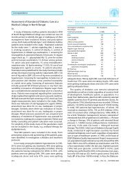
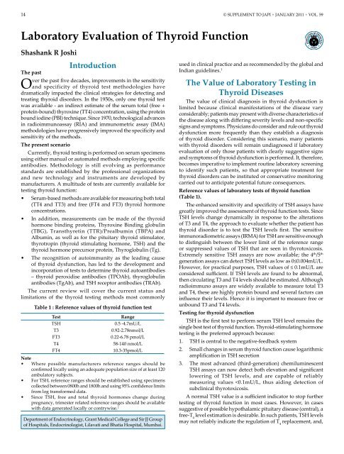
Table 1 : Reference values <strong>of</strong> thyroid function test<br />
Test Range<br />
TSH 0.5 -4.7mU/L<br />
T3 0.92-2.78nmol/L<br />
FT3 0.22-6.78 pmol/L<br />
T4 58-140 nmol/L<br />
FT4 10.3-35pmol/L<br />
Note<br />
• Where possible manufacturers reference ranges should be<br />
confirmed locally using an adequate population size <strong>of</strong> at least 120<br />
ambulatory subjects.<br />
• For TSH, reference ranges should be established using specimens<br />
collected between 0800h and 1800h and using 95% confidence limits<br />
from log transformed data.<br />
• Since TSH, free and total thyroid hormones change during<br />
pregnancy, trimester related reference ranges should be available<br />
with data generated locally or contrywise. 2<br />
Department <strong>of</strong> Endocrinology, Grant Medical College and Sir JJ Group<br />
<strong>of</strong> Hospitals, Endocrinologist, Lilavati and Bhatia Hospital, Mumbai.<br />
used in clinical practice and as recommended by <strong>the</strong> global and<br />
Indian guidelines. 1<br />
The Value <strong>of</strong> <strong>Laboratory</strong> Testing in<br />
<strong>Thyroid</strong> Diseases<br />
The value <strong>of</strong> clinical diagnosis in thyroid dysfunction is<br />
limited because clinical manifestations <strong>of</strong> <strong>the</strong> disease vary<br />
considerably; patients may present with diverse characteristics <strong>of</strong><br />
<strong>the</strong> disease along with differing severity levels and non-specific<br />
signs and symptoms. Physicians do consider and rule out thyroid<br />
dysfunction more frequently than <strong>the</strong>y establish a diagnosis<br />
<strong>of</strong> thyroid disorder. Considering this scenario, many patients<br />
with thyroid disorders will remain undiagnosed if laboratory<br />
evaluation <strong>of</strong> only those patients with clearly suggestive signs<br />
and symptoms <strong>of</strong> thyroid dysfunction is performed. It, <strong>the</strong>refore,<br />
becomes imperative to implement routine laboratory screening<br />
to identify such patients, so that appropriate treatment for<br />
thyroid disorders can be instituted or conservative monitoring<br />
carried out to anticipate potential future consequences.<br />
Reference values <strong>of</strong> laboratory tests <strong>of</strong> thyroid function<br />
(Table 1).<br />
The enhanced sensitivity and specificity <strong>of</strong> TSH assays have<br />
greatly improved <strong>the</strong> assessment <strong>of</strong> thyroid function tests. Since<br />
TSH levels change dynamically in response to <strong>the</strong> alterations<br />
<strong>of</strong> T3 and T4, <strong>the</strong> approach to evaluate whe<strong>the</strong>r <strong>the</strong> patient has<br />
thyroid disorder is to test <strong>the</strong> TSH levels first. The sensitive<br />
immunoradiometric assays (IRMA) for TSH are sensitive enough<br />
to distinguish between <strong>the</strong> lower limit <strong>of</strong> <strong>the</strong> reference range<br />
or suppressed values <strong>of</strong> TSH that are seen in thyrotoxicosis.<br />
Extremely sensitive TSH assays are now available; <strong>the</strong> 4th /5th generation assays can detect TSH levels as low as 0≤0.004mU/L.<br />
However, for practical purposes, TSH values <strong>of</strong> ≤ 0.1mU/L are<br />
considered sufficient. If TSH levels are found to be abnormal,<br />
<strong>the</strong>n circulating T3 and T4 levels should be estimated. Although<br />
radioimmuno assays are widely available to measure total T3<br />
and T4, <strong>the</strong>se are highly protein bound and several factors can<br />
influence <strong>the</strong>ir levels. Hence it is important to measure free or<br />
unbound T3 and T4 levels.<br />
Testing for thyroid dysfunction<br />
TSH is <strong>the</strong> first test to perform serum TSH level remains <strong>the</strong><br />
single best test <strong>of</strong> thyroid function. <strong>Thyroid</strong>-stimulating hormone<br />
testing is <strong>the</strong> preferred approach because:<br />
1. TSH is central to <strong>the</strong> negative-feedback system<br />
2. Small changes in serum thyroid function cause logarithmic<br />
amplification in TSH secretion<br />
3. The most advanced (third-generation) chemiluminescent<br />
TSH assays can now detect both elevation and significant<br />
lowering <strong>of</strong> TSH levels, and are capable <strong>of</strong> reliably<br />
measuring values
© SUPPLEMENT TO JAPI • JANUAry 2011 • VOL. 59 15<br />
<strong>the</strong>refore, may require estimation <strong>of</strong> free T 4 levels. 1<br />
TSH testing should be commonly carried out in <strong>the</strong> following<br />
settings:<br />
• In patients presenting with suspected goitres: Serum TSH levels<br />
must be measured.<br />
• As screening for congenital hypothyroidism: A heel-prick blood<br />
specimen is used for determining serum TSH levels. This is<br />
an established screening test for congenital hypothyroidism<br />
and has been adopted as a routine screening measure<br />
in many countries. The practice <strong>of</strong> routine screening for<br />
congenital hypothyroidism by <strong>the</strong> TSH test should be more<br />
widely adopted and continued. The low cost filterpaper TSH<br />
methods will be available in India soon.<br />
• In patients with atrial fibrillation, dyslipidaemia, osteoporosis,<br />
and infertility: Serum TSH levels should be measured at<br />
presentation.<br />
As screening for thyroid disorders in patients with unclear<br />
diagnoses: Serum TSH test should be carried out in all patients<br />
who have non-specific manifestations, are asymptomatic, and<br />
in whom <strong>the</strong> diagnosis is not clear. The high-sensitivity TSH<br />
test should be performed in such cases (where <strong>the</strong>re’s a low<br />
pre-test probability <strong>of</strong> <strong>the</strong> disease). The advantage <strong>of</strong> this test is<br />
that its negative predictive value is very high and a vast majority<br />
<strong>of</strong> <strong>the</strong> results come out negative. Measurement <strong>of</strong> serum TSH<br />
alone can suffice during sequential follow-up visits (after <strong>the</strong><br />
first investigation has been carried out) in patients who have<br />
not received treatment for thyroid disorders and for those who<br />
may be at risk <strong>of</strong> developing thyroid dysfunction.<br />
Important considerations for <strong>the</strong> clinician if TSH is<br />
abnormal (Table 2)<br />
• In patients with abnormal TSH concentrations, a focused<br />
history, physical examination (in particular thyroid gland<br />
examination), repeat TSH test, serum T and T level<br />
3 4<br />
determination and occasionally imaging studies need to be<br />
carried out.<br />
• It is not uncommon to see that many patients with high<br />
TSH values are informed that <strong>the</strong>y need to take thyroid<br />
medication life-long and after having been prescribed T4, no fur<strong>the</strong>r workup or explanation is undertaken.<br />
• In patients with goitrous changes or <strong>the</strong> presence <strong>of</strong> thyroid<br />
nodules, TSH concentration may be in <strong>the</strong> normal range<br />
because <strong>of</strong> an unaltered thyroid function. This warrants<br />
Table 2 : Some causes <strong>of</strong> abnormal serum TSH concentrations<br />
TSH below normal TSH above normal<br />
• Primary hyperthyroidism • Primary hypothyroidism<br />
• Pituitary/hypothalamic disease with central hypothyroidism (TSH<br />
unreliable)<br />
• Prolonged thyrotroph cell suppression after recent hyperthyroidism<br />
in euthyroid or hypothyroid patient<br />
• Old age • Old age<br />
• Drugs, e.g., glucocorticoids, dopamine • Drugs, e.g., amiodarone<br />
• Pituitary thyrotroph adenoma; Pituitary resistance to thyroid<br />
hormone (central hyperthyroidism) TSH, unreliable.<br />
• Generalized thyroid hormone resistance<br />
• Thyrotoxicosis from overly rapid correction <strong>of</strong> severe hypothyroidism<br />
with parenteral T4 • Problems with T treatment : Overdosage in treatment for fatigue or • Problems with T treatment : Underdosage based on misleadingly<br />
4 4<br />
, overweight, Altered gastrointestinal absorption because <strong>of</strong> drugs or high total T Altered gastrointestinal absorption because <strong>of</strong> drugs or<br />
4<br />
disease, Altered T clearance because <strong>of</strong> drugs, Patient compliance disease, Altered T clearance because <strong>of</strong> drugs, Patient compliance<br />
4 4<br />
problems, Prescription error, Testing too soon after T dose decrease problems, Prescription error, Testing too soon after T dose increase<br />
4 4<br />
• Many severe systemic illnesses (Sick Enthyrid State) • Recovery phase after severe systemic illness (Sick Enthyrid State)<br />
• Combination <strong>of</strong> pulsatile TSH secretion and analytical precision limits • Combination <strong>of</strong> pulsatile TSH secretion and analytical precision limits<br />
Antibody in patient serum against antibody in TSH assay, causing<br />
analytical artefact<br />
complete patient evaluation including testing for antithyroid<br />
antibodies, imaging ultrasound and fine-needle<br />
aspiration cytology.<br />
• In patients whose TSH levels are abnormal, T and T levels<br />
3 4<br />
should be determined. Free T and T level estimation is<br />
3 4<br />
preferred over total T and T estimation because <strong>the</strong>se<br />
3 4<br />
hormones are extensively (>99%) bound to plasma proteins<br />
and only <strong>the</strong> unbound forms are active. 3<br />
Inappropriate TSH<br />
This is a biochemical diagnosis in which elevation in circulating<br />
FT4 and/or FT3 is associated with an “inappropriately” detectable<br />
or elevated serum TSH concentration. If this biochemical<br />
picture is observed <strong>the</strong>n assay artefact/laboratory error should<br />
be considered first. Once <strong>the</strong> laboratory has excluded such<br />
explanations <strong>the</strong>n <strong>the</strong> cause <strong>of</strong> “true” inappropriate TSH should<br />
be considered. The differential diagnosis are a TSH secreting<br />
pituitary tumour (TSH-oma) or a syndrome <strong>of</strong> thyroid hormone<br />
resistance. The finding <strong>of</strong> an elevated serum sex hormone<br />
binding globulin (SHBG) and circulating free a subunit may<br />
support <strong>the</strong> diagnosis <strong>of</strong> TSH-oma, as may <strong>the</strong> finding <strong>of</strong> hyper<br />
or hypo-secretion <strong>of</strong> o<strong>the</strong>r pituitary hormones. Pituitary imaging<br />
usually confirms <strong>the</strong> diagnosis but should not be undertaken<br />
until <strong>the</strong> appropriate biochemical confirmation has been made.<br />
A syndrome <strong>of</strong> thyroid hormone resistance can be confirmed by<br />
family history; sequencing <strong>of</strong> <strong>the</strong> β thyroid hormone receptor<br />
confirms <strong>the</strong> diagnosis. When an ‘inappropriately’ detectable<br />
or elevated serum TSH is found in association with elevated<br />
circulating free T 3 and/or T 4 concentrations, <strong>the</strong> TSH is termed<br />
‘inappropriate’. Such cases may occur due to assay artefacts or<br />
laboratory errors and this should be considered first. However,<br />
if on repeat determination, TSH is still found to be inappropriate,<br />
o<strong>the</strong>r common explanations for apparent elevation <strong>of</strong> FT4<br />
should be considered. These include <strong>the</strong> presence <strong>of</strong> binding<br />
protein abnormalities (such as familial dysalbuminaemic<br />
hyperthyroxinaemia) or assay dependent antibody interference<br />
in <strong>the</strong> measurements <strong>of</strong> FT4, FT3 or TSH. To distinguish between<br />
TSHomas and thyroid hormone resistance, estimations <strong>of</strong> SHBG,<br />
a subunit and o<strong>the</strong>r anterior pituitary hormones may be carried<br />
out.<br />
Total T4<br />
Several laboratories measure <strong>the</strong> total T 4 and total T 3 which
16 © SUPPLEMENT TO JAPI • JANUAry 2011 • VOL. 59<br />
is not a true reflection <strong>of</strong> <strong>the</strong> thyroid status <strong>of</strong> an individual.<br />
This is because thyroid hormones circulate in <strong>the</strong> body largely<br />
in <strong>the</strong> inactive form, bound to carrier proteins (thyroid binding<br />
globulin (TBG), transthyretin and albumin) while only <strong>the</strong><br />
small unbound fraction is metabolically active. Moreover, in<br />
some clinical conditions, particularly those in which <strong>the</strong>re is an<br />
alteration <strong>of</strong> <strong>the</strong> amount <strong>of</strong> carrier proteins, <strong>the</strong> total T and total<br />
3<br />
T may be elevated while <strong>the</strong> thyroid functional state (free T and<br />
4 3<br />
T levels) may be normal. Such conditions include:<br />
4<br />
1. Hereditary abnormalities <strong>of</strong> binding proteins: These include<br />
TBG deficiency or TBG excess, abnormal albumin levels and<br />
abnormal transthyretin levels.<br />
2. Acquired deficiency <strong>of</strong> binding proteins: Conditions such<br />
as nephrotic syndrome may cause protein loss from <strong>the</strong><br />
body. In severe liver disease, <strong>the</strong>re’s impaired production <strong>of</strong><br />
proteins, and <strong>the</strong>rapy with androgens or anabolic steroids<br />
may alter <strong>the</strong> levels <strong>of</strong> carrier proteins.<br />
3. Drug-induced alterations in T4 binding to TBG: Therapy<br />
with salicylates, phenytoin, phenylbutazone may alter<br />
T4-TBG binding.<br />
4. Presence <strong>of</strong> T4 antibodies.<br />
The development <strong>of</strong> newer immunoassay methods for<br />
determining free T3 and T4 has overcome many <strong>of</strong> <strong>the</strong>se<br />
problems. Radioimmunoassay measurement <strong>of</strong> total serum T4<br />
levels is highly sensitive in reflecting <strong>the</strong> hyperthyroid (85-95%)<br />
and <strong>the</strong> hypothyroid status (80-90%) <strong>of</strong> patients.<br />
Total T3<br />
Currently routine measurement <strong>of</strong> serum T3 is not carried out<br />
(only T4 is measured) in patients suspected <strong>of</strong> having thyroid<br />
disorders. About 25% <strong>of</strong> patients with hypothyroidism have low<br />
normal T3 values. Free T3/ total T3 measurements, however,<br />
should be performed in <strong>the</strong> following settings:<br />
1. In patients suspected <strong>of</strong> having T3 thyrotoxicosis.<br />
2. In patients taking drugs that inhibit <strong>the</strong> peripheral<br />
conversion <strong>of</strong> T4 to T3 (such as dexamethasone, propranolol,<br />
propylthiouracil, amiodarone, and iodine-containing<br />
contrast media). 5<br />
Testing both TSH and FT4<br />
There are certain clinical situations where TSH testing<br />
must be coupled with testing <strong>the</strong> FT4 levels. Clinical situations<br />
where measurement <strong>of</strong> both serum TSH and FT4 is required<br />
are principally disorders where <strong>the</strong> pituitary-thyroid axis is not<br />
intact or is unstable. These situations include:<br />
• Optimising thyroxine <strong>the</strong>rapy in newly diagnosed patients<br />
with hypothyroidism.<br />
• Diagnosing and monitoring thyroid disorders in pregnancy.<br />
• Monitoring patients with hyperthyroidism in <strong>the</strong> early<br />
months after treatment.<br />
• Diagnosis and monitoring treatment for central<br />
hypothyroidism.<br />
• End-organ thyroid hormone resistance.<br />
• Sick Euthyroid State.<br />
• TSH-secreting pituitary adenomas.<br />
• Women with type I diabetes should have <strong>the</strong>ir thyroid<br />
function, including serum TSH, FT4 and thyroid peroxidase<br />
antibody status, established preconception, at booking when<br />
pregnant and at 3 months post-partum.<br />
• Possible subclinical hypothyroidism: If screening is<br />
performed, and a high serum TSH concentration is found,<br />
and <strong>the</strong> FT4 is normal, <strong>the</strong> measurement should be repeated<br />
3-6 months later, along with measurement <strong>of</strong> serum FT4,<br />
after excluding non-thyroidal illness and drug interference.<br />
• Overtly hypothyroid patients (who have serum TSH greater<br />
than 10 mU/L and low FT4 concentrations) should be treated<br />
with thyroxine.<br />
In patients with a high serum TSH level and normal FT4<br />
concentrations (possible subclinical hypothyroidism), TSH<br />
measurements and FT4 should be repeated 3 to 6 months later,<br />
after precluding non-thyroid disorders and drug interference. In<br />
cases <strong>of</strong> doubt in identifying specimens in which both serum TSH<br />
and FT4 should be carried out, it is prudent to test all specimens<br />
for TSH and FT4, ra<strong>the</strong>r than test for TSH alone. 6<br />
Testing TSH and FT4 and FT3<br />
In hospital inpatient ICU: In <strong>the</strong> absence <strong>of</strong> an abnormal<br />
thyroid gland by careful physical examination, a hospital<br />
inpatient with a mild or moderate (
© SUPPLEMENT TO JAPI • JANUAry 2011 • VOL. 59 17<br />
Table 3 : Characterization <strong>of</strong> thyroid disorders according to results <strong>of</strong> thyroid function tests<br />
Disorder TSH T 4 T 3 FT 4 Tg TBG rT 3 ATPO ATG TBII TSI TBA<br />
Primary hypothyroidism ↑ ↓ N or ↓ ↓ N or ↓ N ↓ N or ↑ N or ↑ N or ↑ n n or ↑<br />
Transient neonatal hypothyroidism ↑ ↓ ↓ ↓ N or ↓ N ↓ N N ↑ n ↑<br />
Hashimoto thyroiditis hypothyroidism ↑ N or ↓ N or ↓ N or ↓ N or ↓ N ↓ ↑ ↑ n or ↑ n n or ↑<br />
Graves’ disease ↓ ↑ ↑ ↑ ↑ N ↑ ↑ ↑ ↑ ↑ n or ↑<br />
Neonatal Graves’ disease ↓ ↑ ↑ ↑ ↑ N ↑ n or ↑ n or ↑ ↑ ↑ n or ↑<br />
TSH deficiency N or ↓ ↓ ↓ ↓ ↓ N ↓ n N n n n<br />
<strong>Thyroid</strong> dishormonogenesis ↑ ↓ ↓ ↓ N, ↓<br />
or ↑<br />
N ↑ n N n n n<br />
<strong>Thyroid</strong> hormone resistance N or ↑ ↑ ↑ ↑ ↑ N ↑ n N n n n<br />
TSH-dependent hyperthyroidism ↑ ↑ ↑ ↑ ↑ N ↑ n N n n n<br />
T protein-binding abnormalities 4 [*] N V V N N V + V n N n n n<br />
Nonthyroidal illness V N or ↓ ↓ V N N N or ↑ n N n n n<br />
Subacute thyroiditis [†] ↓ or ↑ ↑ or ↓ ↑ or ↓ ↑ or ↓ ↑ or ↓ N ↑ or ↓ n n n n n<br />
TSH = thyroid-stimulating hormone; T 4 = thyroxine; T 3 = triiodothyronine; FT 4 = free thyroxine; Tg = thyroglobulin; TBG = thyroxine-binding<br />
globulin; rT 3 = reverse T 3 ; ATPO = antithyroidperoxidase; ATG = antithyroglobulin; TBII = TSH-binding inhibiting immunoglobulin; TSI = thyroidstimulating<br />
immunoglobulin; TBA = TSH receptor-blocking antibody; N = normal; n = negative; V = variable.<br />
* The spectrum <strong>of</strong> binding protein abnormalities includes increased or decreased TBG binding, increased or decreased transthyretin binding, and ↑<br />
albumin binding.<br />
† Subacute thyroiditis involves a transient period <strong>of</strong> hyperthyroidism followed by a transient hypothyroid state.<br />
(Reprinted from Fisha DA (ed) : Disorders <strong>of</strong> <strong>Thyroid</strong> <strong>Function</strong>, Quest Diagnostic Manual. 3rd Editor, p 268.)<br />
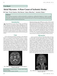
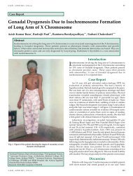
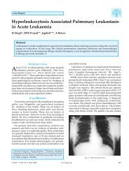
High<br />
TSH<br />
Primary<br />
Hypothyroidism<br />
• ES<br />
• TT<br />
• Central<br />
Hypothyroidism<br />
• ES<br />
• TT<br />
• Subclinical<br />
Hypothyroidism<br />
• ES*<br />
• TT*<br />
• Inappropriate TSH<br />
secretion<br />
• ES<br />
• TT<br />
Normal • ES<br />
• TT<br />
• Subclinical<br />
Hyperthyroidism<br />
• ES<br />
• TT<br />
Overt<br />
Hyperthyroidism<br />
Low<br />
Low Free T4 * ES, euthyroid sick; TT, thyroid in transition<br />
High<br />
Fig. 1a : <strong>Thyroid</strong> <strong>Function</strong> Test Algorithm<br />
<strong>Thyroid</strong> Autoimmunity <strong>Thyroid</strong>specific<br />
Autoantibodies (TPOAb,<br />
TGAb AnD TRAb)<br />
Tests for antibodies against thyroid-specific antigens, antithyroid<br />
peroxidase (TPO), thyroglobulin (Tg) and TSH receptors<br />
are used in <strong>the</strong> diagnosis <strong>of</strong> autoimmune thyroid disorders. Over<br />
<strong>the</strong> last five decades, antibody measurement techniques have<br />
evolved from semi-quantitative agglutination and complement<br />
fixation tests and whole animal bioassays to specific ligand<br />
assays using recombinant antigens and cell culture systems<br />
transfected with <strong>the</strong> human TSH receptor. Unfortunately, <strong>the</strong><br />
diagnostic and prognostic value <strong>of</strong> <strong>the</strong>se thyroid autoantibody<br />
measurements is hampered by differences in <strong>the</strong> sensitivity and<br />
specificity <strong>of</strong> current methods. Although autoantibody tests have<br />
inherent clinical utility in a number <strong>of</strong> clinical situations, <strong>the</strong>se<br />
tests should be selectively employed.<br />
<strong>Thyroid</strong> peroxidase autoantibodies (TPOAb)<br />
Originally, thyroid peroxidase autoantibodies (TPOAb) were<br />
detected as thyroid microsomal antibodies by semi-quantitative<br />
complement fixation and tanned erythrocyte hemagglutination<br />
techniques and were labeled antimicrosomal antibodies (AMA).<br />
The principal antigen in <strong>the</strong> thyroid microsomes was recently<br />
discovered to be <strong>the</strong> thyroid peroxidase enzyme (TPO), a 100kD<br />
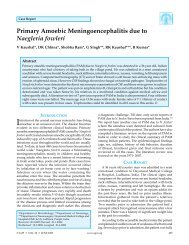
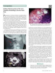
TSH<br />
High Normal Low<br />
Free T4 Free T4<br />
Low Normal Normal High<br />
Hypothyroidism Hyperthyroidism<br />
Subclinical Hypothyroidism Subclinical Hyperthyroidism<br />
Fig. 1b : Algorithm for <strong>the</strong> Diagnosis <strong>of</strong> <strong>Thyroid</strong> Dysfunction<br />
glycosylated protein. Currently, automated tests are replacing<br />
<strong>the</strong> older manual agglutination tests. These new tests are more<br />
specific TPOAb immunoassays or immunometric assay methods,<br />
and are based on purified or recombinant TPO.<br />
Clinical Use <strong>of</strong> TPOAb Tests<br />
An abnormal TPOAb is detected in 15 to 20 percent <strong>of</strong><br />
“healthy” euthyroid subjects and even higher percentages<br />
<strong>of</strong> patients with various non-thyroid autoimmune disorders.<br />
Approximately 70-80 % <strong>of</strong> patients with Graves’ disease and<br />
virtually all patients with Hashimoto’s, atrophic thyroiditis<br />
or post-partum thyroiditis have TPOAb detected. In fact,<br />
TPOAb is implicated as a cytotoxic agent in <strong>the</strong> destructive<br />
thyroiditic process. TPO antibodies are involved in <strong>the</strong> tissue<br />
destructive processes associated with <strong>the</strong> hypothyroidism<br />
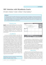
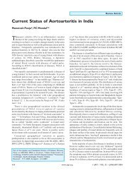
observed in Hashimoto’s thyroiditis (Fig. 2). In <strong>the</strong> future,<br />
TPOAb measurement may be used as a prognostic indicator for<br />
thyroid dysfunction. Although <strong>the</strong> appearance <strong>of</strong> TPOAb usually<br />
precedes <strong>the</strong> development <strong>of</strong> thyroid dysfunction, recent studies<br />
suggest that a hypoechoic ultrasound pattern may precede a<br />
biochemical TPOAb abnormality, as shown in Figure 2. The<br />
paradoxical absence <strong>of</strong> TPOAb in some patients with unequivocal<br />
TSH abnormalities likely reflects <strong>the</strong> suboptimal sensitivity and/<br />
or specificity <strong>of</strong> current TPOAb tests or non-autoimmune thyroid<br />
failure (atrophic thyroiditis). Although changes in autoantibody<br />
concentrations <strong>of</strong>ten reflect a change in disease activity, serial
18 © SUPPLEMENT TO JAPI • JANUAry 2011 • VOL. 59<br />
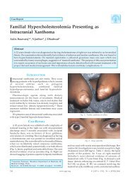
Prevalence <strong>of</strong> <strong>Thyroid</strong> Antidodies in <strong>the</strong> General Population<br />
NHANES III (n =16,869)<br />
15<br />
14.7<br />
10<br />
%<br />
Prevalence<br />
5<br />
0<br />
Thayroid<br />
Ab<br />
Odds Ratio for Hypothyroidism<br />
(Low TT4+ TSH > 4.5 mU/L)<br />
Fig. 2 : <strong>Thyroid</strong> Autoantibody Prevalence and Associations with<br />
Hypothyroidism (Reprinted from Hollowell JG, Staehling NW,<br />
Hannon WH, Flanders WD, Gunter EW, Spencer CA, and Braverman<br />
LE. Serum thyrotropin, thyroxine, and thyroid antibodies in <strong>the</strong><br />
United States population (1988 to 1994): NHANES III. 2002;J Clin<br />
Endocrinol Metab 2002,87:489-99)<br />
thyroid autoantibody measurements are not recommended<br />
for monitoring treatment for AITD. The prevalence <strong>of</strong> TPOAb<br />
is higher in patients with non-thyroid autoimmune diseases<br />
such as type 1diabetes and pernicious anemia. Aging is also<br />
associated with higher prevalence <strong>of</strong> TPOAb that parallel <strong>the</strong><br />
increased prevalence seen in both subclinical (mild) and clinical<br />
hypothyroidism. A euthyroid subject with detectable TPOAb is<br />
at increased risk <strong>of</strong> development <strong>of</strong> hypothyroidism. Detectable<br />
level <strong>of</strong> TPOAb typically precedes <strong>the</strong> development <strong>of</strong> an<br />
elevated TSH and is <strong>the</strong>refore a risk factor for hypothyroidism.<br />
Moreover, reproductive complications (such as miscarriage,<br />
infertility, IVF failure, fetal death, pre-eclampsia, preterm<br />
delivery and post-partum thyroiditis and depression) have been<br />
associated with <strong>the</strong> presence <strong>of</strong> TPOAb. The enhanced sensitivity<br />
and specificity <strong>of</strong> <strong>the</strong> TPO immunoassay methods make <strong>the</strong>m a<br />
more cost-effective option over <strong>the</strong> older semi-quantitative AMA<br />
agglutination tests, since <strong>the</strong>y obviate <strong>the</strong> need for additional<br />
TgAb measurements in <strong>the</strong> routine diagnosis <strong>of</strong> autoimmune<br />
thyroid disorders. 7<br />
Thyroglobulin autoantibodies (TgAb)<br />
Antithyroglobulin autoantibodies (TgAb) were <strong>the</strong><br />
first thyroid antibodies to be recognized to circulate in<br />
patients with autoimmune thyroid disorders. The first TgAb<br />
methods were based on tanned red cell hemagglutination.<br />
Subsequently, methodologies have evolved in parallel with<br />
TPOAb methodology from semi-quantitative techniques, to<br />
more sensitive ELISA and RIA methods and more recently<br />
chemiluminescent immunoassays. Unfortunately, <strong>the</strong> intermethod<br />
variability <strong>of</strong> current TgAb assays is even greater than<br />
that <strong>of</strong> <strong>the</strong> TPOAb tests discussed above.<br />
Clinical Use <strong>of</strong> TgAb Tests<br />
Auto antibodies against Tg are encountered in autoimmune<br />
thyroid conditions, usually in association with TPOAb. However,<br />
<strong>the</strong> recent NHANES III study found that 3 % <strong>of</strong> subjects<br />
with no risk factors for thyroid disease had detectable TgAb<br />
without TPOAb. In <strong>the</strong>se subjects with only TgAb detected,<br />
no association with TSH abnormalities was found so that <strong>the</strong><br />
clinical significance <strong>of</strong> an isolated TgAb abnormality remains to<br />
be established. This suggests that it is unnecessary to measure<br />
6.9<br />
TPOAb<br />
+TgAb<br />
5.7<br />
TPOAb<br />
+Alone<br />
3.1<br />
TgAb<br />
Alone<br />
34.7 6.1 0.6<br />
both TPOAb and TgAb for a routine evaluation <strong>of</strong> thyroid<br />
autoimmunity. According to <strong>the</strong> current guidelines, all sera<br />
should be prescreened for TgAb by a sensitive immunoassay<br />
method prior to Tg testing. Therefore, TgAb is primarily used<br />
as an adjunct test for serum Tg estimation. TgAb is detected<br />
in approximately 20% <strong>of</strong> patients with differentiated thyroid<br />
carcinoma compared with 10% <strong>of</strong> normal subjects by <strong>the</strong><br />
immunoassay methods. The threshold TgAb concentration above<br />
normal that precludes TgAb interference is ei<strong>the</strong>r not known or<br />
does not appear to exist. False positives may occur due to assay<br />
artifacts or illegitimate transcription while false negatives results<br />
may be seen in patients with metastatic disease.<br />
TSH receptor autoantibodies (TRAb)<br />
TSH Receptor Antibodies (TRAb) were first recognized as<br />
long-acting thyroid stimulator (LATS) using mouse bioassays.<br />
These autoantibodies are directed against epitopes on <strong>the</strong><br />
ectodomain <strong>of</strong> <strong>the</strong> TSH receptor. Methods for measuring TRAb<br />
are even more varied than for TPOAb and TgAb. Two classes<br />
<strong>of</strong> TRAb can be associated with autoimmune thyroid disorders<br />
– (a) thyroid stimulating autoantibodies (TSAb) that cause<br />
Graves’ hyperthyroidism and (b) thyroid stimulation-blocking<br />
antibodies (TBAb) which block receptor binding <strong>of</strong> TSH. Each<br />
class <strong>of</strong> TRAb (TSAb and TBAb) may be detected alone or in<br />
combination in Graves’ disease and Hashimoto’s thyroiditis. The<br />
relative concentrations <strong>of</strong> <strong>the</strong> two classes <strong>of</strong> TRAb may modulate<br />
<strong>the</strong> severity <strong>of</strong> Graves’ hyperthyroidism and may change in<br />
response to <strong>the</strong>rapy or pregnancy.<br />
Clinical Use <strong>of</strong> TRAb Tests<br />
TRAb tests are used in <strong>the</strong> differential diagnosis <strong>of</strong><br />
hyperthyroidism, <strong>the</strong> prediction <strong>of</strong> fetal and neonatal thyroid<br />
dysfunction due to transplacental passage <strong>of</strong> maternal TRAb<br />
and prediction <strong>of</strong> <strong>the</strong> course <strong>of</strong> Graves’ disease treated with<br />
antithyroid drugs. Although TBII assays do not directly<br />
measure <strong>the</strong> stimulating antibodies, <strong>the</strong>se tests have comparable<br />
diagnostic sensitivity to TSAb bioassays (70-95%) for diagnosing<br />
Graves’ hyperthyroidism or detecting a relapse or response<br />
to <strong>the</strong>rapy. The second generation assays employing human<br />
recombinant TSH receptor are now becoming available and<br />
are reported to have superior diagnostic sensitivity for Graves’<br />
disease. Current tests are manual and expensive and vary in<br />
precision, sensitivity, specificity and reference ranges. However,<br />
<strong>the</strong> TBII tests are important for evaluating pregnant patients<br />
with a history <strong>of</strong> autoimmune thyroid disease, in whom <strong>the</strong>re<br />
is a risk <strong>of</strong> transplacental passage <strong>of</strong> TRAb to <strong>the</strong> infant . The<br />
lack <strong>of</strong> specificity <strong>of</strong> <strong>the</strong> TBII methods is actually an advantage<br />
in this clinical situation, since a TBII test will detect both <strong>the</strong><br />
stimulating and blocking classes <strong>of</strong> TRAb that can produce<br />
transient hyper- or hypothyroidism, respectively, in <strong>the</strong> fetus<br />
and newborn. TRAb plays an uncertain role in thyroid-associated<br />
ophthalmopathy (TAO), which appears to be exacerbated by<br />
radioiodine <strong>the</strong>rapy. Since TRAb and o<strong>the</strong>r thyroid antibodies<br />
levels increase acutely significantly after radioiodine <strong>the</strong>rapy,<br />
a TRAb measurement prior to radioiodine <strong>the</strong>rapy may be<br />
useful to predict risk <strong>of</strong> TAO. However, prospective studies are<br />
needed to establish <strong>the</strong> clinical utility <strong>of</strong> TRAb measurement in<br />
this context. Patients with very high circulating concentrations<br />
<strong>of</strong> hCG due to choriocarcinoma or hydatiform mole, as well<br />
as a small number <strong>of</strong> pregnant patients, may have misleading<br />
positive results using TSAb assays.<br />
Thyroglobulin (Tg) methods<br />
Serum Tg measurement is used as a tumor marker in <strong>the</strong>
© SUPPLEMENT TO JAPI • JANUAry 2011 • VOL. 59 19<br />
management <strong>of</strong> patients with differentiated thyroid carcinomas<br />
(DTC). Current Tg methods are based ei<strong>the</strong>r on IMA or RIA<br />
techniques. There is a trend for non-isotopic IMA methods<br />
to replace RIA methods because IMA methods are easier to<br />
automate, have shorter turn around times, wider working ranges<br />
and use reagents with a longer shelf life. 4<br />
<strong>Thyroid</strong> <strong>Function</strong> Tests in Special<br />
Patient Populations<br />
Patients with atrial fibrillation, hyperlipidaemia,<br />
osteoporosis, infertility<br />
Patients presenting with atrial fibrillation, hyperlipidemia,<br />
subfertility and osteoporosis, should undergo serum TSH<br />
estimations as assessment <strong>of</strong> thyroid function because:<br />
• Atrial fibrillation may be secondary to thyrotoxicosis in<br />
about 5-10% <strong>of</strong> patients.<br />
• Osteoporosis may be secondary to hyperthyroidism and<br />
can be corrected by treating <strong>the</strong> underlying cause.<br />
• Both hyper as well as hypothyroidism may be contributing<br />
factors in menstrual cycle disorders, fetal loss and infertility.<br />
Women with type 1 diabetes<br />
Type 1 diabetes in women raises <strong>the</strong>ir likelihood <strong>of</strong> developing<br />
post-partum thyroid dysfunction by three times. Women with<br />
type 1 diabetes should have <strong>the</strong>ir thyroid function (including<br />
TSH, FT4 and thyroid peroxidise antibody status) assessed at<br />
preconception, at <strong>the</strong> time <strong>of</strong> registration for pregnancy and at<br />
three months post-partum.<br />
Women with a past history <strong>of</strong> post-partum thyroiditis<br />
In women with post-partum thyroiditis, <strong>the</strong>re is an increased<br />
long-term risk <strong>of</strong> developing hypothyroidism and its recurrence<br />
in subsequent pregnancies. Therefore, all women with a history<br />
<strong>of</strong> post-partum thyroiditis should be recommended to have a<br />
yearly thyroid function test, and also prior to and at 6 to 8 weeks<br />
after <strong>the</strong>ir subsequent pregnancies.<br />
Patients with diabetes<br />
The frequency <strong>of</strong> patients with type 1 diabetes and<br />
asymptomatic thyroid dysfunction is high. These patients<br />
should have a yearly thyroid function test. In patients with type<br />
2 diabetes, thyroid function should be assessed at diagnosis,<br />
however, annual thyroid function assessment may not be<br />
recommended.<br />
Down syndrome and Turner’s syndrome<br />
Patients <strong>of</strong> Down syndrome as well as Turner’s syndrome are<br />
recommended to undergo thyroid function assessment annually,<br />
keeping in mind <strong>the</strong> high incidence <strong>of</strong> hypothyroidism seen in<br />
<strong>the</strong>se patients.<br />
Patients receiving Amiodarone and Lithium<br />
Therapy with amiodarone is associated with iodide-induced<br />
thyroid dysfunction (hypothyroidism or hyperthyroidism)<br />
because <strong>of</strong> <strong>the</strong> presence <strong>of</strong> 75 mg iodine per each 200 mg tablet.<br />
Patients on amiodarone treatment should have thyroid function<br />
assessment at <strong>the</strong> time <strong>of</strong> beginning <strong>of</strong> amiodarone <strong>the</strong>rapy and<br />
<strong>the</strong>reafter every 6 months during treatment and till 12 months<br />
after cessation <strong>of</strong> <strong>the</strong>rapy. Lithium <strong>the</strong>rapy (for bipolar disorder)<br />
is associated with mild to overt hypothyroidism in up to 34%<br />
to 16% <strong>of</strong> patients respectively, which can occur abruptly even<br />
many years after cessation <strong>of</strong> <strong>the</strong>rapy. Thyrotoxicosis can also<br />
occur due to long-term treatment with lithium but is relatively<br />
rare. Therefore, all patients on lithium <strong>the</strong>rapy should have a<br />
thyroid function assessment before commencement <strong>of</strong> treatment<br />
and <strong>the</strong>reafter every 6-12 months during lithium <strong>the</strong>rapy.<br />
Post neck irradiation<br />
Patients who undergo surgery or external radiation <strong>the</strong>rapy<br />
<strong>of</strong> <strong>the</strong> neck, or both, for head and neck cancer (including<br />
lymphoma) have a high incidence (up to 50%) <strong>of</strong> hypothyroidism.<br />
The incidence is particularly high in patients who undergo<br />
surgery and receive high doses <strong>of</strong> radiation because <strong>the</strong> effect<br />
is dose-dependent. The onset <strong>of</strong> overt hypothyroidism due to<br />
surgery or irradiation is gradual and may precede subclinical<br />
hypothyroidism for many years. In such patients, thyroid<br />
function assessment should be carried out annually.<br />
Following destructive treatment for thyrotoxicosis by ei<strong>the</strong>r<br />
radioiodine or surgery<br />
Patients treated with radioiodine or those who<br />
undergo thyroidectomy should be screened indefinitely<br />
for <strong>the</strong> development <strong>of</strong> hypothyroidism or recurrence <strong>of</strong><br />
hyperthyroidism. Assessment <strong>of</strong> thyroid function in <strong>the</strong>se<br />
patients should be done four to eight weeks after treatment,<br />
followed by quarter yearly assessments for <strong>the</strong> subsequent year<br />
and annually <strong>the</strong>reafter.<br />
Treatment <strong>of</strong> thyrotoxicosis with anti-thyroid drugs<br />
Antithyroid drugs used in <strong>the</strong> management <strong>of</strong> thyrotoxicosis,<br />
carbimazole and propylthiouracil, decrease thyroid hormone<br />
secretion. <strong>Thyroid</strong> function assessment should be carried<br />
out every 1-3 months to determine whe<strong>the</strong>r stable hormonal<br />
concentrations have been reached when antithyroid <strong>the</strong>rapy is<br />
instituted and annually <strong>the</strong>reafter if long-term treatment is used.<br />
Patients on thyroxine <strong>the</strong>rapy<br />
In patients undergoing thyroxine <strong>the</strong>rapy regardless <strong>of</strong> <strong>the</strong><br />
cause, long-term follow-up with annual measurements <strong>of</strong> serum<br />
TSH are recommended. This helps to check compliance, verify<br />
<strong>the</strong> dosage and take account <strong>of</strong> variations in dosage requirements<br />
due to concomitant medications. In pregnant women, <strong>the</strong> dose<br />
may need to be increased by a minimum <strong>of</strong> 50 μg per day to<br />
maintain normal serum TSH levels. The TSH levels should be<br />
tested in each trimester. 8<br />
Interferences with<br />
<strong>Thyroid</strong> Test Methodologies<br />
There are four categories <strong>of</strong> interferences in competitive<br />
immunoassays (IMA) as well as non-competitive IMAs:<br />
1. Cross reactivity interferences<br />
2. Endogenous analyte antibodies<br />
3. Heterophilic antibodies<br />
4. Drug interactions<br />
1. Cross reactivity interferences<br />
Early TSH RIA methods had <strong>the</strong> limitation <strong>of</strong> cross-reactivity<br />
with glycoprotein hormones (such as LH, hCG). Currently,<br />
this problem has been almost completely overcome by using<br />
monoclonal antibodies for TSH IMA methods. Occasionally,<br />
however, unusual cross-reacting is<strong>of</strong>orms <strong>of</strong> TSH may be<br />
encountered while using <strong>the</strong> current assays.<br />
2. Endogenous analyte antibodies<br />
Robbins et al were <strong>the</strong> first to report an unusual thyroxine<br />
binding globulin in <strong>the</strong> serum in 1956. Subsequently,<br />
autoantibodies against T3, T4 and TSH have been identified<br />
in <strong>the</strong> sera <strong>of</strong> patients with autoimmune thyroid disorders
20 © SUPPLEMENT TO JAPI • JANUAry 2011 • VOL. 59<br />
as well as non-thyroid disorders. A number <strong>of</strong> reports have<br />
shown interference due to T3, T4 and TSH autoantibodies<br />
leading to anomalous free and total thyroid hormone levels<br />
and TSH values. However, <strong>the</strong> currently used methods<br />
rarely have this interference problem. Characteristics <strong>of</strong><br />
interference due to endogenous autoantibodies may lead<br />
to falsely low or falsely high values, depending upon <strong>the</strong><br />
type <strong>of</strong> assay and its composition.<br />
3. Heterophilic antibodies<br />
Heterophilic antibodies (particularly HAMA) may affect<br />
IMA methods more than competitive immunoassays by<br />
causing <strong>the</strong> formation <strong>of</strong> a bridge between <strong>the</strong> signal and<br />
capture antibodies. This creates a false signal resulting<br />
in a high value artifact. Moreover, <strong>the</strong> result may not be<br />
abnormal; it may be inappropriately normal. A potential for<br />
influencing results <strong>of</strong> neonatal screening also exists because<br />
antibodies are able to cross <strong>the</strong> placenta. Interference due to<br />
heterophilic antibodies can be classified into two categories:<br />
i. HAMA (or human anti-mouse antibodies) are relatively<br />
weak, polyreactive, multispecific antibodies that are<br />
frequently IgM. The presence <strong>of</strong> HAMA can alter <strong>the</strong><br />
total as well as free T3, T4 and TSH results due to<br />
interference. Use <strong>of</strong> Fab fragments and heterospecies<br />
assay configurations can be employed as approaches<br />
to reduce this kind <strong>of</strong> interference.<br />
ii. HAAA. Specific human anti-animal antibodies<br />
(HAAA) are produced in response to well-defined<br />
specific antigens after exposure to <strong>the</strong>rapeutic agents<br />
containing animal antigens (such as murine antibody)<br />
or by coincidental immunization through workplace<br />
contact (such as that which occurs in animal handlers).<br />
Though assays for HAMA have been developed, <strong>the</strong>re<br />
are large inter-method differences and <strong>the</strong>refore <strong>the</strong><br />
reliability <strong>of</strong> <strong>the</strong>se tests is questioned.<br />
4. Drug interferences<br />
In vitro and in vivo effects may occur due to drug<br />
interferences. When <strong>the</strong> specimen contains a sufficient<br />
concentration <strong>of</strong> an interfering <strong>the</strong>rapeutic or diagnostic<br />
agent, it may lead to methodologic interference resulting<br />
in in-vitro effects. An example is that <strong>of</strong> heparin which, in<br />
<strong>the</strong> specimen can cause in-vitro stimulation <strong>of</strong> lipoprotein<br />
lipase; free fatty acids are liberated that inhibit T4 binding<br />
to serum proteins. On <strong>the</strong> o<strong>the</strong>r hand, when results are<br />
altered due to administration <strong>of</strong> an interfering <strong>the</strong>rapeutic<br />
agent, <strong>the</strong>n it is termed in-vivo effect. An example is that <strong>of</strong><br />
furosemide which competitively inhibits thyroid hormone<br />
binding to <strong>the</strong> specimen, <strong>the</strong>reby causing an abnormal value<br />
(low) thyroid hormone result. Interference may also be<br />
secondary to certain pathologic conditions. For instance, in<br />
uraemia, abnormal serum constituents such as indole acetic<br />
acid may accumulate and cause interference. In addition,<br />
<strong>the</strong> presence <strong>of</strong> fluorophor-related <strong>the</strong>rapeutic or diagnostic<br />
agents in <strong>the</strong> specimen may alter <strong>the</strong> results <strong>of</strong> thyroid tests<br />
that employ fluorescent signals. 1<br />
Conclusion<br />
• <strong>Thyroid</strong> disorders have diverse clinical manifestations<br />
<strong>the</strong>refore, on part <strong>of</strong> vigilant clinician every suspected case<br />
<strong>of</strong> thyroid disease needs to be evaluated with laboratory<br />
investigations.<br />
• Thereby appropriate treatment for thyroid disorders can<br />
be instituted or conservative monitoring carried out to<br />
anticipate potential future consequences.<br />
• The enhanced sensitivity and specificity <strong>of</strong> TSH assays have<br />
greatly improved <strong>the</strong> assessment <strong>of</strong> thyroid function tests.<br />
Since TSH levels change dynamically in response to <strong>the</strong><br />
alterations <strong>of</strong> T3 and T4, <strong>the</strong> approach to evaluate whe<strong>the</strong>r<br />
<strong>the</strong> patient has thyroid disorder is to test <strong>the</strong> TSH levels first.<br />
• When hypothyroidism is suspected, a free-T4 estimate<br />
is appropriate because total-T3 and free-T3 tests have<br />
inadequate sensitivity and specificity in this setting.<br />
• When hyperthyroidism is suspected, <strong>the</strong> combination<br />
<strong>of</strong> a free-T4 estimate and a total- or free-T3estimate<br />
provides <strong>the</strong> most complete assessment <strong>of</strong> <strong>the</strong> severity <strong>of</strong><br />
hyperthyroidism and identifies cases <strong>of</strong> “T3-toxicosis”, i.e.<br />
a selective increase <strong>of</strong> <strong>the</strong> serum T3 concentration.<br />
References<br />
1. Spencer C. <strong>Thyroid</strong> <strong>Function</strong> Tests: Assay <strong>of</strong> <strong>Thyroid</strong> Hormones<br />
and Related Substances, www.thyroidmanager.com. 2010.<br />
2. Supit EJ, Peiris AN. Interpretation <strong>of</strong> <strong>Laboratory</strong> <strong>Thyroid</strong> <strong>Function</strong><br />
Tests: Selection and Interpretation. Sou<strong>the</strong>rn Medical <strong>Journal</strong>.<br />
2002;95:481-85.<br />
3. Werner SC, Ingbar SH. Werner & Ingbar’s The <strong>Thyroid</strong>: a<br />
fundamental and clinical text, 9th edition. Lippincott Williams &<br />
Wilkins.<br />
4. Mascarenhas JMA. RxPG AIPG 2004 Book, 2006.<br />
5. Daniels GH, Amiodarone-Induced Thyrotoxicosis, The J Clinical<br />
Endo & Metab 2000;86:3-8.<br />
6. Walfish PG. Triiodothyronine and thyroxine interrelationships in<br />
health and disease. Can Med Assoc J 1976;115:338–42.<br />
7. Düsünsel R, Poyrazoglu HM, Gündüz Z et al. Evidence <strong>of</strong> central<br />
hypothyroidism in children on continuous ambulatory peritoneal<br />
dialysis. Adv Perit Dial. 1999;15:262-8.<br />
8. UK Guidelines for <strong>the</strong> Use <strong>of</strong> <strong>Thyroid</strong> <strong>Function</strong> Tests, 2006.