

Biogenesis of the sperm head perinuclear theca during human ...
Biogenesis of the sperm head perinuclear theca during human ...
Biogenesis of the sperm head perinuclear theca during human ...

Create successful ePaper yourself
Turn your PDF publications into a flip-book with our unique Google optimized e-Paper software.
<strong>Biogenesis</strong> <strong>of</strong> <strong>the</strong> <strong>sperm</strong> <strong>head</strong> <strong>perinuclear</strong> <strong>the</strong>ca <strong>during</strong><br />
<strong>human</strong> <strong>sperm</strong>iogenesis<br />
Cristian Alvarez Sedo, M.S., a,b Richard Oko, Ph.D., c Peter Sutovsky, Ph.D., d Hector Chemes, M.D.,<br />
Ph.D., b and Vanesa Y. Rawe, M.S., Ph.D. a<br />
a Centro de Estudios en Ginecologıa y Reproduccion (CEGyR), Buenos Aires, and b Laboratory <strong>of</strong> Testicular Physiology and<br />
Pathology, Center for Research in Endocrinology, National Research Council (CONICET), Endocrinology Division, Buenos<br />
Aires Children’s Hospital, Buenos Aires, Argentina; c Department <strong>of</strong> Anatomy and Cell Biology, Queen’s University,<br />
Kingston, Ontario, Canada; and d Division <strong>of</strong> Animal Sciences and Department <strong>of</strong> Obstetrics, Gynecology and Women’s<br />
Health, University <strong>of</strong> Missouri, Columbia, Missouri<br />
We analyzed <strong>the</strong> appearance and localization <strong>of</strong> <strong>the</strong> sub-acrosomal <strong>perinuclear</strong> <strong>the</strong>ca (PT) <strong>during</strong> <strong>human</strong><br />
<strong>sperm</strong>iogenesis. The PT is tightly associated with acrosomal biogenesis. (Fertil SterilÒ 2009;-:-–-. Ó2009 by<br />
American Society for Reproductive Medicine.)<br />
Key Words: Perinuclear <strong>the</strong>ca, acrosome, <strong>sperm</strong>iogenesis<br />
The <strong>perinuclear</strong> <strong>the</strong>ca (PT) has been studied in animal models<br />
for more than two decades. Its composition and localization as<br />
well as its important function <strong>during</strong> oocyte activation and fertilization<br />
have been well described mostly in bovine <strong>sperm</strong>atogenesis<br />
(1) or after <strong>sperm</strong> penetration in cattle (1),pigs(2),and<br />
rhesus monkeys (3). The biogenesis <strong>of</strong> PT in relation to acrosomal<br />
biogenesis in <strong>human</strong> <strong>sperm</strong>atogenic cells has not been explored.<br />
For this purpose, we have used antiacrosin antibodies<br />
(C5F10) as an acrosomal marker toge<strong>the</strong>r with antibodies<br />
against <strong>the</strong> subacrosomal PT (PT427) in <strong>sperm</strong>atogenic cells<br />
<strong>of</strong> <strong>human</strong> testicular biopsies isolated by chemical digestion<br />
(4). The accurate determination <strong>of</strong> <strong>the</strong> developmental stages<br />
<strong>of</strong> <strong>the</strong> germ cells was based on <strong>the</strong> morphology <strong>of</strong> <strong>the</strong> developing<br />
acrosome and chromatin condensation revealed by phase<br />
contrast and epifluorescence microscopy.<br />
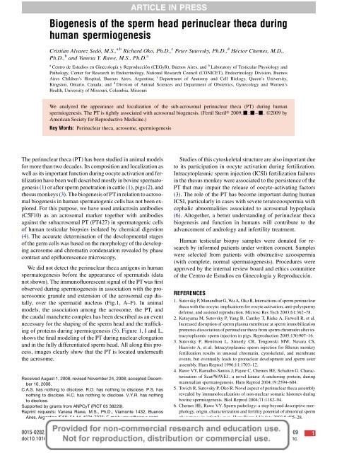
We did not detect <strong>the</strong> <strong>perinuclear</strong> <strong>the</strong>ca antigens in <strong>human</strong><br />
<strong>sperm</strong>atogenesis before <strong>the</strong> appearance <strong>of</strong> <strong>sperm</strong>atids (data<br />
not shown). The immun<strong>of</strong>luorescent signal <strong>of</strong> <strong>the</strong> PT was first<br />
observed <strong>during</strong> <strong>sperm</strong>iogenesis in association with <strong>the</strong> proacrosomic<br />
granule and extension <strong>of</strong> <strong>the</strong> acrosomal cap distally,<br />
over <strong>the</strong> <strong>sperm</strong>atid nucleus (Fig.1, A–F). In animal<br />
models, <strong>the</strong> association among <strong>the</strong> acrosome, <strong>the</strong> PT, and<br />
<strong>the</strong> caudal manchette complex has been described as an event<br />
necessary for <strong>the</strong> shaping <strong>of</strong> <strong>the</strong> <strong>sperm</strong> <strong>head</strong> and <strong>the</strong> trafficking<br />
<strong>of</strong> proteins <strong>during</strong> <strong>sperm</strong>iogenesis (5). Figure 1, I and L,<br />
shows <strong>the</strong> final modeling <strong>of</strong> <strong>the</strong> PT <strong>during</strong> nuclear elongation<br />
and in <strong>the</strong> fully differentiated <strong>sperm</strong> <strong>head</strong>. All along this process,<br />
images clearly show that <strong>the</strong> PT is located underneath<br />
<strong>the</strong> acrosome.<br />
Received August 1, 2008; revised November 24, 2008; accepted December<br />
10, 2008.<br />
C.A.S. has nothing to disclose. R.O. has nothing to disclose. P.S. has<br />
nothing to disclose. H.C. has nothing to disclose. V.Y.R. has nothing<br />
to disclose.<br />
Supported by grants from ANPCyT (PICT 05 38229).<br />
Reprint requests: Vanesa Rawe, M.S., Ph.D., Viamonte 1432, Buenos<br />
Aires, Argentina (FAX: 54-11-4371-7275; E-mail: vrawe@cegyr.com).<br />
ARTICLE IN PRESS<br />
Studies <strong>of</strong> this cytoskeletal structure are also important due<br />
to its participation in oocyte activation <strong>during</strong> fertilization.<br />
Intracytoplasmic <strong>sperm</strong> injection (ICSI) fertilization failures<br />
in <strong>the</strong> rhesus monkey were associated to <strong>the</strong> persistence <strong>of</strong> <strong>the</strong><br />
PT that may impair <strong>the</strong> release <strong>of</strong> oocyte-activating factors<br />
(3). The role <strong>of</strong> <strong>the</strong> PT has become important <strong>during</strong> <strong>human</strong><br />
ICSI, particularly in cases with severe teratozoo<strong>sperm</strong>ia with<br />
cephalic abnormalities associated to acrosomal hypoplasia<br />
(6). Altoge<strong>the</strong>r, a better understanding <strong>of</strong> <strong>perinuclear</strong> <strong>the</strong>ca<br />
biogenesis and function in <strong>human</strong>s will contribute to <strong>the</strong><br />
advancement <strong>of</strong> andrology and infertility treatment.<br />
Human testicular biopsy samples were donated for research<br />
by informed patients under written consent. Samples<br />
were selected from patients with obstructive azoo<strong>sperm</strong>ia<br />
(with complete, normal <strong>sperm</strong>atogenesis). Procedures were<br />
approved by <strong>the</strong> internal review board and ethics committee<br />
<strong>of</strong> <strong>the</strong> Centro de Estudios en Ginecologıa y Reproduccion.<br />
REFERENCES<br />
1. Sutovsky P, Manandhar G, Wu A, Oko R. Interactions <strong>of</strong> <strong>sperm</strong> <strong>perinuclear</strong><br />
<strong>the</strong>ca with <strong>the</strong> oocyte: implications for oocyte activation, anti-poly<strong>sperm</strong>y<br />
defense, and assisted reproduction. Microsc Res Tech 2003;61:362–78.<br />
2. Katayama M, Sutovsky P, Yang B, Cantley T, Rieke A, Farwell R, et al.<br />
Increased disruption <strong>of</strong> <strong>sperm</strong> plasma membrane at <strong>sperm</strong> immobilization<br />
promotes dissociation <strong>of</strong> <strong>perinuclear</strong> <strong>the</strong>ca from <strong>sperm</strong> chromatin after intracytoplasmic<br />
<strong>sperm</strong> injection in pigs. Reproduction 2005;130:907–16.<br />
3. Sutovsky P, Hewitson L, Simerly CR, Tengowski MW, Navara CS,<br />
Haavisto A, et al. Intracytoplasmic <strong>sperm</strong> injection for Rhesus monkey<br />
fertilization results in unusual chromatin, cytoskeletal, and membrane<br />
events, but eventually leads to pronuclear development and <strong>sperm</strong> aster<br />
assembly. Hum Reprod 1996;11:1703–12.<br />
4. Rawe VY, Ramalho-Santos J, Payne C, Chemes HE, Schatten G. Characterization<br />
<strong>of</strong> Scar/WAVE1, a novel kinase A-anchoring protein, <strong>during</strong><br />
mammalian <strong>sperm</strong>atogenesis. Hum Reprod 2004;19:2594–604.<br />
5. Tovich R, Sutovsky P, Oko R. Novel aspect <strong>of</strong> <strong>perinuclear</strong> <strong>the</strong>ca assembly<br />
revealed by immunolocalization <strong>of</strong> non-nuclear somatic histones <strong>during</strong><br />
bovine <strong>sperm</strong>iogenesis. Biol Reprod 2004;71:1182–94.<br />
6. Chemes HE, Rawe VY. Sperm pathology: a step beyond descriptive morphology.<br />
origin, characterization and fertility potential <strong>of</strong> abnormal <strong>sperm</strong><br />
phenotypes in infertile men. Hum Reprod Update 2003;9:405–28.<br />
0015-0282/09/$36.00 Fertility and Sterility â Vol. -, No. -, - 2009 1<br />
doi:10.1016/j.fertnstert.2008.12.051 Copyright ª2009 American Society for Reproductive Medicine, Published by Elsevier Inc.
FIGURE 1<br />
Immun<strong>of</strong>luorescence <strong>of</strong> different stages <strong>of</strong> <strong>human</strong> <strong>sperm</strong>iogenesis showing acrosin (antibody C5F10, red, left<br />
panels), <strong>the</strong> PT (PT427 antiserum, green, central panels), and merged views (yellow) over contrast phase images<br />
<strong>of</strong> <strong>the</strong> same cells (right panels). (A–C) The biogenesis and initial distribution <strong>of</strong> <strong>the</strong> PT and its early association with<br />
<strong>the</strong> acrosomic vesicle and proacrosomic granule in round <strong>sperm</strong>atids. (D–F) During cap phase and initial<br />
<strong>sperm</strong>atid elongation, <strong>the</strong> acrosome extends distally on <strong>the</strong> apical pole <strong>of</strong> <strong>the</strong> <strong>sperm</strong>atid nucleus with <strong>the</strong> PT<br />
associated underneath. In this stage, we can also clearly recognize <strong>the</strong> process <strong>of</strong> DNA condensation. (G–I) Front<br />
views and (J–L) side views <strong>of</strong> a completely elongated <strong>sperm</strong>atid with a fully formed acrosome depict <strong>the</strong> PT in<br />
a subacrosomal location. Isolated cells <strong>of</strong> <strong>human</strong> <strong>sperm</strong>atogenesis were examined using an epifluorescence<br />
microscope (Nikon BX40), under ultraviolet light with specific filters for <strong>the</strong> desired wavelengths. (Images edited<br />
using Adobe Photoshop 7.0.)<br />
Alvarez Sedo. Perinuclear <strong>the</strong>ca <strong>during</strong> <strong>human</strong> <strong>sperm</strong>iogenesis. Fertil Steril 2009.<br />
ARTICLE IN PRESS<br />
2 Alvarez Sedo et al. Perinuclear <strong>the</strong>ca <strong>during</strong> <strong>human</strong> <strong>sperm</strong>iogenesis Vol. -, No. -, - 2009
1 <strong>Biogenesis</strong> <strong>of</strong> <strong>the</strong> <strong>sperm</strong> <strong>head</strong> <strong>perinuclear</strong> <strong>the</strong>ca<br />
<strong>during</strong> <strong>human</strong> <strong>sperm</strong>iogenesis<br />
C. Alvarez Sedo, R. Oko, P. Sutovsky, H. Chemes, and<br />
V. Y. Rawe<br />
Buenos Aires, Argentina; Ontario, Canada; and<br />
Columbia, Missouri<br />
To explore <strong>the</strong> biogenesis <strong>of</strong> <strong>the</strong> subacrosomal <strong>perinuclear</strong><br />
<strong>the</strong>ca <strong>during</strong> <strong>human</strong> <strong>sperm</strong>iogenesis, we analyzed<br />
<strong>the</strong> presence and localization <strong>of</strong> this structure<br />
in relation to <strong>the</strong> development <strong>of</strong> <strong>the</strong> acrosome.<br />
Fertility and Sterility â<br />
ARTICLE IN PRESS