Monitor the Lipid Profile - South Jersey Heart Group
Monitor the Lipid Profile - South Jersey Heart Group
Monitor the Lipid Profile - South Jersey Heart Group

Create successful ePaper yourself
Turn your PDF publications into a flip-book with our unique Google optimized e-Paper software.
44<br />
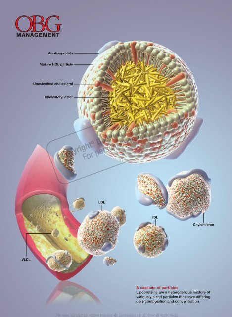
VLDL<br />
Apolipoprotein<br />
Mature HDL particle<br />
Unesterifi ed cholesterol<br />
Cholesteryl ester<br />
Copyright ® Dowden Health Media<br />
For personal use only<br />
LDL<br />
Chylomicron<br />
A cascade of particles<br />
Lipoproteins are a heterogenous mixture of<br />
variously sized particles that have differing<br />
core composition and concentration<br />
OBG Management | December 2008 | Vol. 20 No. 12<br />
For mass reproduction, content licensing and permissions contact Dowden Health Media.<br />
IDL
GARY CARLSON FOR OBG MANAGEMENT<br />
YOU HAVE A NEW JOB<br />
<strong>Monitor</strong> <strong>the</strong><br />
lipid profile<br />
Th e standard panel provides more details than you might<br />
imagine about your patient’s risk of cardio vascular disease<br />
Thomas Dayspring, MD<br />
Alan Helmbold, DO<br />
Dr. Dayspring is Director of <strong>the</strong><br />
North <strong>Jersey</strong> Institute of Menopausal<br />
<strong>Lipid</strong>ology in Wayne, NJ, and a<br />
Diplomate of <strong>the</strong> American Board<br />
of Internal Medicine and American<br />
Board of Clinical <strong>Lipid</strong>ology.<br />
Dr. Helmbold is a Cardiology Fellow<br />
at Brooke Army Medical Center at Fort<br />
Sam Houston, Texas, and a Diplomate<br />
of <strong>the</strong> American Board of Internal<br />
Medicine and American Board of<br />
Clinical <strong>Lipid</strong>ology.<br />
Dr. Dayspring serves on <strong>the</strong> advisory<br />
board for LipoScience. Dr. Helmbold<br />
reports no fi nancial relationships<br />
relevant to this article.<br />
›› SHARE YOUR COMMENTS<br />
Are you ready to monitor your<br />
patients’ risk of a<strong>the</strong>rosclerosis?<br />
Or does this new duty make it<br />
harder to provide focused<br />
ObGyn care? Let us know:<br />
E-MAIL obg@dowdenhealth.com<br />
FAX 201-391-2778<br />
Add ano<strong>the</strong>r item to your ever-growing list of responsibilities:<br />
monitoring your patients’ risk of<br />
a<strong>the</strong>rosclerosis.<br />
Th is task used to be <strong>the</strong> purview of internists and<br />
cardiologists but, because gynecologists are increasingly<br />
serving as a primary care provider, you need to learn to<br />
recognize and diagnose <strong>the</strong> many clinical expressions of<br />
a<strong>the</strong>rosclerosis in your aging patients.<br />
A crucial part of that knowledge is a thorough understanding<br />
of each and every lipid concentration parameter<br />
reported within <strong>the</strong> standard lipid profi le. Th is article reviews<br />
those parameters, explains how to interpret <strong>the</strong>m<br />
individually and in combination, and introduces a new<br />
paradigm: <strong>the</strong> analysis of lipoprotein particle concentrations<br />
as a more precise way to determine risk.<br />
If used in its entirety, <strong>the</strong> lipid profi le provides a signifi -<br />
cant amount of information about <strong>the</strong> presence or absence<br />
of pathologic lipoprotein concentrations. Far too many clinicians<br />
focus solely on low-density lipoprotein cholesterol<br />
(LDL-C) and ignore <strong>the</strong> rest of <strong>the</strong> profi le. Failure to consider<br />
<strong>the</strong> o<strong>the</strong>r variables is one reason why a<strong>the</strong>rosclerotic<br />
disease is underdiagnosed and undertreated in <strong>the</strong> United<br />
States in many patients—especially women. 1<br />
Why lipoproteins are important<br />
Th ere is only one absolute in a<strong>the</strong>rosclerosis: Sterols—predominantly<br />
cholesterol—enter <strong>the</strong> artery wall, where <strong>the</strong>y<br />
IN THIS<br />
ARTICLE<br />
How to read<br />
a lipid panel in<br />
6 quick steps<br />
page 46<br />
Desirable lipid<br />
values for women<br />
page 47<br />
Similar lipid<br />
panels, different<br />
levels of risk<br />
page 49<br />
obgmanagement.com Vol. 20 No. 12 | December 2008 | OBG Management 45
<strong>Monitor</strong>ing <strong>the</strong> lipid profi le<br />
6 tests to assess ovarian reserve in <strong>the</strong> offi ce<br />
How to read a lipid panel<br />
in 6 quick steps<br />
1. Look at <strong>the</strong> triglyceride (TG) level. If it is >500 mg/dL, treatment<br />
is indicated, and TG reduction takes precedence over all o<strong>the</strong>r lipid<br />
concentrations. If TG is 190 mg/dL, drug <strong>the</strong>rapy is indicated regardless of o<strong>the</strong>r fi ndings.<br />
At lower levels, <strong>the</strong> need for <strong>the</strong>rapy is based on <strong>the</strong> patient’s<br />
overall risk of cardiovascular disease (CVD). Therapeutic lifestyle<br />
recommendations are always indicated.<br />
3. Look at high-density lipoprotein cholesterol (HDL-C). Increased<br />
risk is present if it is 4.0.<br />
5. Calculate <strong>the</strong> non-HDL-C level (TC minus HDL-C). If it is >130<br />
mg/dL (or >100 mg/dL in very-high-risk women), <strong>the</strong>rapy is warranted.<br />
Newer data reveal that this calculation is always equal to,<br />
or better than, LDL-C at predicting CVD risk. Non-HDL-C is less<br />
valuable if TG is >500 mg/dL.<br />
6. Calculate <strong>the</strong> TG/HDL-C ratio to estimate <strong>the</strong> size of LDL. If <strong>the</strong><br />
ratio is >3.8, <strong>the</strong> likelihood of small LDL is 80%. (Small LDL usually<br />
has very high LDL-P.)<br />
46<br />
are oxidized, internalized by macrophages,<br />
and transformed into foam cells, <strong>the</strong> histologic<br />
hallmark of a<strong>the</strong>rosclerosis. With <strong>the</strong> accumulation<br />
of foam cells, fatty streaks develop<br />
and, ultimately, so does complex plaque.<br />
<strong>Lipid</strong>s associated with cardiovascular<br />
disease (CVD) include:<br />
• cholesterol<br />
• noncholesterol sterols such as sitosterol,<br />
campesterol, and o<strong>the</strong>rs of mostly plant<br />
or shellfi sh origin<br />
• triacylglycerol, or triglycerides (TG)<br />
• phospholipids.<br />
Because lipids are insoluble in aqueous<br />
solutions such as plasma, <strong>the</strong>y must be “traffi<br />
cked” within protein-enwrapped particles<br />
called lipoproteins. Th e surface proteins that<br />
provide structure and solubility to lipoproteins<br />
are called apolipoproteins. A key con-<br />
OBG Management | December 2008 | Vol. 20 No. 12<br />
cept is that, with <strong>the</strong>ir surface apolipoproteins<br />
and cholesterol core, certain lipoproteins are<br />
potential agents of a<strong>the</strong>rogenesis in that <strong>the</strong>y<br />
transport sterols into <strong>the</strong> artery wall. 2<br />
Estimation of <strong>the</strong> risk of CVD involves<br />
careful analysis of all standard lipid concentrations<br />
and <strong>the</strong>ir various ratios, and prediction<br />
of <strong>the</strong> potential presence of a<strong>the</strong>rogenic<br />
lipoproteins. Successful prevention or treatment<br />
of a<strong>the</strong>rosclerosis entails limiting <strong>the</strong><br />
presence of a<strong>the</strong>rogenic lipoproteins.<br />
A new paradigm is on its way<br />
Th e a<strong>the</strong>rogenicity of lipoprotein particles is<br />
determined by particle concentration as well<br />
as o<strong>the</strong>r variables, including particle size,<br />
lipid composition, and distinct surface apolipoproteins.<br />
Lipoproteins smaller than 70 nm in diameter<br />
are driven into <strong>the</strong> arterial intima<br />
primarily by concentration gradients, regardless<br />
of lipid composition or particle<br />
size. 3 A recent Consensus Statement from<br />
<strong>the</strong> American Diabetes Association and <strong>the</strong><br />
American College of Cardiology observed<br />
that quantitative analysis of <strong>the</strong>se potentially<br />
a<strong>the</strong>rogenic lipoproteins is one of <strong>the</strong> best<br />
lipid/lipoprotein-related determinants of<br />
CVD risk. 4 Lipoprotein particle concentrations<br />
have emerged not only as superb predictors<br />
of risk, but also as goals of <strong>the</strong>rapy. 5–7<br />
Because of cost, third-party reimbursement,<br />
varying test availability, and lack of interpretive<br />
knowledge, few clinicians routinely<br />
order lipoprotein quantifi cation. Historically,<br />
CVD risk and goals of <strong>the</strong>rapy have been<br />
based on lipid concentrations (<strong>the</strong> amount<br />
of lipids traffi cked within lipoprotein cores)<br />
reported in <strong>the</strong> lipid profi le. Guidelines from<br />
<strong>the</strong> National Cholesterol Education Program,<br />
Adult Treatment Panel III (NCEP ATP-III) 8,9<br />
and <strong>the</strong> American <strong>Heart</strong> Association (AHA)<br />
CVD Prevention in Women 10,11 use lipid concentrations<br />
such as total cholesterol (TC),<br />
LDL-C, high-density lipoprotein cholesterol<br />
(HDL-C), and TG as estimates or surrogates<br />
of lipoprotein concentrations (TABLE 1).<br />
Th e day is rapidly approaching, however,<br />
when lipoprotein concentrations may replace
TABLE 1 Desirable lipid values<br />
for women<br />
<strong>Lipid</strong> Level (mg/dL)<br />
Total cholesterol
<strong>Monitor</strong>ing <strong>the</strong> lipid profi le<br />
6 tests to assess ovarian reserve<br />
lomicrons,<br />
in<br />
1<br />
<strong>the</strong><br />
hour;<br />
offi<br />
VLDL,<br />
ce<br />
2–6 hours; IDL,<br />
1–2 hours; LDL, 2–3 days), <strong>the</strong> great majority<br />
(90% to 95%) of apoB-containing particles<br />
are LDL. Although apoB measurement yields<br />
quantifi cation of all beta-lipoproteins, it is<br />
primarily a surrogate of LDL particle (LDL-<br />
P) concentration. 15<br />
Individual particle concentrations, deter<br />
mined by NMR spectroscopy, are reported<br />
as VLDL-P, IDL-P, LDL-P, and HDL-P (see<br />
<strong>the</strong> “Glossary” on page 50). 14<br />
Several epidemiologic studies that enrolled<br />
both genders found <strong>the</strong> best predictors<br />
of risk to be:<br />
• elevated levels of apoB or LDL-P and reduced<br />
levels of apoA-I or HDL-P<br />
• a high apoB/apoA-I ratio or LDL-P/<br />
HDL-P ratio. 6,13,14<br />
After adjustment for lipoprotein concentration<br />
data (apoB or LDL-P), o<strong>the</strong>r lipoprotein<br />
characteristics such as particle<br />
lipid content, size, or composition, for <strong>the</strong><br />
most part, had no statistically signifi cant<br />
relationship with <strong>the</strong> risk of cardiovascular<br />
disease. 16,17<br />
Using lipid measurements to<br />
estimate lipoproteins<br />
Total cholesterol represents <strong>the</strong> cholesterol<br />
content within all lipoproteins in 1 dL of<br />
plasma. Because beta-lipoproteins are considerably<br />
larger than alpha-lipoproteins, approximately<br />
75% of total cholesterol is carried<br />
in <strong>the</strong> apoB-containing particles, making TC<br />
an apoB surrogate.<br />
VLDL-C, an often ignored variable, is<br />
not measured but calculated using <strong>the</strong> Friedewald<br />
formula, dividing TG by fi ve. Th is<br />
calculation assumes—often erroneously<br />
as TG levels rise—that TG consists only of<br />
VLDL particles and that VLDL composition<br />
contains fi ve times more TG than cholesterol<br />
molecules.<br />
A desirable TG level is
emnant particles. 18 However, because <strong>the</strong><br />
vast majority of beta-lipoproteins are LDL,<br />
LDL-C (especially if elevated) is a better<br />
apoB surrogate than VLDL-C and is <strong>the</strong> primary<br />
CVD risk factor and goal of <strong>the</strong>rapy in<br />
every current guideline.<br />
LDL-C is usually a calculated value using<br />
<strong>the</strong> formula:<br />
LDL-C = TC – (HDL-C + VLDL-C)<br />
Upon special order, laboratories can directly<br />
measure LDL-C. Th is option is most<br />
useful when TG levels are high, rendering<br />
<strong>the</strong> Friedewald formula less accurate<br />
(TABLE 2, page 51). 19 For population cut<br />
points and desirable goals of <strong>the</strong>rapy for<br />
lipid and lipoprotein concentrations, see <strong>the</strong><br />
FIGURE (page 51).<br />
HDL-C, apoA-I are inversely related<br />
to cardiovascular risk<br />
Th e epidemiologic data strongly indicate<br />
that both HDL-C and apoA-I are strongly and<br />
inversely related to CVD risk. 6 HDL particles<br />
are a heterogenous collection of:<br />
• unlipidated apoA-I<br />
• very small pre-beta HDL<br />
• more mature, lipidated HDL3 and HDL2<br />
species (HDL3 smaller than HDL2).<br />
NMR nomenclature identifi es <strong>the</strong> smaller<br />
HDL species as H1 and H2 and <strong>the</strong> larger<br />
HDL species as H4 and H5. 14 Th e smaller<br />
HDL species also contain apoA-II.<br />
Although HDL can acquire cholesterol<br />
from any cell, including arterial-wall foam<br />
cells, <strong>the</strong> majority of HDL lipidation occurs<br />
in <strong>the</strong> liver or proximal small intestine, after<br />
which it is traffi cked to steroidogenic tissue,<br />
adipocytes, or back to <strong>the</strong> liver. Normally,<br />
HDL carries little TG. 20 Th e only lipid concentration<br />
that can serve as a surrogate of apoA-I<br />
or HDL-P is HDL-C, where <strong>the</strong> assumption is<br />
that higher HDL-C indicates higher apoA-I,<br />
and vice versa.<br />
In reality, <strong>the</strong> correlation between apoA-I<br />
and HDL-C varies because each HDL particle<br />
can have from two to four apoA-I molecules,<br />
and <strong>the</strong> volume of cholesterol within <strong>the</strong> particle<br />
is a function of particle size and its TG<br />
content. For <strong>the</strong> most part, total HDL-C is indicative<br />
of <strong>the</strong> cholesterol carried in <strong>the</strong> larg-<br />
Two patients, similar lipid profi les:<br />
Why is only one at heightened risk?<br />
Two premenopausal women undergo assessment of <strong>the</strong>ir basic<br />
lipid panel, with <strong>the</strong>se results:<br />
LIPID PATIENT 1 PATIENT 2<br />
Total cholesterol (TC) 180 180<br />
LDL-C 100 100<br />
HDL-C 60 40<br />
VLDL-C 20 40<br />
Triglycerides (TG) 100 200<br />
Non-HDL-C 120 160<br />
TC/HDL-C ratio 3.0 4.5<br />
TG/HDL-C ratio<br />
LDL-C, low-density lipoprotein cholesterol<br />
HDL-C, high-density lipoprotein cholesterol<br />
1.6 5.0<br />
VLDL-C, very-low-density lipoprotein cholesterol<br />
Both patients have <strong>the</strong> same desirable TC and LDL-C values. However,<br />
fur<strong>the</strong>r analysis reveals an abnormal TC/HDL-C ratio and an<br />
abnormal non-HDL-C level in patient 2. This fi nding indicates a<br />
higher risk of CVD.<br />
In addition, <strong>the</strong> TG/HDL-C ratio of 5.0 in patient 2 is highly suggestive<br />
of small-LDL phenotype B. That designation means that<br />
this patient will have 40% to 70% more LDL particles to traffi c her<br />
LDL-C than patient 1, who appears to have LDL of normal size. 27<br />
The elevated VLDL-C of patient 2 indicates <strong>the</strong> presence of VLDL<br />
remnants, which predict risk above that conveyed by LDL-C. 7<br />
The typical clinician, looking only at TC or LDL-C, would miss<br />
<strong>the</strong> increased risk (high apoB) in patient 2. Obvious clues to her<br />
lipoprotein pathology are <strong>the</strong> elevated TG and reduced HDL-C<br />
(TG-HDL axis disorder). Beyond elevated TG and reduced HDL-C,<br />
patient 2 is also likely to have increased waist size, subtle hypertension,<br />
and possibly impaired fasting glucose—three additional<br />
parameters of metabolic syndrome. 7,10,25<br />
er, mature HDL2 (H4, H5) particles; patients<br />
with low HDL-C typically lack <strong>the</strong>se mature,<br />
lipidated HDL particles.<br />
Because HDL rapidly and repeatedly<br />
lipidates and <strong>the</strong>n delipidates, <strong>the</strong>re is no relationship<br />
between <strong>the</strong> HDL-C level and <strong>the</strong><br />
complex dynamic process termed reverse<br />
cholesterol transport process. Nei<strong>the</strong>r HDL-<br />
C, nor apoA-I, nor HDL-P, nor HDL size is<br />
consistently related to HDL particle func-<br />
obgmanagement.com Vol. 20 No. 12 | December 2008 | OBG Management 49
<strong>Monitor</strong>ing <strong>the</strong> lipid profi le<br />
6 tests to assess ovarian reserve<br />
<strong>Lipid</strong>s and<br />
in <strong>the</strong><br />
lipoproteins:<br />
offi ce<br />
A glossary<br />
The best lipid<br />
concentration<br />
estimate of<br />
apoB is <strong>the</strong><br />
calculated non-<br />
HDL-C value<br />
50<br />
Variable What is it?<br />
Triglycerides (TG) The triacylglycerol concentration within all of <strong>the</strong> TG-traffi cking<br />
lipoproteins in 100 mL or 1 dL of plasma<br />
Total cholesterol (TC) Cholesterol content of all lipoproteins in 1 dL of plasma<br />
Low-density lipoprotein Cholesterol content of all intermediate-density lipoprotein (IDL)<br />
(LDL) cholesterol and LDL particles in 1 dL of plasma<br />
High-density lipoprotein Cholesterol content of all HDL particles in 1 dL of plasma<br />
(HDL) cholesterol<br />
Very-low-density lipoprotein Cholesterol content of all VLDL particles in 1 dL of plasma<br />
(VLDL) cholesterol<br />
Remnant-C Cholesterol content of all remnants in 1 dL of plasma<br />
Lipoprotein (a) [Lp(a)] Cholesterol content of LDL particles that have apo(a) attached<br />
cholesterol<br />
Lp(a) concentration Concentration of apo(a) in 1 dL of plasma<br />
Non-HDL cholesterol Cholesterol within all apoB particles in 1 dL of plasma<br />
LDL-P Number of LDL particles in 1 L of plasma (expressed in nmol/L).<br />
This represents LDL particles of all sizes<br />
Small LDL-P Number of small and intermediate LDL particles in 1 L of<br />
plasma (nmol/L)<br />
HDL-P Number of HDL particles in 1 L of plasma (μmol/L). HDL-P is<br />
also reported as large, intermediate, and small HDL-P (μmol/L)<br />
VLDL-P Number of VLDL particles in 1 L of plasma (nmol/L)<br />
IDL-P Number of IDL particles in 1 L of plasma (nmol/L)<br />
LDL size Diameter of <strong>the</strong> predominant LDL species:<br />
• Pattern or phenotype A refers to predominantly large,<br />
buoyant LDL particles<br />
• Pattern or phenotype B refers to predominantly small, dense<br />
LDL particles<br />
tionality—i.e., <strong>the</strong> ability of HDL to lipidate<br />
or delipidate, appropriately traffi c cholesterol,<br />
or perform numerous o<strong>the</strong>r nonlipid<br />
antia<strong>the</strong>rogenic functions. 20,21<br />
Focus on lipoprotein particle<br />
concentrations<br />
To most accurately predict lipid-related CVD<br />
risk, you must determine which patients<br />
OBG Management | December 2008 | Vol. 20 No. 12<br />
have elevated numbers of a<strong>the</strong>rogenic lipoproteins<br />
using actual particle concentrations.<br />
In most practices, lipoprotein particle<br />
numbers must be estimated by scrutinizing<br />
all of <strong>the</strong> lipid concentrations and ratios (not<br />
simply LDL-C).<br />
TC and, especially, LDL-C are apoB and<br />
LDL-P surrogates, but <strong>the</strong> best lipid concentration<br />
estimate of apoB is <strong>the</strong> calculated<br />
non-HDL-C value. By subtracting HDL-C
from TC, it is possible to identify <strong>the</strong> cholesterol<br />
not in <strong>the</strong> HDL particles but in all of <strong>the</strong><br />
potentially a<strong>the</strong>rogenic apoB particles. In essence,<br />
non-HDL-C is VLDL-C plus LDL-C.<br />
Th is equation yields a better apoB or LDL-P<br />
proxy, compared with LDL-C alone. 18 If a patient<br />
has reached her LDL-C goal but still has<br />
a high non-HDL-C level, we can assume that<br />
<strong>the</strong>re are still too many apoB particles and<br />
that <strong>the</strong>y are contributing to residual risk.<br />
Because LDL is <strong>the</strong> predominant apoB<br />
species, non-HDL-C is <strong>the</strong> best lipid concentration<br />
predictor of LDL-P. 15 Because nei<strong>the</strong>r<br />
TC nor HDL-C assays require a patient to fast,<br />
non-HDL-C is accurate in nonfasting patients,<br />
making it a very practical way to screen<br />
for CVD risk. 8 In <strong>the</strong> Women’s Health Study,<br />
which involved mostly healthy women, non-<br />
HDL-C predicted <strong>the</strong> risk of coronary heart<br />
disease as well as apoB did, but not as well as<br />
LDL-P. 22,23 In independent, separately published<br />
analyses from <strong>the</strong> Framingham Off -<br />
spring Study, LDL-P was a better predictor of<br />
risk than LDL-C and apoB. 15,24<br />
NCEP ATP-III guidelines introduced<br />
non-HDL-C as a secondary goal of <strong>the</strong>rapy<br />
in patients with TG >200 mg/dL. Subsequent<br />
data indicate that non-HDL-C is always a<br />
better predictor of risk than LDL-C is, regardless<br />
of TG levels. 18<br />
Th e AHA Women’s Guideline was <strong>the</strong><br />
fi rst to set a desired non-HDL-C level (130<br />
mg/dL) independent of <strong>the</strong> TG value. 10 Because<br />
a normal VLDL-C concentration is 30<br />
mg/dL, <strong>the</strong> non-HDL-C goal is 30 mg/dL<br />
above <strong>the</strong> desired LDL-C goal. For example,<br />
if <strong>the</strong> desired LDL-C value is 100 mg/dL, <strong>the</strong><br />
non-HDL-C goal is 130 mg/dL. If <strong>the</strong> desired<br />
LDL-C goal is 70 mg/dL—as it is in a patient<br />
at very high risk—<strong>the</strong> non-HDL-C goal would<br />
be 100 mg/dL (FIGURE). 9,11<br />
Insulin resistance diminishes<br />
accuracy of lipid profi le<br />
Th e ability to predict lipoprotein particle concentrations<br />
using <strong>the</strong> lipid profi le becomes<br />
far less accurate in situations associated with<br />
insulin resistance and metabolic syndrome<br />
in patients who have TG-HDL axis disorders.<br />
CONTINUED ON PAGE 52<br />
TABLE 2<br />
How lipid concentrations are determined<br />
TC = apoA-I-C + apoB-C<br />
TC = HDL-C + LDL-C + VLDL-C + IDL-C + Chylomicron-C + Lp(a)-C +<br />
Remnant-C<br />
In a fasting patient under normal circumstances, <strong>the</strong>re are no<br />
chylomicrons and remnants (smaller chylomicrons or VLDL particles)<br />
and very few, if any, IDL particles. These are postprandial lipoproteins.<br />
Most patients do not have Lp(a) pathology. Therefore, <strong>the</strong> lipid<br />
concentration formula simplifi es:<br />
TC = HDL-C + LDL-C + VLDL-C<br />
VLDL-C is estimated by TG/5 (assumes that all TG is in VLDL and that<br />
VLDL TG:cholesterol composition is 5:1). Therefore:<br />
TC = HDL-C + LDL-C + TG/5<br />
LDL-C = TC – (HDL-C + TG/5)<br />
Non-HDL-C = TC – HDL-C<br />
In actuality, <strong>the</strong> calculated or directly measured LDL-C values in <strong>the</strong><br />
standard lipid panel represent LDL-C + IDL-C + Lp(a)-C. However,<br />
because labs do not usually separate IDL and Lp(a) particles from<br />
LDL (without signifi cant added expense), only total LDL-C is reported.<br />
FIGURE Population percentile cut<br />
points and goals for LDL-C, LDL-P, ApoB,<br />
and non-HDL-C<br />
Goal for<br />
very-high-risk<br />
patients<br />
LDL-C<br />
mg/dL<br />
LDL-P<br />
nmol/L<br />
ApoB<br />
mg/dL<br />
Goal for<br />
high-risk<br />
patients<br />
Goal for<br />
low-risk<br />
patients<br />
20 th 50 th 80 th<br />
20 th 50 th 80 th<br />
20 th 50 th 80 th<br />
LDL-C<br />
Population cut points<br />
(NCEP ATP III)<br />
obgmanagement.com Vol. 20 No. 12 | December 2008 | OBG Management 51<br />
LDL-P<br />
Population cut points<br />
(MESA Trial)<br />
ApoB<br />
Population cut points<br />
(NHANES)<br />
Non-HDL -C mg/dL Non-HDL -C
<strong>Monitor</strong>ing <strong>the</strong> lipid profi le<br />
6 tests to assess ovarian reserve in<br />
<strong>Lipid</strong><br />
<strong>the</strong> offi<br />
markers<br />
ce<br />
TABLE 3<br />
of<br />
small low-density lipoproteins<br />
Both NCEP ATP-III<br />
and AHA Women’s<br />
Guidelines use <strong>the</strong><br />
total cholesterol/<br />
HDL ratio as<br />
a powerful risk<br />
predictor<br />
52<br />
High-density lipoprotein cholesterol<br />
(HDL-C) 130–150 mg/dL<br />
Total cholesterol/HDL-C ratio >4.0 with normal<br />
low-density lipoprotein cholesterol (LDL-C)<br />
TG/HDL-C ratio >3.8 in women<br />
Unremarkable LDL-C but elevated non-HDL-C<br />
TABLE 4 <strong>Lipid</strong> markers of<br />
remnant lipoproteins<br />
Triglyceride (TG) >150–200 mg/dL<br />
Very-low-density lipoprotein cholesterol<br />
>30 mg/dL<br />
Unremarkable low-density lipoprotein cholesterol<br />
with elevated non-high-density lipoprotein<br />
cholesterol (HDL-C)<br />
Low HDL-C in insulin-resistant patients<br />
Elevated total cholesterol/HDL-C ratio and<br />
TG >150 mg/dL<br />
In women, <strong>the</strong>se disorders are typifi ed by an<br />
elevation of TG >150 mg/dL and a decrease<br />
in HDL-C 3.8), which are indicative<br />
of too many small LDL particles (high apoB,<br />
LDL-P) and reduced number of HDL particles<br />
(high apoB/A-I ratio). 26,27<br />
Such a scenario, typical of TG-HDL axis<br />
disorders, explains much of <strong>the</strong> risk associated<br />
with rising TG levels and is very common<br />
in premenopausal women who have insulin-resistant<br />
states such as type 2 diabetes<br />
or polycystic ovary syndrome and in menopausal<br />
women who have insulin resistance<br />
and coronary artery disease. 1<br />
LDL-C and LDL-P do not<br />
always correlate<br />
Because <strong>the</strong> volume of a lipoprotein is a<br />
function of its radius cubed (V = 4/3πr 3 ), 14 a<br />
patient who has small LDL will require up<br />
to 40% to 70% more LDL particles to traffi c<br />
a given amount of LDL-C. In such a patient,<br />
<strong>the</strong>re is often little correlation between LDL-<br />
C and LDL-P or apoB values. Regardless of<br />
<strong>the</strong> LDL-C, <strong>the</strong> apoB, LDL-P, or non-HDL-C<br />
is often elevated. 28 Th is risk, which cannot<br />
be predicted by looking only at LDL-C, is <strong>the</strong><br />
main reason guidelines advocate <strong>the</strong> use of<br />
non-HDL-C or <strong>the</strong> TC/HDL-C ratio. 8,11 (See<br />
<strong>the</strong> case studies on page 49.)<br />
In summary, a large part of <strong>the</strong> risk of<br />
CVD seen in patients who have low HDL-<br />
C derives from <strong>the</strong> associated increase in<br />
<strong>the</strong> number of apoB particles, mostly composed<br />
of small LDL, as well as an increase<br />
in remnant particles. 15,21,28 Th is crucial point<br />
explains why treatment of low HDL-C states
should always fi rst target apoB or LDL-P<br />
(LDL-C and non-HDL-C), ra<strong>the</strong>r than apoA-<br />
I or HDL-C (TABLES 3 and 4). 8,9<br />
A few words of advice<br />
Th e driving forces of a<strong>the</strong>rogenesis are increased<br />
numbers of apoB-containing lipoproteins<br />
and impaired endo<strong>the</strong>lial integrity.<br />
ApoB and LDL-P are <strong>the</strong> available lab assays<br />
that most accurately quantify a<strong>the</strong>rogenic<br />
particle number.<br />
Th e lipid-concentration surrogates that<br />
you should be using to better predict apoB<br />
and CVD risk are:<br />
• TC (unless HDL-C is very high)<br />
• LDL-C<br />
• Non-HDL-C<br />
• TC/HDL-C ratio<br />
• TG/HDL-C ratio.<br />
Because LDL is by far <strong>the</strong> most numerous<br />
of <strong>the</strong> apoB particles present in plasma, it<br />
References<br />
1. Lloyd-Jones DM, O’Donnell CJ, D’Agostino RB, et<br />
al. Applicability of cholesterol-lowering primary prevention<br />
trials to a general population. Th e Framingham<br />
<strong>Heart</strong> Study. Arch Intern Med. 2001;161:949–954.<br />
2. Biggerstaff KD, Wooten JS. Understanding lipoproteins<br />
as transporters of cholesterol and o<strong>the</strong>r lipids.<br />
Adv Physiol Educ. 2004;28:105–106.<br />
3. Nordestgaard BG, Wooten R, Lewis B. Selective<br />
retention of VLDL, IDL and LDL in <strong>the</strong> arterial intima<br />
of genetically hyperlipidemic rabbits in vivo. Molecular<br />
size as a determinant of fractional loss from <strong>the</strong><br />
intima-inner media. Arterioscler Th romb Vasc Biol.<br />
1995;15:534–542.<br />
4. Brunzell JD, Davidson M, Furberg CD, et al. Lipoprotein<br />
management in patients with cardiometabolic<br />
risk. Consensus statement from <strong>the</strong> American Diabetes<br />
Association and <strong>the</strong> American College of Cardiology<br />
Foundation. Diabetes Care. 2008;31:811–822.<br />
5. Barter PJ, Ballantyne CM, Carmena R, et al. ApoB<br />
versus cholesterol in estimating cardiovascular risk<br />
and in guiding <strong>the</strong>rapy: report of <strong>the</strong> thirty-person/<br />
ten-country panel. J Intern Med. 2006;259:247–258.<br />
6. Walldius G, Jungner I, Holme I, Aastveit AH,<br />
Kolar W, Steiner E. High apolipoprotein B, low apolipoprotein<br />
A-I, and improvement in <strong>the</strong> prediction<br />
of fatal myocardial infarction (AMORIS study): a prospective<br />
study. Lancet. 2001;358:2026–2033.<br />
7. Mudd JO, Borlaug BA, Johnson PV, et al. Beyond<br />
low-density lipoprotein cholesterol: defi ning <strong>the</strong> role<br />
of low-density lipoprotein heterogeneity in coronary<br />
artery disease. J Am Coll Cardiol. 2007;50:1735–1741.<br />
8. Executive Summary of <strong>the</strong> Th ird Report of <strong>the</strong> National<br />
Cholesterol Education Program (NCEP) Expert<br />
Panel on Detection, Evaluation, and Treatment of High<br />
Blood Cholesterol in Adults (Adult Treatment Panel<br />
III). JAMA. 2001;285:2486–2497.<br />
9. Grundy SM, Cleeman JI, Merz CN, et al. Implica-<br />
is <strong>the</strong> primary agent of a<strong>the</strong>rogenesis. However,<br />
apoB and LDL-P do not correlate with<br />
LDL-C when LDL particles are small, are TGrich<br />
and cholesterol-poor, or simply cholesterol-poor<br />
(seen in some patients who have<br />
low LDL-C levels). 7,15<br />
Both NCEP ATP-III and AHA Women’s<br />
Guidelines use <strong>the</strong> TC/HDL ratio as a powerful<br />
risk predictor. However, as a goal of<br />
<strong>the</strong>rapy, <strong>the</strong>se guidelines recommend normalizing<br />
LDL-C and <strong>the</strong>n non-HDL-C. 8,11 In<br />
reality, normalization of non-HDL-C takes<br />
care of LDL-C as well. For example, say a patient<br />
has LDL-C 130 mg/dL or TC/HDL-C ratio >4. Th ese<br />
readings indicate residual risk and suggest<br />
that an elevated number of apoB particles is<br />
present. Th erapy to normalize non-HDL-C<br />
or, better yet, apoB/LDL-P, is warranted. Th e<br />
clue that residual risk is present even when<br />
LDL-C is normal is <strong>the</strong> reduction of HDL-C<br />
and elevation of TG and non-HDL-C.<br />
tions of recent clinical trials for <strong>the</strong> National Cholesterol<br />
Education Program Adult Treatment Panel III<br />
Guidelines. Circulation. 2004;110:227–239.<br />
10. Mosca L, Appel LJ, Benjamin EJ, et al. Evidencebased<br />
guidelines for cardiovascular disease prevention<br />
in women. Circulation. 2004;109:672–693.<br />
11. Mosca L, Banka CL, Benjamin EJ, et al. Evidencebased<br />
guidelines for cardiovascular disease prevention<br />
in women: 2007 update. Circulation. 2007;115:1481.<br />
12. Sniderman AD. Apolipoprotein B versus nonhigh-density<br />
lipoprotein cholesterol. And <strong>the</strong> winner<br />
is... Circulation. 2005;112:3366–3367.<br />
13. Sniderman AD, Marcovina SM. Apolipoprotein A-<br />
I and B. Clin Lab Med. 2006;26:733–750.<br />
14. Jeyarajah EJ, Cromwell WC, Otvos JD. Lipoprotein<br />
particle analysis by nuclear magnetic resonance spectroscopy.<br />
Clin Lab Med. 2006;26:847–870.<br />
15. Cromwell WC, Otvos JD, Keyes MJ, et al. LDL particle<br />
number and risk of future cardiovascular disease<br />
in <strong>the</strong> Framingham Off spring Study—implications for<br />
LDL management. J Clin <strong>Lipid</strong>ol. 2007;1:583–592.<br />
16. El Harchaoui K, van der Steeg WA, Stroes ES, et<br />
al. Value of low-density lipoprotein particle number<br />
and size as predictors of coronary artery disease in<br />
apparently healthy men and women: <strong>the</strong> EPIC-Norfolk<br />
Prospective Population Study. J Am Coll Cardiol.<br />
2007;49:547–553.<br />
17. Mora S, Szklo M, Otvos JD, et al. LDL particle subclasses,<br />
LDL particle size, and carotid a<strong>the</strong>rosclerosis<br />
in <strong>the</strong> Multi-Ethnic Study of A<strong>the</strong>rosclerosis (MESA).<br />
A<strong>the</strong>rosclerosis. 2007;192:211–217.<br />
18. Liu J, Sempos CT, Donahue RP, et al. Non-highdensity<br />
lipoprotein and very-low-density lipoprotein<br />
cholesterol and <strong>the</strong>ir predictive risk values in coronary<br />
heart disease. Am J Cardiol. 2006;98:1363–1368.<br />
19. National Cholesterol Education Program. Recommendations<br />
on lipoprotein measurement from <strong>the</strong><br />
Working <strong>Group</strong> on Lipoprotein Measurement. National<br />
Institutes of Health. National <strong>Heart</strong>, Lung, and Blood<br />
Institute. NIH Publication No. 95-3044. Be<strong>the</strong>sda, Md:<br />
September 1995.<br />
20. Dayspring T. High density lipoproteins: emerging<br />
knowledge. J Cardiometabol Syndr. 2007;2:59–62.<br />
21. Cromwell WC. High-density lipoprotein associations<br />
with coronary heart disease: does measurement<br />
of cholesterol content give <strong>the</strong> best result? J Clin <strong>Lipid</strong>ol.<br />
2007;1:57–64.<br />
22. Ridker PM, Rifai N, Cook NR, et al. Non-HDL cholesterol,<br />
apolipoproteins A-I and B100, standard lipid<br />
measures, lipid ratios, and CRP as risk factors for cardiovascular<br />
disease in women. JAMA. 2005;294:326.<br />
23. Blake GJ, Otvos JD, Rifai N, Ridker PM. Low-density<br />
lipoprotein particle concentration and size as determined<br />
by nuclear magnetic resonance spectroscopy as<br />
predictors of cardiovascular disease in women. Circulation.<br />
2002;106:1930–1937.<br />
24. Ingelsson E, Schaefer EJ, Contois JH, et al. Clinical<br />
utility of diff erent lipid measures for prediction of<br />
coronary heart disease in men and women. JAMA.<br />
2007;298:776–785.<br />
25. Szapary PO, Rader DJ. Th e triglyceride–high-density<br />
lipoprotein axis: an important target of <strong>the</strong>rapy. Am<br />
<strong>Heart</strong> J. 2004;148:211–221.<br />
26. Davidson MH, Yannicelli D. New concepts in dyslipidemia<br />
in <strong>the</strong> metabolic syndrome and diabetes.<br />
Metab Syndr Relat Disord. 2006;4:299–314.<br />
27. Hanak V, Munoz J, Teague J, Stanley A Jr, Bittner V.<br />
Accuracy of <strong>the</strong> triglyceride to high-density lipoprotein<br />
cholesterol ratio for prediction of <strong>the</strong> low-density lipoprotein<br />
phenotype B. Am J Cardiol. 2004;94:219–222.<br />
28. Kathiresan S, Otvos JD, Sullivan LM, et al. Increased<br />
small low-density lipoprotein particle number:<br />
a prominent feature of <strong>the</strong> metabolic syndrome in <strong>the</strong><br />
Framingham <strong>Heart</strong> Study. Circulation. 2006;113:20–29.<br />
obgmanagement.com Vol. 20 No. 12 | December 2008 | OBG Management 53