Ch 15 Brain and Cranial Nerves.pdf - Faculty.piercecollege.edu
Ch 15 Brain and Cranial Nerves.pdf - Faculty.piercecollege.edu
Ch 15 Brain and Cranial Nerves.pdf - Faculty.piercecollege.edu

You also want an ePaper? Increase the reach of your titles
YUMPU automatically turns print PDFs into web optimized ePapers that Google loves.
<strong>Ch</strong>apter 1 <strong>15</strong> Lecture<br />
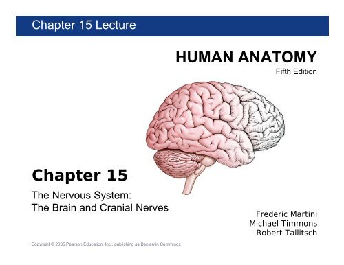
<strong>Ch</strong>apter <strong>15</strong><br />
The Nervous System:<br />
The <strong>Brain</strong> <strong>and</strong> <strong>Cranial</strong> <strong>Nerves</strong><br />
Copyright © 2005 Pearson Education, Inc., publishing as Benjamin Cummings<br />
HUMAN ANATOMY<br />
Fifth Edition<br />
Frederic Martini<br />
Michael Timmons<br />
Robert Tallitsch
The Ventricles of the <strong>Brain</strong><br />
Figure <strong>15</strong>.2a Lateral View Figure <strong>15</strong>.2c Anterior View
The <strong>Cranial</strong> Meninges<br />
Figure <strong>15</strong>.3a Lateral View Figure <strong>15</strong>.3b Superior View
The <strong>Cranial</strong> Meninges<br />
Figure <strong>15</strong>.3c Midsagittal View
The <strong>Cranial</strong> Meninges<br />
Figure <strong>15</strong>.4a Superior Cut away
The <strong>Cranial</strong> Meninges<br />
Figure <strong>15</strong>.4b,c Coronal Section
The Cerebrospinal Fluid<br />
<strong>and</strong> <strong>Ch</strong>oroid Plexus<br />
Figure <strong>15</strong>.5b The <strong>Ch</strong>oroid Plexus <strong>and</strong> Blood–<strong>Brain</strong> Barrier
The Cerebrospinal Fluid<br />
<strong>and</strong> <strong>Ch</strong>oroid Plexus<br />
Figure <strong>15</strong>.6 Circulation of Cerebrospinal Fluid
The Cerebrum<br />
Figure <strong>15</strong>.8a,b The Cerebral Hemispheres, Superior <strong>and</strong> Anterior Views
The Cerebrum<br />
Figure <strong>15</strong>.8c Posterior View Figure <strong>15</strong>.9a Lateral View
Functions of the Cerebrum<br />
Table <strong>15</strong>.2 The Cerebral Cortex
Motor <strong>and</strong> Sensory Areas<br />
of the Cerebral Cortex<br />
Figure <strong>15</strong>.9b Functional Areas of the Cortex
Central White Matter of the <strong>Brain</strong><br />
Figure <strong>15</strong>.10a Lateral View Figure <strong>15</strong>.10b Anterior View
Basal Nuclei<br />
Figure <strong>15</strong>.11b,c Coronal View
Basal Nuclei<br />
Figure <strong>15</strong>.11d,e Horizontal View
The Limbic System<br />
Figure <strong>15</strong>.12a Lateral View Figure <strong>15</strong>.12b Close up
Sectional View Inside the <strong>Brain</strong>:<br />
The Diencephalon<br />
Figure <strong>15</strong>.13a Midsagittal View
Sectional View Inside the <strong>Brain</strong>:<br />
The Diencephalon<br />
Figure <strong>15</strong>.13b Coronal Section
The Diencephalon: Thalamus<br />
Figure <strong>15</strong>.14 The Thalamus, Lateral <strong>and</strong> Deep Views
The Diencephalon: Hypothalamus<br />
Figure <strong>15</strong>.<strong>15</strong>b Deep View Table <strong>15</strong>.7 The Hypothalamus
The Mesencephalon<br />
Figure <strong>15</strong>.16a Lateral View Figure <strong>15</strong>.16c Posterior View
The Pons<br />
Figure <strong>15</strong>.18 The Pons
The Cerebellum<br />
Figure <strong>15</strong>.19a Posterior, Superior Surface
The Cerebellum<br />
Figure <strong>15</strong>.19b Sagittal Section of Cerebellum
The M<strong>edu</strong>lla Oblongata<br />
Figure <strong>15</strong>.20a Anterior View Figure <strong>15</strong>.20b Posterior View
The <strong>Cranial</strong> <strong>Nerves</strong><br />
• <strong>Cranial</strong> nerves are components of the<br />
peripheral nervous system that connect to<br />
the brain rather than to the spinal cord.<br />
– There are twelve pairs of cranial nerves.<br />
– <strong>Cranial</strong> nerves are numbered using Roman<br />
numerals.<br />
• Each cranial nerve attaches to the brain<br />
near the associated sensory or motor<br />
nuclei.
The Origins of the <strong>Cranial</strong> <strong>Nerves</strong><br />
Figure <strong>15</strong>.21a Inferior View
The Olfactory Nerve (N I)<br />
• Primary function:<br />
– Special sensory (smell)<br />
• Origin:<br />
– Receptors of olfactory epithelium<br />
Figure <strong>15</strong>.22 The Olfactory Nerve
The Olfactory Nerve (N I)<br />
• Passes through:<br />
– Cribriform plate of ethmoid<br />
• Destination:<br />
– Olfactory bulbs<br />
Figure <strong>15</strong>.22 The Olfactory Nerve
The Optic Nerve (N II)<br />
• Primary function:<br />
– Special sensory (vision)<br />
• Origin:<br />
– Retina of eye<br />
• Passes through:<br />
– Optic canal of sphenoid<br />
• Destination:<br />
– Diencephalon by way of the optic chiasm
The Optic Nerve (N II)<br />
Figure <strong>15</strong>.23 The Optic Nerve
The Oculomotor Nerve (N III)<br />
• Primary function:<br />
– Motor, eye movements<br />
• Origin:<br />
– Mesencephalon<br />
• Passes through:<br />
– Superior orbital fissure of sphenoid
The Oculomotor Nerve (N III)<br />
• Destination:<br />
– Somatic motor:<br />
• Superior, inferior, <strong>and</strong> medial rectus muscles; the<br />
inferior oblique muscle; the levator palpebrae<br />
superioris muscle<br />
– Visceral motor:<br />
• Intrinsic eye muscles
The Oculomotor Nerve (N III)<br />
Figure <strong>15</strong>.24 The Oculomotor Nerve
The Trochlear Nerve (N IV)<br />
• Primary function:<br />
– Motor, eye movements<br />
• Origin:<br />
– Mesencephalon<br />
• Passes through:<br />
– Superior orbital fissure of sphenoid<br />
• Destination:<br />
– Superior oblique muscle
The Trochlear Nerve (N IV)<br />
Figure <strong>15</strong>.24 The Trochlear Nerve
The Trigeminal Nerve (N V)<br />
• Primary function:<br />
– Mixed (sensory <strong>and</strong> motor)<br />
– Ophthalmic <strong>and</strong> maxillary branches sensory<br />
– M<strong>and</strong>ibular branch mixed
The Trigeminal Nerve (N V)<br />
• Origin:<br />
– Ophthalmic branch (sensory):<br />
• Orbital structures, nasal cavity, skin of forehead,<br />
superior eyelid, eyebrow, <strong>and</strong> part of the nose<br />
– Maxillary branch (sensory):<br />
• Inferior eyelid, upper lip, gums, <strong>and</strong> teeth; cheek;<br />
nose, palate, <strong>and</strong> part of the pharynx<br />
– M<strong>and</strong>ibular branch (mixed):<br />
• Sensory from lower gums, teeth, <strong>and</strong> lips; palate<br />
<strong>and</strong> tongue (part); motor from motor nuclei of pons
The Trigeminal Nerve (N V)<br />
• Passes through:<br />
– Ophthalmic branch through superior orbital<br />
fissure<br />
– Maxillary branch through foramen rotundum<br />
– M<strong>and</strong>ibular branch through foramen ovale<br />
• Destination:<br />
– Ophthalmic, maxillary, <strong>and</strong> m<strong>and</strong>ibular<br />
branches to sensory nuclei in the pons<br />
– M<strong>and</strong>ibular branch also innervates muscles of<br />
mastication
The Trigeminal Nerve (N V)<br />
Figure <strong>15</strong>.25 The Trigeminal Nerve
The Abducens Nerve (N VI)<br />
• Primary function:<br />
– Motor, eye movements<br />
• Origin:<br />
– Pons<br />
• Passes through:<br />
– Superior orbital fissure of sphenoid<br />
• Destination:<br />
– Lateral rectus muscle
The Abducens Nerve (N VI)<br />
Figure <strong>15</strong>.2 The Abducens Nerve
The Facial Nerve (N VII)<br />
• Primary function:<br />
– Mixed (sensory <strong>and</strong> motor)<br />
• Origin:<br />
– Sensory from taste receptors on anterior twothirds<br />
of tongue<br />
– Motor from motor nuclei of pons<br />
• Passes through:<br />
– Internal acoustic meatus of temporal bone,<br />
along facial canal to reach stylomastoid<br />
foramen
The Facial Nerve (N VII)<br />
• Destination:<br />
– Sensory to sensory nuclei of pons<br />
– Somatic motor: muscles of facial expression<br />
– Visceral motor: lacrimal (tear) gl<strong>and</strong> <strong>and</strong> nasal<br />
mucous gl<strong>and</strong>s via pterygopalatine ganglion;<br />
subm<strong>and</strong>ibular <strong>and</strong> sublingual salivary gl<strong>and</strong>s<br />
via subm<strong>and</strong>ibular ganglion
The Facial Nerve (N VII)<br />
Figure <strong>15</strong>.26 The Facial Nerve
The Vestibulocochlear Nerve (N VIII)<br />
• Primary function:<br />
– Special sensory:<br />
• Origin:<br />
• Balance <strong>and</strong> equilibrium (vestibular branch) <strong>and</strong><br />
hearing (cochlear branch)<br />
– Receptors of the inner ear (vestibule <strong>and</strong><br />
cochlea)<br />
• Passes through:<br />
– Internal acoustic meatus of the temporal bone
The Vestibulocochlear Nerve (N VIII)<br />
• Destination:<br />
– Vestibular <strong>and</strong> cochlear nuclei of pons <strong>and</strong><br />
m<strong>edu</strong>lla oblongata
The Vestibulocochlear Nerve (N VIII)<br />
Figure <strong>15</strong>.27 The Vestibulocochlear Nerve
The Glossopharyngeal Nerve (N IX)<br />
• Primary function:<br />
– Mixed (sensory <strong>and</strong> motor)<br />
• Origin:<br />
– Sensory from posterior one-third of the<br />
tongue, part of the pharynx <strong>and</strong> palate, the<br />
carotid arteries of the neck<br />
– Motor from motor nuclei of m<strong>edu</strong>lla oblongata<br />
• Passes through:<br />
– Jugular foramen between occipital <strong>and</strong><br />
temporal bones
The Glossopharyngeal Nerve (N IX)<br />
• Destination:<br />
– Sensory fibers to sensory nuclei of m<strong>edu</strong>lla<br />
oblongata<br />
– Somatic motor:<br />
• Pharyngeal muscles involved in swallowing<br />
– Visceral motor:<br />
• Parotid salivary gl<strong>and</strong>, after synapsing in the otic<br />
ganglion
The Glossopharyngeal Nerve (N IX)<br />
Figure <strong>15</strong>.28 The Glossopharyngeal Nerve
The Vagus Nerve (N X)<br />
• Primary function:<br />
– Mixed (sensory <strong>and</strong> motor)<br />
• Origin:<br />
– Visceral sensory from pharynx (part), auricle,<br />
external acoustic meatus, diaphragm, <strong>and</strong><br />
visceral organs in thoracic <strong>and</strong><br />
abdominopelvic cavities<br />
– Visceral motor from motor nuclei in the<br />
m<strong>edu</strong>lla oblongata
The Vagus Nerve (N X)<br />
• Passes through:<br />
– Jugular foramen between occipital <strong>and</strong><br />
temporal bones<br />
• Destination:<br />
– Sensory fibers to sensory nuclei <strong>and</strong><br />
autonomic centers of m<strong>edu</strong>lla oblongata<br />
– Somatic motor to muscles of the palate <strong>and</strong><br />
pharynx<br />
– Visceral motor to respiratory, cardiovascular,<br />
<strong>and</strong> digestive organs in the thoracic <strong>and</strong><br />
abdominal cavities
The Vagus Nerve (N X)<br />
Figure <strong>15</strong>.29 The Vagus Nerve
The Accessory Nerve (N XI)<br />
• Primary function:<br />
– Motor<br />
• Origin:<br />
– Motor nuclei of spinal cord <strong>and</strong> m<strong>edu</strong>lla<br />
oblongata<br />
• Passes through:<br />
– Jugular foramen between occipital <strong>and</strong><br />
temporal bones
The Accessory Nerve (N XI)<br />
• Destination:<br />
– Internal branch innervates voluntary muscles<br />
of palate, pharynx, <strong>and</strong> larynx<br />
– External branch controls sternocleidomastoid<br />
<strong>and</strong> trapezius muscles
The Accessory Nerve (N XI)<br />
Figure <strong>15</strong>.30 The Accessory Nerve
The Hypoglossal Nerve (N XII)<br />
• Primary function:<br />
– Motor, tongue movements<br />
• Origin:<br />
– Motor nuclei of the m<strong>edu</strong>lla oblongata<br />
• Passes through:<br />
– Hypoglossal canal of occipital bone<br />
• Destination:<br />
– Muscles of the tongue
The Hypoglossal Nerve (N XII)<br />
Figure <strong>15</strong>.30 The Hypoglossal Nerve